
Systems and methods for assisting cardiac valve coaptation

Inactive Publication Date: 2006-01-05
ETHICON INC
View PDF23 Cites 98 Cited by
  • Summary
  • Abstract
  • Description
  • Claims
  • Application Information

AI Technical Summary

Benefits of technology

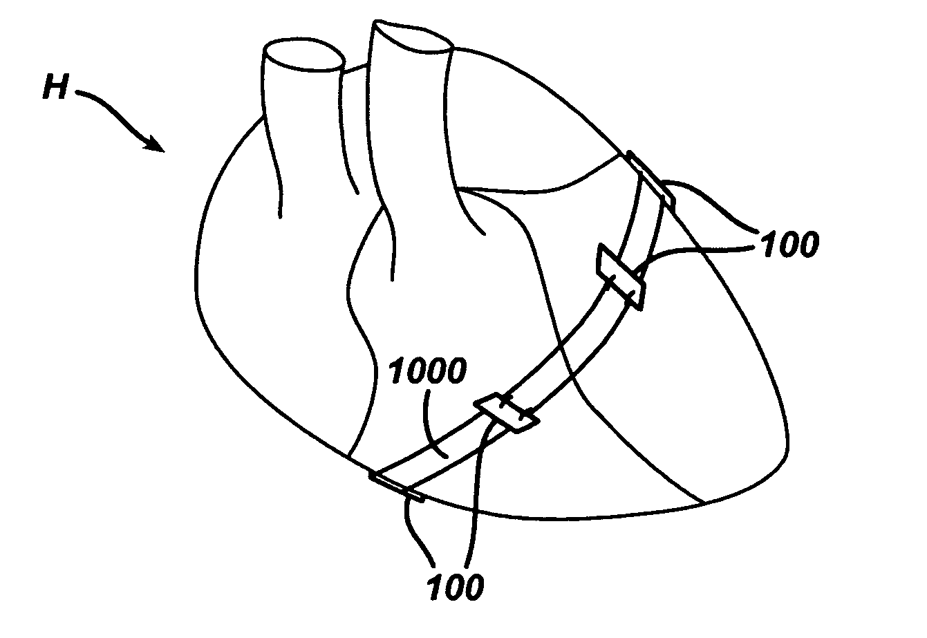
[0015] In some embodiments of the systems and methods of the invention, the cardiac assist device, for example, is a strap that wraps around a circumference of a designated portion of the heart whereat distention is to be reduced. Once in the desired position about the heart, ends of the strap are pulled to tighten the strap. The tightened strap is then secured to one or more of the anchoring devices to maintain the strap in the desired position. Tightening and securing the strap in this manner supports the heart at a specified location that helps to reduce the propensity of that portion of the heart to distend. At the same time, the tightened and secured strap aids the alignment and coaptation of one or more of the cardiac valves of the heart due to the localized position of the strap around the heart. In various embodiments of the systems and methods of the invention, the ends of the strap are secured to one another in addition to being secured to one or more of the anchoring devices. The artisan should appreciate that additional straps may be used in similar manner to reduce distention of other portions of the heart and to further aid alignment and coaptation of one or more of the cardiac valves.
[0016] In other embodiments of the systems and methods of the invention, the cardiac assist device is comprised of a bio-compatible material as disclosed in co-pending U.S. patent application Ser. Nos. ______ (Attorney Docket Nos. 17385, 17386, respectively) referenced above. For example, the material of the cardiac assist device may comprise an open first end with a seam about its periphery and a second end opposite the first end as disclosed in co-pending U.S. patent application Ser. No. ______ (Attorney Docket No. 17385) referenced above. The second end may be closed to form a sack-like cardiac assist device, or open to form a tubular-like cardiac device. In either case, the heart is received in the material of the device until an apex of the heart approaches the second end of the cardiac assist device and the seam at the first end of the material is positioned around the circumference of that portion of the heart whereat distention is to be reduced. The seam is then tightened and secured to one or more of the anchoring devices already secured to the heart. The tightening and securing of the seam in this manner reduces distention of that portion of the heart and aids the alignment and coaptation of one or more of the cardiac valves in that localized area of the heart. Increased cardiac efficiency and output may be achieved as a result.
[0017] In other embodiments of the systems and methods of the invention, the seam may instead comprise a drawstring. The drawstring may be incorporated into the material of the cardiac assist device, for example, as in co-pending U.S. patent application Ser. No. (Attorney Docket No. 17385) referenced above. The drawstring is tightened and secured to one or more of the anchoring devices to aid the alignment and coaptation of one or more of the cardiac valves of the heart due to the localized position of the drawstring around the heart. In this manner, distention is decreased and better coaptation of the intended one or more cardiac valves tends to be achieved
[0020] The anchoring devices are separately secured to the heart using the instruments and visualization means inserted through the plurality of ports, or openings, in the chest wall of the patient or subject. Thereafter, the cardiac assist device is manipulated to a desired position about portions of the heart. The cardiac assist device is then tightened and secured to one or more of the anchoring devices in order to maintain the cardiac assist device in the specified location about the heart. By tightening and securing the cardiac assist device at a localized portion of the heart, distention of that portion of the heart may be reduced and alignment of a targeted one or more of the cardiac valves in that portion of the heart may be achieved. Increased cardiac output and efficiency tends to occur as a result.
[0022] The various embodiments of the system and methods of the invention described herein provide several advantages over prior known devices and methods of applying a cardiac assist device to a heart. The various embodiments of the system and methods according to the invention minimize the time and cumbersome efforts previously required to fit and suture a cardiac assist device around an entire circumference of the heart. The various embodiments of the systems and methods according to the invention target a localized area of the heart to reduce the propensity of the heart to distend in that area. As a result, the systems and methods of the invention simultaneously aids alignment and coaptation of cardiac valves when the cardiac assist device is tightened and secured to one or more of the anchoring devices that are separately secured to the heart. Increased cardiac output and efficiency is rendered more likely as a result. Further, the various embodiments of the systems and methods of the invention may be performed endoscopically, even during beating heart procedures, which renders a precise fit of the cardiac assist device and optimal alignment of the intended cardiac valves more likely.

Problems solved by technology

The disease of heart failure is common, lethal, and expensive to treat.
Heart failure is also the most common cause of hospitalization for patients 65 years and older in the United States.
The mortality rate is 50% at five years for patients diagnosed with heart failure, and to date, there are limited treatment alternatives available.
Heart failure typically occurs when a weakened heart cannot pump an adequate amount of blood to meet the demands of the body's other organs and tissues.
Malfunction of any of the valves within the heart can cause problems effecting the entire circulatory system.
A leaky valve, for example, does not close properly and permits blood to flow backward as the heart contracts.
Overworking the heart in this manner damages the muscle of the heart and often masks underlying valvular disfunction.
Of the cardiac valves, the mitral valve is most often compromised as the powerful pumping action of the left ventricle tends to cause systolic outward bulging of an ischemic region of the left ventricle free wall.
Though not as prevalent, the tri-cuspid valve is also susceptible to misalignment and malfunction due to the less aggressive pumping action of the right ventricle, which pumps blood to the lungs.
In either case, coaptation of the leaflets of the cardiac valves is compromised, and cardiac output and efficiency is decreased.
Ultimately, a complex process of damaging structural and functional changes to the heart results.
Any damage or impairment in function of any of these components can render the valve structure incompetent.
While being generally successful and routine in surgical practice today, these procedures are also costly, highly invasive and have high morbidity and mortality rates associated therewith.
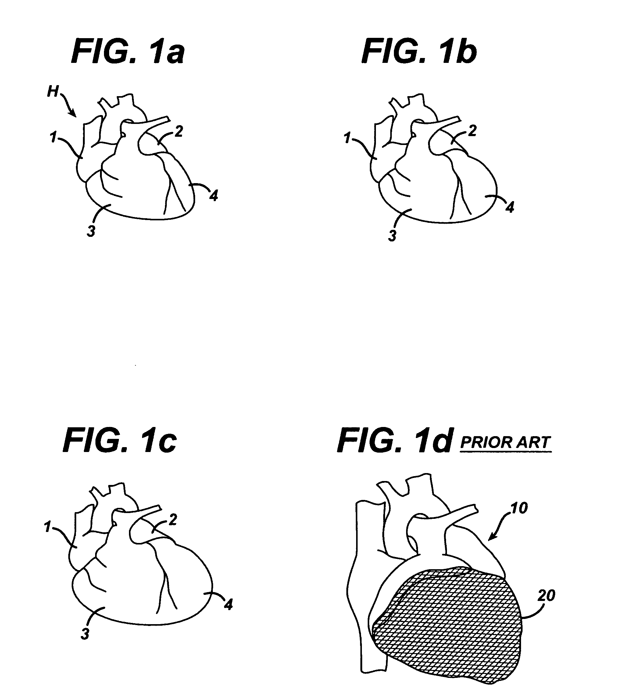
The jacket, however, does not address localized valve mis-alignment and coaptation concerns that often occur due to myocardial over-stretch and wall stress.
Moreover, suturing the jacket 20 around a circumference of the heart is a time-consuming and cumbersome procedure.
Further still, precise positioning and securing of the jacket about the heart using endoscopic tools and techniques as the heart is beating has proved challenging.

Method used


Image

  • Systems and methods for assisting cardiac valve coaptation
  • Systems and methods for assisting cardiac valve coaptation
  • Systems and methods for assisting cardiac valve coaptation

Examples


Embodiment Construction

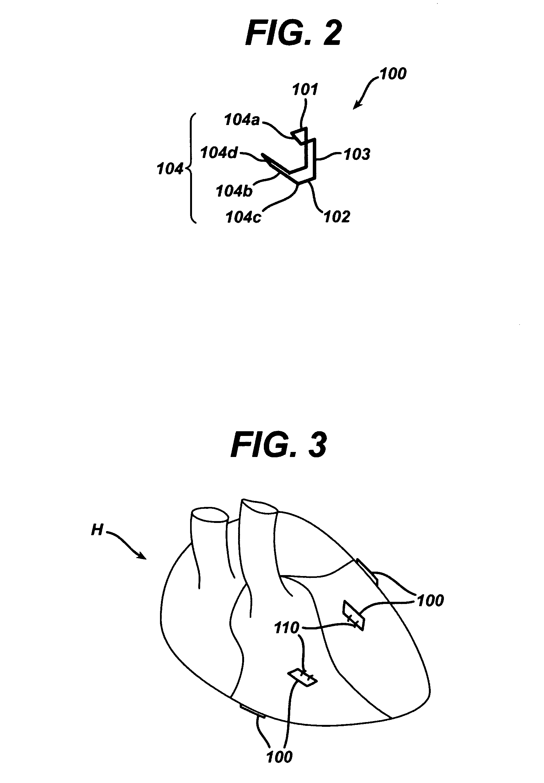
[0036]FIGS. 2-4 illustrate anchoring devices 100 according to the systems and methods of the invention. The anchoring devices 100 are placed on and secured to the heart at designated locations. The anchoring devices may be secured to the heart using sutures 110, u-clips, staples, or other securing means known in the art. The anchoring devices are similar to those anchoring devices disclosed in co-pending U.S. patent application Ser. Nos. ______ (Attorney Docket Nos. 17385 and 17386) referenced above.

[0037] As shown most clearly in FIG. 2, each anchoring device 100 is a frame-like member comprised of a top 101, a bottom 102, a back 103, and a front 104. The front 104 is further comprised of a lip 104a, and pivotable flange 104b. The pivotable flange 104b has a hinged end 104c attached to the bottom 102 of the anchoring device, and a free end 104d opposite the hinged end. The pivotable flange 104b engages the lip 104a, when the pivotable flange 104b is rotated about the hinged end 10...


PUM


Abstract

Systems and methods for reducing distention in designated portions of a heart muscle while assisting cardiac valve alignment and coaptation. Anchoring devices are secured to the heart muscle and a bio-compatible cardiac assist device placed over portions of the heart muscle is separately secured to the anchoring devices. The cardiac assist device may be a material that wraps portions of the heart muscle, or may be straps that wrap portions of the heart muscle. Tightening and securing the cardiac assist device in place helps to reduce distention in chambers of the heart muscle and aids alignment and coaptation of cardiac valves.

Description

BACKGROUND OF THE INVENTION [0001] 1. Field of the Invention [0002] The invention generally relates to systems and methods for increasing cardiac output and efficiency. More specifically, the invention relates to systems and methods for reducing distention of one or more chambers of the heart and / or aligning one or more cardiac valves for better closure and coaptation thereof. [0003] 2. Related Art [0004] Heart failure syndrome is a highly debilitating and degenerative disorder resulting from damage to the heart muscle. The damage to the heart muscle may be caused by a number of conditions, including coronary artery disease, long standing hypertension, leaky heart valve(s), and infections. [0005] The disease of heart failure is common, lethal, and expensive to treat. An estimated 5.1 million Americans have heart failure with approximately 500,000 new cases diagnosed each year. In 1999, an estimated $20.3 billion in directs costs were spent for the care of heart failure patients. Hea...

Claims


Application Information

Patent Timeline
IPC IPC(8): A61F13/00A61B17/08
CPCA61B17/00234A61B17/0401A61B17/0487A61B17/06166A61B17/0682A61F2/2481A61B2017/00243A61B2017/0458A61B2017/0461A61B2017/0464A61B2017/06176A61B17/083
Inventor KUTE, STEPHANIEWU, STEVEN ZUNG-HONG
Owner ETHICON INC
Learn More