Method and device for surgical ventricular repair

a ventricular repair and ventricular valve technology, applied in the field of surgical ventricular repair, can solve the problems of ischemic muscle no longer being able to contract, affecting the limited pumping action available, and blood pressure tending to develop a bulge or expansion of the chamber, so as to reduce stress on the heart muscle, improve the surgical outcome, and inhibit the remodeling of akinetic and/or dyskinetic heart tissu

Inactive Publication Date: 2006-02-02
CAPITAL SOUTHWEST
View PDF6 Cites 250 Cited by
  • Summary
  • Abstract
  • Description
  • Claims
  • Application Information

AI Technical Summary

Benefits of technology

[0024] The procedure, by excluding much, if not all, of the akinetic and dyskinetic tissue while allowing the surgeon to create the proper shape with an apex, significantly reduces stress on the heart muscle and improves surgical outcome. The procedure, by being done with a precise device allows the surgeon to make the procedure repeatable and reliable. The device takes the variation out of the procedure.
[0025] In some embodiments, a system and/or method may be directed to supporting and or to reinforcing myocardial tissue in the region of an infarct site. The system and/or method may

Problems solved by technology

The ischemic muscle is no longer capable of contracting so it may not contribute to either squeezing or twisting motion required to pump blood.
In severe cases the akinetic tissue, which is not capable of contracting, is in fact elastic so that blood pressure tends to develop a bulge or expansion of the chamber.
This is particularly detrimental to the limited pumping action available, as the heart loses even more of its energy to pumping the bulge instead of the blood.
As a consequence, the fibers are poorly oriented to accomplish even the squeezing action, as the lines of force become less perpendicular to the heart wall.
The spherical shape further reduces pumping efficiency since the fibers which normally encircle the apex facilitate writhing are changed to a

Method used


Image

  • Method and device for surgical ventricular repair
  • Method and device for surgical ventricular repair
  • Method and device for surgical ventricular repair

Examples


Embodiment Construction

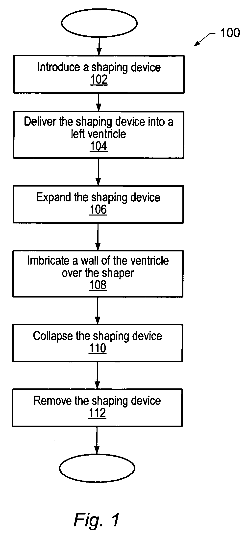
[0094] Turning to FIG. 1, there is presented an overview method 100 for performing and using an embodiment. Method 100 may use the following components: a shaping device, a patch, and / or a stapling device.

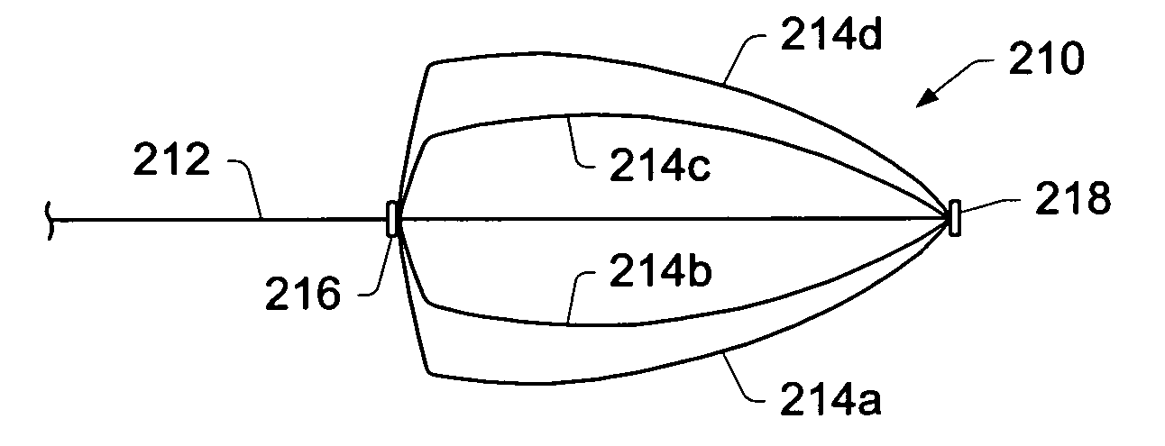
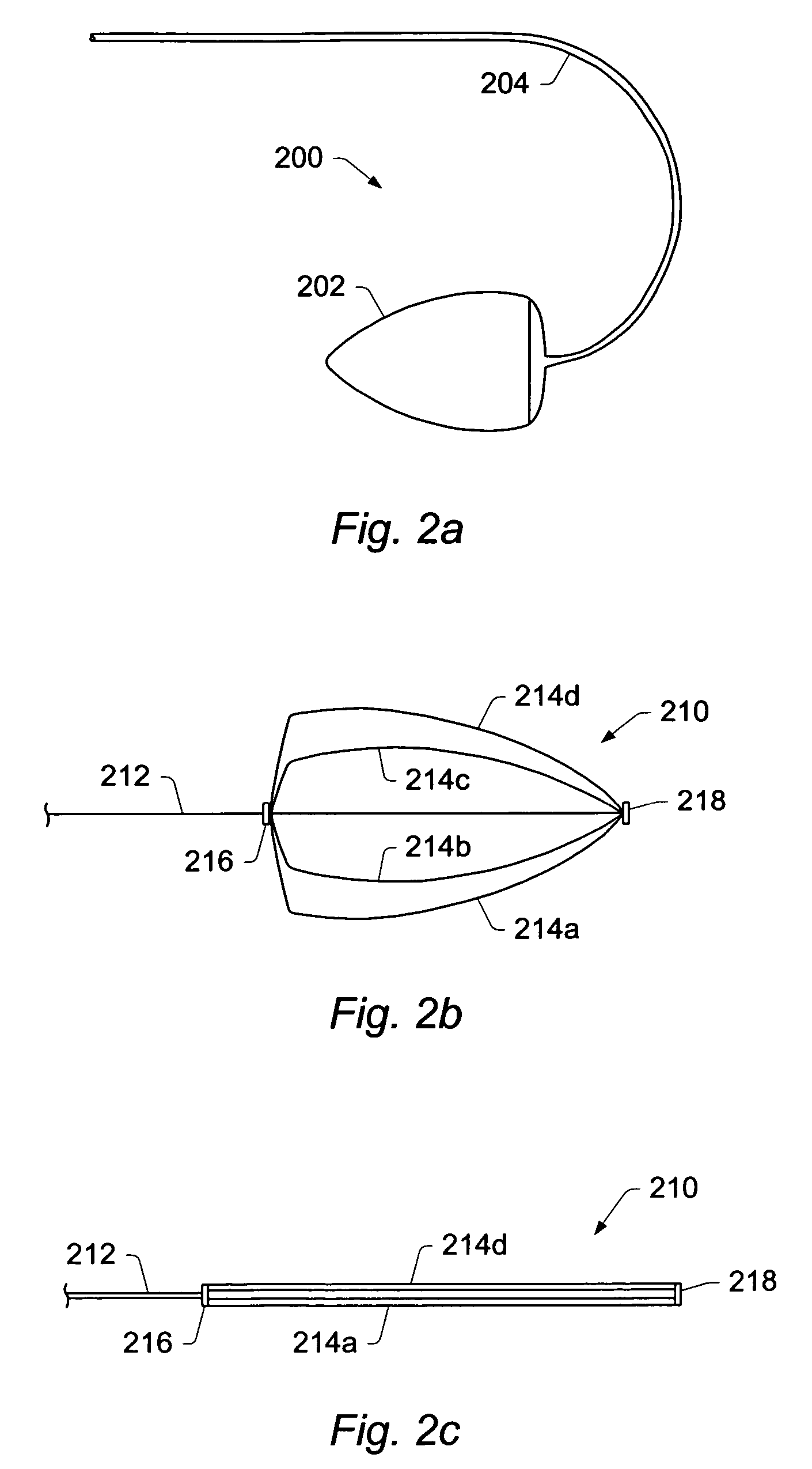
[0095] In some embodiments, a shaping device may be pre-shaped to generally model the appropriate volume and shape of the left ventricle, as is depicted in FIG. 2a. Shaping device 200 may be used as a guide in reforming the left ventricle so that the reconstructed heart may be formed closer to the size and shape of the pre-enlarged heart. Consequently, the heart performs better post operatively than with conventional methods. As illustrated in FIG. 2a, shaping device 200 may be conical or “tear drop” in shape. The length of shaping device 200 may vary with each patient and will typically be a function of the volume selected for the shaping device. The size, shape, and / or volume of shaping device 200 may vary according to individual patient specific needs. Shaping device 200 may be...


PUM


Abstract

Embodiments disclose a method for repairing a heart of a human. A method may include introducing a collapsed reinforcing element through the skin into the vascular system of the human. The method may include delivering the reinforcing element into a left ventricle through the arteries. Once inside the left ventricle, the reinforcing element may be expanded to an expanded shape. In certain embodiments, a reinforcing element may be used to structurally reinforce a portion of an endocardial surface of a heart. The reinforcing element may include a preshaped patch and/or a plurality of preshaped flexible conduits. The method may include deploying the reinforcing element soon after a myocardial infarction to inhibit naturally occurring remodeling of the heart. The reinforcing element may be deployed with or without the use of a shaper. In some embodiments, a reinforcement element may be positioned on/coupled to an external surface of a human heart. In some embodiments, a reinforcing element may include an externally positioned apparatus configured to substantially reshape a portion of an interior chamber of a heart.

Description

PRIORITY CLAIM [0001] This application is a continuation-in-part of U.S. patent application Ser. No. 10 / 790,669 entitled “METHOD AND DEVICE FOR PERCUTANEOUS SURGICAL VENTRICULAR REPAIR” filed on Mar. 1, 2004, which is a continuation-in-part of U.S. patent application Ser. No. 10 / 235,295 entitled “METHOD AND DEVICE FOR PERCUTANEOUS SURGICAL VENTRICULAR REPAIR” filed on Sep. 5, 2002, which claims priority to U.S. Provisional Patent Application Ser. No. 60 / 317,197 entitled “DEVICE AND METHOD FOR ENDOSCOPIC SURGICAL VENTRICULAR REPAIR” filed on Sep. 5, 2001, and U.S. Provisional Patent Application Ser. No. 60 / 327,221 entitled “METHOD AND DEVICE FOR CLOSED CHEST PLACEMENT OF SEPTUM” filed on Oct. 5, 2001, the disclosures of which are hereby incorporated by reference.BACKGROUND OF THE INVENTION [0002] 1. Field of the Invention [0003] This invention relates generally to surgical methods and apparatuses for performing surgical ventricular repair endoscopically and / or through a minimally inv...

Claims


Application Information

Patent Timeline
IPC IPC(8): A61M29/00
CPCA61B17/0057A61B2017/00243A61B2017/00575A61F2/2487A61B2017/00615A61B2017/00632A61F2/2481A61B2017/00579
Inventor SURESH, MITTA
Owner CAPITAL SOUTHWEST
Why Eureka
Try Eureka