Methods of classifying, diagnosing, stratifying and treating cancer patients and their tumors

a cancer patient and tumor technology, applied in the field of cancer patients and tumors, can solve the problems of inability to detect diagnostic tools, inability to predict the clinical behavior of breast tumors, and serious limitations in the approach

Inactive Publication Date: 2006-02-23
APPL GENOMICS INC +1
View PDF2 Cites 25 Cited by
  • Summary
  • Abstract
  • Description
  • Claims
  • Application Information

AI Technical Summary

Benefits of technology

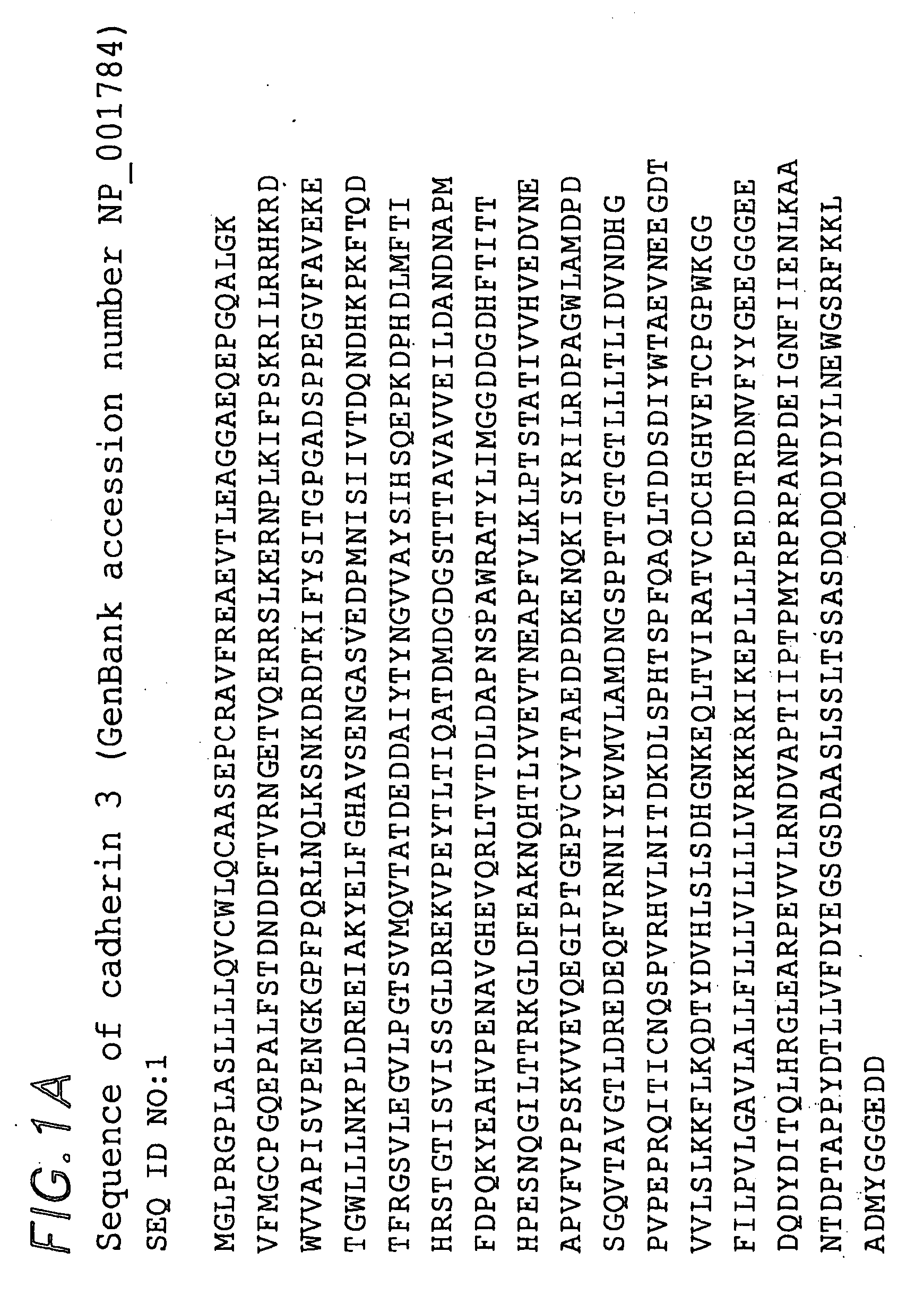
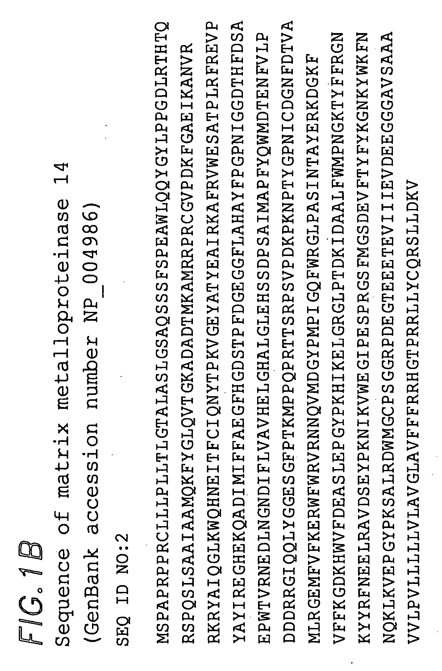
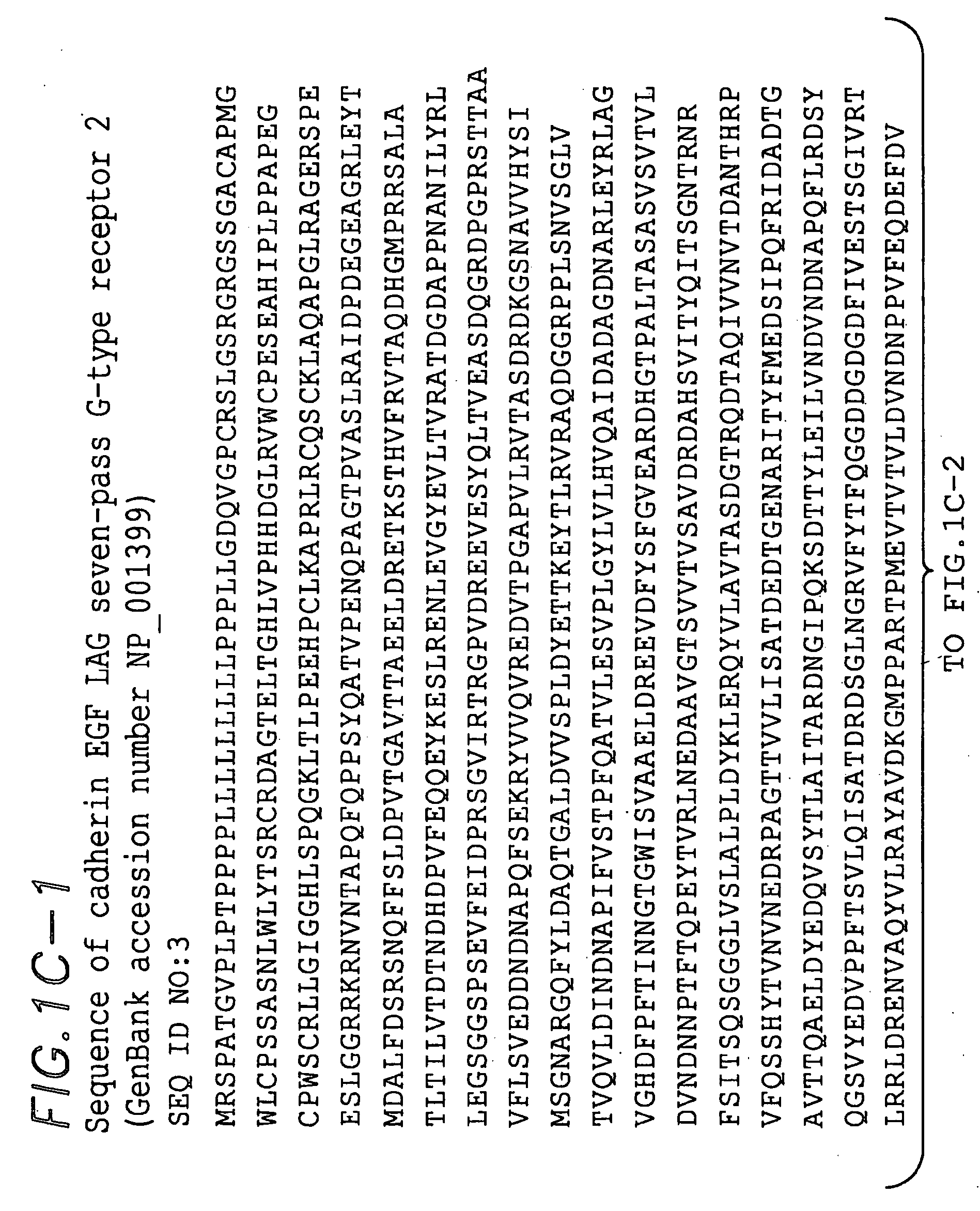
[0009] The present invention relates to the identification of markers that are useful in classifying tumors, particularly breast tumors. The markers identify a class of tumors whose cells have characteristics of basal cells of normal breast lactation ducts. The markers were identified based on their expression profiles in human breast tumor samples, normal breast tissue, and cell lines as assessed using cDNA microarrays. In particular, the basal cell markers of the present invention were identified based on the similarity of their mRNA expression patterns to the expression patterns of markers previously known to identify breast duct basal cells, e.g., cytokeratin 5 and cytokeratin 17, across a set of breast tumor samples. The basal markers include the three genes known as cadherin 3 or P-cadherin (SEQ ID NO:1; GenBank protein accession number NP—001784; GenBank cDNA accession number NM—001793), matrix metalloproteinase 14 (SEQ ID NO:2; GenBank protein accession number NP—004986; GenBank cDNA accession number NM—004995); and cadherin EGF LAG seven-pass G-type receptor 2 or EGF-Like Domain, Multiple 2 (SEQ ID NO:3; GenBank protein accession number NP—001399; GenBank cDNA accession number NM—001408). The invention further provides antibodies that specifically bind to the polypeptides encoded by the basal marker genes identified herein. The antibodies recognize basal cells of normal mammary lactation glands.

Problems solved by technology

A major challenge of cancer treatment is to target specific therapies to distinct tumor types in order to maximize efficacy and minimize toxicity.
A related challenge lies in the attempt to provide accurate diagnostic, prognostic, and predictive information.
However, this approach has serious limitations.
Though useful, these analyses only in part predict the clinical behavior of breast tumors.
There is phenotypic diversity present in breast cancers that current diagnostic tools fail to detect.
However, inappropriate activity or expression of oncogenes can lead to the uncontrolled cell proliferation that is a feature of cancer.
Loss of their activity, e.g., due to mutations or decreased expression at the level of mRNA or protein, can lead to unrestrained cell division.
In particular, understanding of the underlying differences in gene expression that may contribute to tumor phenotype is limited.
Rather than specifically killing tumor cells, these agents target any dividing cell, resulting in a variety of adverse effects.
In addition, current therapeutic strategies are of limited efficacy, and the mortality rate of breast cancer remains high.

Method used


Image

  • Methods of classifying, diagnosing, stratifying and treating cancer patients and their tumors
  • Methods of classifying, diagnosing, stratifying and treating cancer patients and their tumors
  • Methods of classifying, diagnosing, stratifying and treating cancer patients and their tumors

Examples


example 1

Preparation of Microarrays Containing 8498 Human cDNAs

[0205] The human cDNA clones used in this study were obtained from Research Genetics (Huntsville Ala., USA) as bacterial colonies in 96-well microtiter plates. The clones were chosen from a set of 15,000 cDNA clones that corresponded to the Research Genetics Human Gene Filters sets GF200-202 (www.resgen.com / ). These clones form part of a set of clones assembled by the I.M.A.G.E. consortium (Lennon, G. G., Auffray, C., Polymeropoulos, M., Soares, M. B. The I.M.A.G.E. Consortium: An Integrated Molecular Analysis of Genomes and their Expression. Genomics 33:151-152, 1996) and are identified by I.M.A.G.E. clone ID numbers. All clones printed on these arrays were sequence validated as part of a product offered at Research Genetics, Inc. We estimate that greater than 97% of the clones on the array are correctly identified.

[0206] A detailed protocol for the production of the cDNA microarrays used in this study is available at cmgm.sta...

example 2

Cell Lines, Breast Tissue, and Breast Tumor Samples for Microarray Analysis and Preparation of mRNA Samples

Common Reference Sample

[0242] Each of the 84 experimental samples tested here was analyzed by a comparative hybridization, using a common reference RNA pool as a standard; this reference sample was composed of equal mixtures of mRNA isolated from 11 established cell lines derived from human tissue (MCF7, Hs578T, OVCAR3, HepG2, NTERA2, MOLT4, RPMI-8226, NB4+ATRA, UACC-62, SW872, and Colo205: also see Table 3 for more details). The 11 cell lines were all grown to 70-90% confluence in RPMI medium, containing 10% Fetal Calf Serum and Penicillin / Streptomycin. The cells were harvested either by scraping or centrifugation, quickly resuspended in RNA lysis buffer and mRNA prepared using the FastTrack™ 2.0 mRNA Isolation Kit (Invitrogen, Carlsbad, Calif.) according to the manufacturer's instructions. In each case, multiple individual mRNA preparations were collected for each cell lin...

example 3

Characterization of Breast Tissue and Tumor Samples

[0246] For all but two of the tumor specimens (i.e. New York 1 and New York 2), the mutational status of the TP53 gene was determined using published methods (Aas, T., et al.).

[0247] A single pathologist (applicant Matt van de Rijn) reviewed hematoxylin and eosin (H&E) sections of each tumor, including all before and after pairs, and made a histological evaluation of each while blinded to the source. Tumors were graded using a modified version of the Bloom-Richardson method (Robbins, P., et al., Hum Pathol, 26, 873-879, 1995). These data are displayed in Appendix H, Table 4. Representative H&E sections of each tumor are posted on Applicants' website at genome-www.stanford.edu / molecularportraits / .

[0248] Immunohistochemistry was performed as described previously (Perou, C., et al., 1999; Bindl, J. and Warnke, R., Am J Clin Pathol, 85, 490-493, 1986, and Natkunam, Y., et al., Am. J. Path., 156 (1), 2000). The antibodies used include...


PUM

PropertyMeasurementUnit
pHaaaaaaaaaa
pHaaaaaaaaaa
diameteraaaaaaaaaa

Abstract

The invention provides a variety of reagents for use in the diagnosis and management of cancer, particularly breast cancer. cDNA microarray technology was used to identify genes whose expression profile across a large group of tumor samples correlates with that of cytokeratin 5 and cytokeratin 17, markers for basal cells of the normal mammary lactation gland. The invention demonstrates that tumors that express cytokeratin 5/6 and/or 17 have a poor prognosis relative to tumors overall. The invention provides basal marker genes and their expression products and uses of these genes for diagnosis of cancer and for identification of therapies for cancer. In particular, the invention provides basal marker genes including cadherin3, matrix metalloproteinase 14, and cadherin EGF LAG seven-pass G-type receptor 2. The invention provides antibodies to the polypeptides expressed by these genes and methods of use thereof.

Description

CROSS-REFERENCE TO RELATED APPLICATIONS [0001] This application is a divisional application of U.S. Ser. No. 09 / 916,849, filed Jul. 26, 2001 which claims priority to provisional application U.S. Ser. No. 60 / 220,967, filed Jul. 26, 2000, each of which is incorporated herein by reference.GOVERNMENT SUPPORT [0002] The U.S. Government has a paid-up license in this invention and the right in limited circumstances to require the patent owner to license others on reasonable terms as provided for by the terms of Grant No. NIH CA 77097 awarded by the National Cancer Institute.REFERENCE TO MATERIAL PRESENTED IN APPENDICES [0003] This patent application refers to material comprising tables and data that were presented as appendices on CD-ROM in parent application U.S. Ser. No. 09 / 916,849, filed Jul. 26, 2001. A duplicate CD-ROM is not being presented with this divisional application; instead the entire contents of Appendices A-H are incorporated herein by reference via parent application U.S. ...

Claims


Application Information

Patent Timeline
Patent Type & Authority Applications(United States)
IPC IPC(8): C12Q1/68G01N33/574A61K38/00A61K39/00C07K14/47C07K16/18
CPCA61K38/00C07K16/18C07K14/4748
Inventor BOTSTEIN, DAVIDBROWN, PATRICKPEROU, CHARLESRING, BRIANROSS, DOUGLASSEITZ, ROBVAN DE RIJN, JAN
Owner APPL GENOMICS INC
Why Eureka
Try Eureka