

Reconstituted human breast tumor model
a human breast tumor and reconstructed technology, applied in the field of molecular biology and oncology, can solve the problems of significant differences in the properties and behavior of xenografted cells, human cells maintained in culture as distinct cell lines, etc., and achieve the effect of propagate the tumor
- Summary
- Abstract
- Description
- Claims
- Application Information
AI Technical Summary
Benefits of technology
Problems solved by technology
Method used
Image

Examples
example 1
Construction of Breast Tumor Model
[0038] Human Tissues and Cell Lines
[0039] Fresh human breast tissue from reduction mammoplasty surgical procedures was obtained from Dr. Andrea Richardson at the Brigham and Women's Hospital, Boston, Mass., in compliance with institutional guidelines and with IRB approval. The fresh tissue was digested overnight at 37° C. using 2.8 mg / ml collagenase and 0.6 mg / ml Hyaluronidase in HMEC medium (F12 medium containing 5% fetal bovine serum, 10 ug / ml insulin, 10 ng / ml EGF, 10 μg / ml Hydrocortisone, 50 U / ml penicillin, 50 ug / ml streptomycin and 0.25 μg / ml Fungizone). The following morning, the digested human breast tissue, consisting of epithelial organoids (clusters of about 10 to 1,000 epithelial cells) and primary fibroblasts, was collected by centrifugation at 1000 rpm for 5 minutes and then washed three times in PBS+5% FBS. The mixture of epithelial organoids and primary fibroblasts were frozen in freezing medium (10% DMSO, 15% fetal bovine serum, 1...
example 2
Efficiency of Lentivirus Infection and RNAi
[0055] To determine the efficiency of lentivirus infection of human mammary epithelial cells, a lentivirus construct was generated in which KRAS was placed behind the CMV promoter and EYFP (enhanced green fluorescent protein) was placed behind the SV40 promoter (pLenti-CMV-KRAS+SV40-EYFP). Human mammary epithelial cell organoids were infected with lentivirus expressing pLenti-CMV-KRAS+SV40-EYFP. The organoids were cultured for 3 more days after infection. Judging by the percentage of green fluorescent cells when observed under UV light, we concluded that about 50% of the human mammary epithelial cells were infected with pLenti-CMV-KRAS+SV40-EYFP lentivirus. Similarly, about 10% of the human mammary epithelial cells were infected with pLenti-CMV-ErbB2+SV40-EYFP lentivirus in a separate experiment. Therefore, depending on the titer of the virus used, about 10% to about 50% of the human mammary epithelial cells were infected with lentiviruses...
example 3
Transduction with p53 RNAi and ErbB2 or KRAS
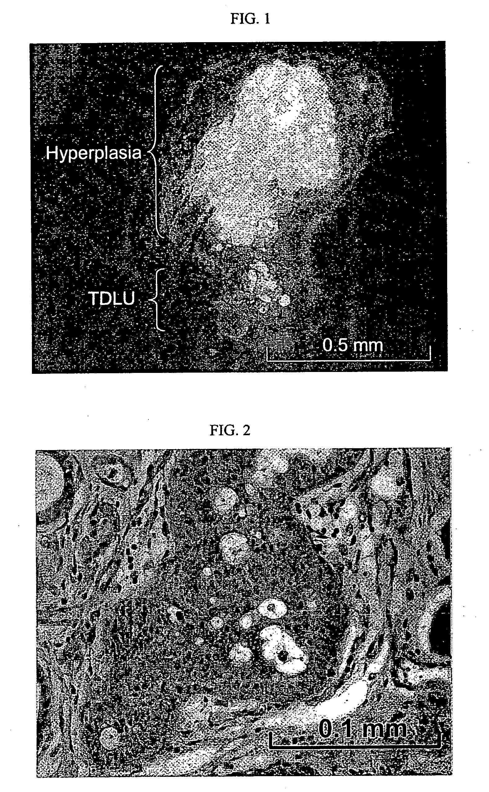
[0057] Human mammary epithelial cells transduced with p53 RNAi and ErbB2 or KRAS developed hyperplasia in the reconstituted human in mouse mammary gland. As a first step towards generating human breast tumor model in mouse, we transduced human mammary epithelial cells with p53RNAi plus either ErbB2V659E or KRASG12V, through lentivirus transduction. The infected human mammary epithelial cells were either mixed with human breast fibroblasts prior to injection or injected alone into fat pads that had been injected with immortalized human fibroblasts two weeks previously. In all combinations, normal, e.g. terminal ductual lobular unit, and hyperplastic human breast structures developed at the implantation sites between one to twelve month after implantation (FIGS. 1 and 2). However, no tumor developed from the transduced human mammary epithelial cells.
PUM
 Abstract
Description
Claims
Application Information