Three dimensional bioengineered smooth muscle tissue and sphincters and methods therefor

a bioengineered smooth muscle and cell ring technology, applied in the field of three dimensional bioengineered smooth muscle cell ring and three dimensional bioengineered sphincter, can solve the problems of slow contractile response of skeletal and smooth muscle of sphincter, lack of cell-cell interaction and cell-matrix interaction potential of cell suspension, and lack of two-dimensional cell culture options to study contractile force production, etc., to achieve the effect of restoring the contractile response, re-

Active Publication Date: 2006-06-22
RGT UNIV OF MICHIGAN
View PDF10 Cites 8 Cited by
  • Summary
  • Abstract
  • Description
  • Claims
  • Application Information

AI Technical Summary

Benefits of technology

[0016] The invention further provides for methods of co-culturing the smooth muscle cells and embryonic nerve cells or stem cells to form bioengineered rings of smooth muscle. For example, co-cultures of colon smooth muscle with embryonic nerve cells using the culture methods of the invention to create bioengineered colon rings that are useful for the identification of treatments of colonic disorders such as Hirschsprung's Disease. In addition, airway smooth muscle cells may be cultured using the methods of the invention to create bioengineered airway smooth muscle rings which will be useful for the identification of treatments for bronchial diseases such as asthma.
[0017] In one aspect, methods of the present invention identified chaperone protein heat shock protein 27 (HSP27) as a stimulator of smooth muscle cell contraction. In particular, phosho-HSP27 reinstates the contractile force of aged smooth muscle cells and bioengineered sphincters generated with aged smooth muscle cells. As described in detail in Example 4, phosho-HSP27 restored contractile response in bioengineered internal anal sphincter (IAS) rings that comprise smooth muscle cells from aged rats. Thus, phosho-HSP27 may be a therapeutic for reinstating the contractile force in sphincters that dysfunction or weaken with increasing age. The invention also provides methods of stimulating or reinstating smooth muscle contractile force comprising administering phosphor-HSP27 in an amount effective to increase or regenerate smooth muscle contractile force to a mammal in need. The invention also provides methods of treating age-related disorders associated with sphincter dysfunction comprising administering phosphor-HSP27 or pharmaceutical compositions comprising phospho-HSP27 in an amount effective to induce or regenerate smooth muscle contraction.

Problems solved by technology

Sphincter malfunction results from sluggish contractile response of the skeletal and smooth muscle of the sphincter.
New York: Wiley-Liss Inc, 1994) However, two-dimensional cell culture provides few options to study contractile force production as a cellular function.
260: G537-G542, 1991) Yet, cell suspensions also lack the potential for cell-cell interactions and cell-matrix interactions which are provided by three-dimensional tissues.
Despite the obvious advantages of tissue / organ explants, many limitations remain.
In addition, explanted tissues do not survive indefinitely and are usually only viable for up to four hours after isolation.
Damage to the tissue and / or release of material from damaged erythrocytes occurring during dissection also inhibits the ability to produce normal functionality and environmental conditions.

Method used


Image

  • Three dimensional bioengineered smooth muscle tissue and sphincters and methods therefor
  • Three dimensional bioengineered smooth muscle tissue and sphincters and methods therefor
  • Three dimensional bioengineered smooth muscle tissue and sphincters and methods therefor

Examples


example 1

Isolation of Internal Anal Sphincter Cells and Determination of Contractility

[0104] Smooth muscle cells were isolated from the internal anal sphincter (IAS) of New Zealand White rabbits as described previously (Bitar et al., Am J Physiol 260: G537-G542, 1991; Bitar et al., Am J Physiol 242: G400-G407, 1982). Briefly, the IAS, consisting of the most distal 3 mm of the circular muscle layer, ending at the junction of skin and mucosa, was removed by sharp dissection. The sphincter was rapidly cleaned and striped of connective tissue in ice cold carbonated Krebs solution containing 2% penicillin-streptomycin. The tissue was cut into small pieces and transferred to a 100 mm plate containing 15 ml of HEPES buffer (pH 7.4; NaCl (115 mM), KCl (5.7 mM), KH2PO4 (2.0 mM), HEPES (24.6 mM), CaCl2 (1.9 mM), MgCl2 (0.6 mM), glucose (5.6 mM), 0.01% soybean trypsin inhibitor, and 0.184 (wt / vol) DMEM) with 0.1% collagenase type II (Worthington Biochemical Corp., Lakewood, N.J.) for digestion in an i...

example 2

Bioengineering a Three-Dimensional Sphincter Ring

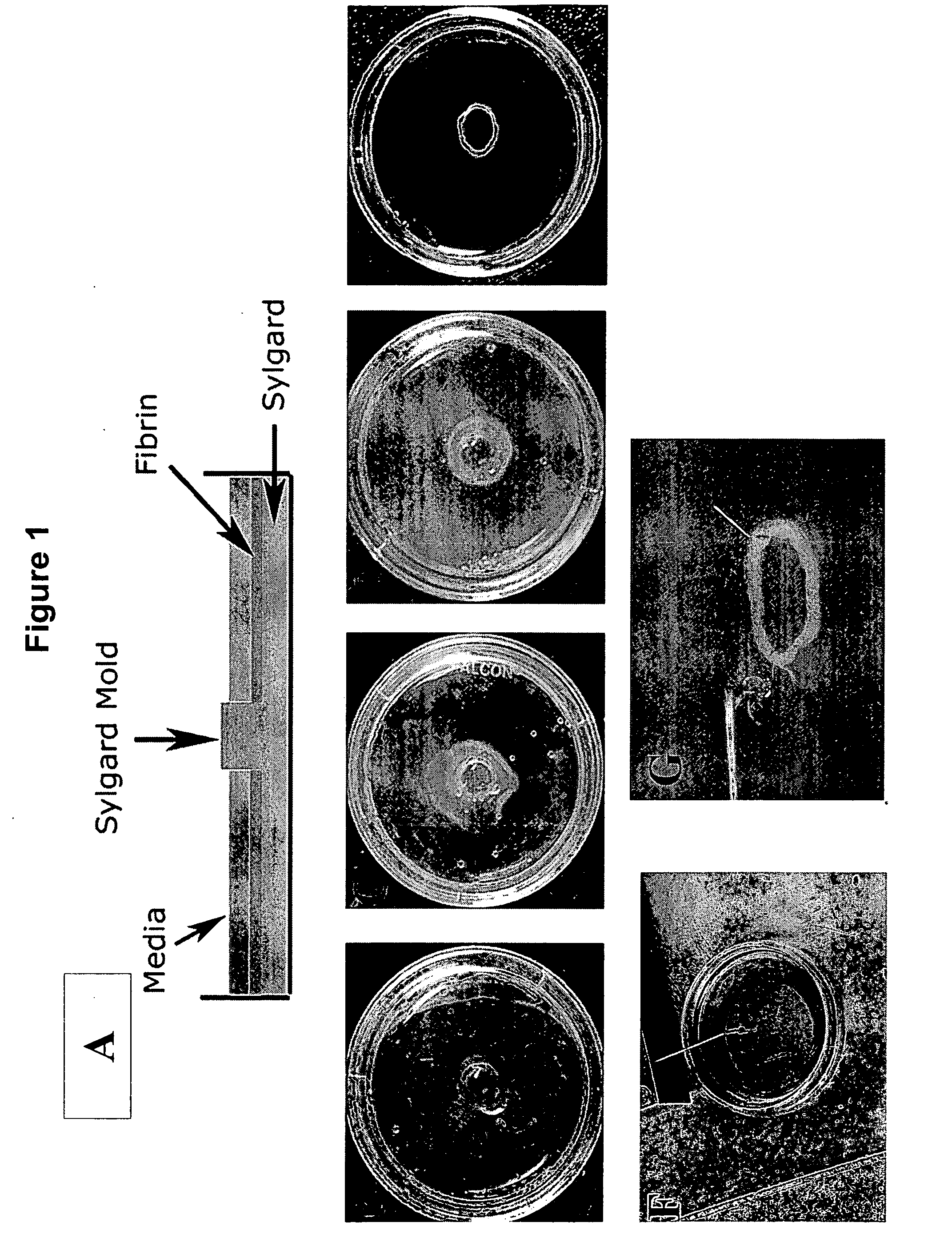
[0107] Culture plates (35 mm) were prepared as described previously (Dennis et al., In Vitro Cellular &Developmental Biology Animal 36: 327-335, 2000). Briefly, 1.5 ml of the polymer SYLGARD™ (polydimethylsiloxane—PDMS) was poured into each plate and allowed to cure for 24 hours. The polymer layer provided an anchor that materials could be easily pinned in place and provided a surface to which, unless coated, the cells could not adhere. A 5 mm diameter cylindrical SYLGARD mold was placed in the center of the dish to provide a luminal space for the engineered sphincter (FIG. 1, Panel A). Adhesion of the mold to the SYLGARD coated culture dish was facilitated by pressing the mold firmly against the bottom of the dish with forceps. The culture dish was sterilized with 70% ethanol for 30 minutes. Ethanol was then aspirated and culture dishes were exposed to UV light for 1 hour. Subsequently, growth media (500 μl) containing 10 U / ml throm...

example 3

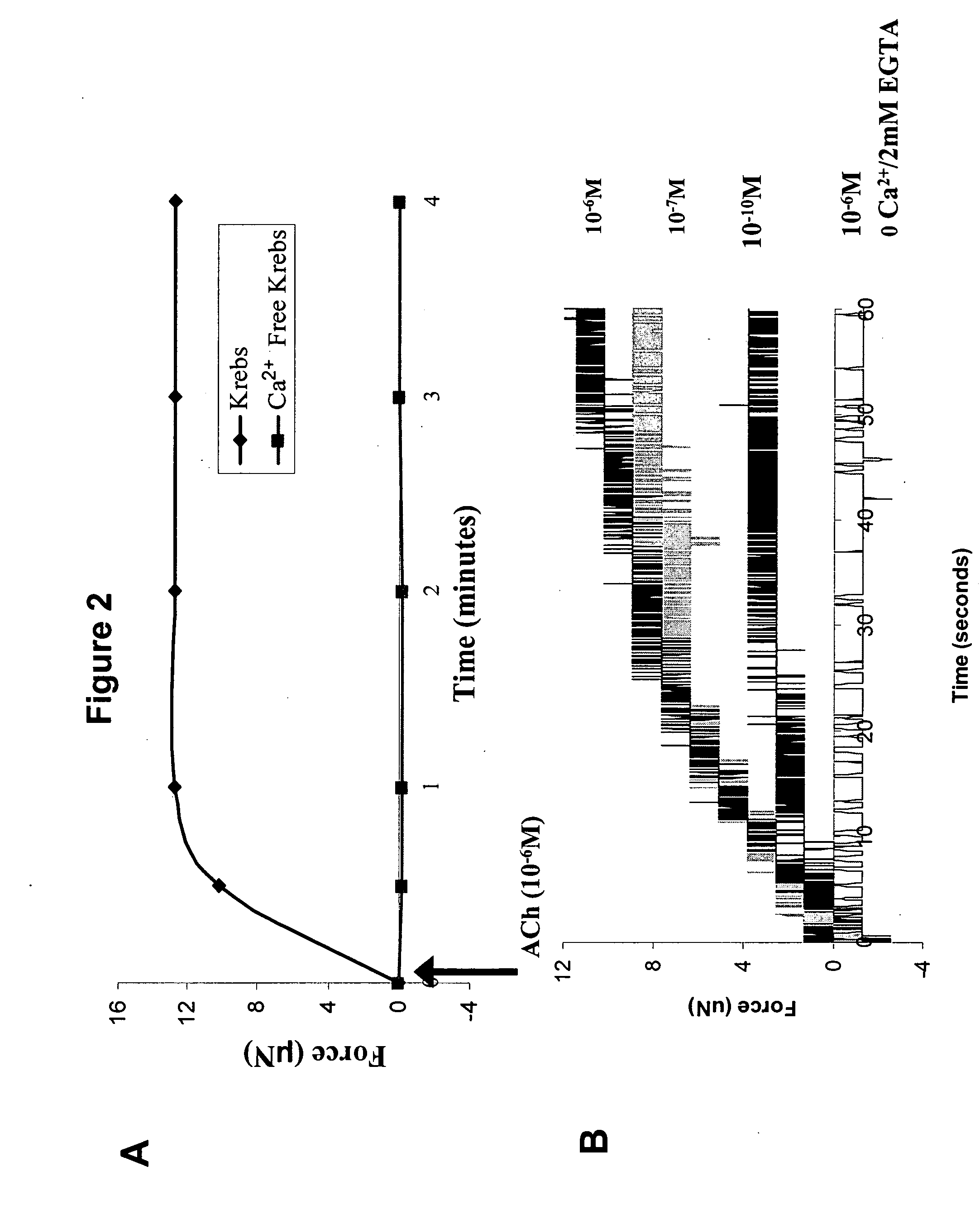
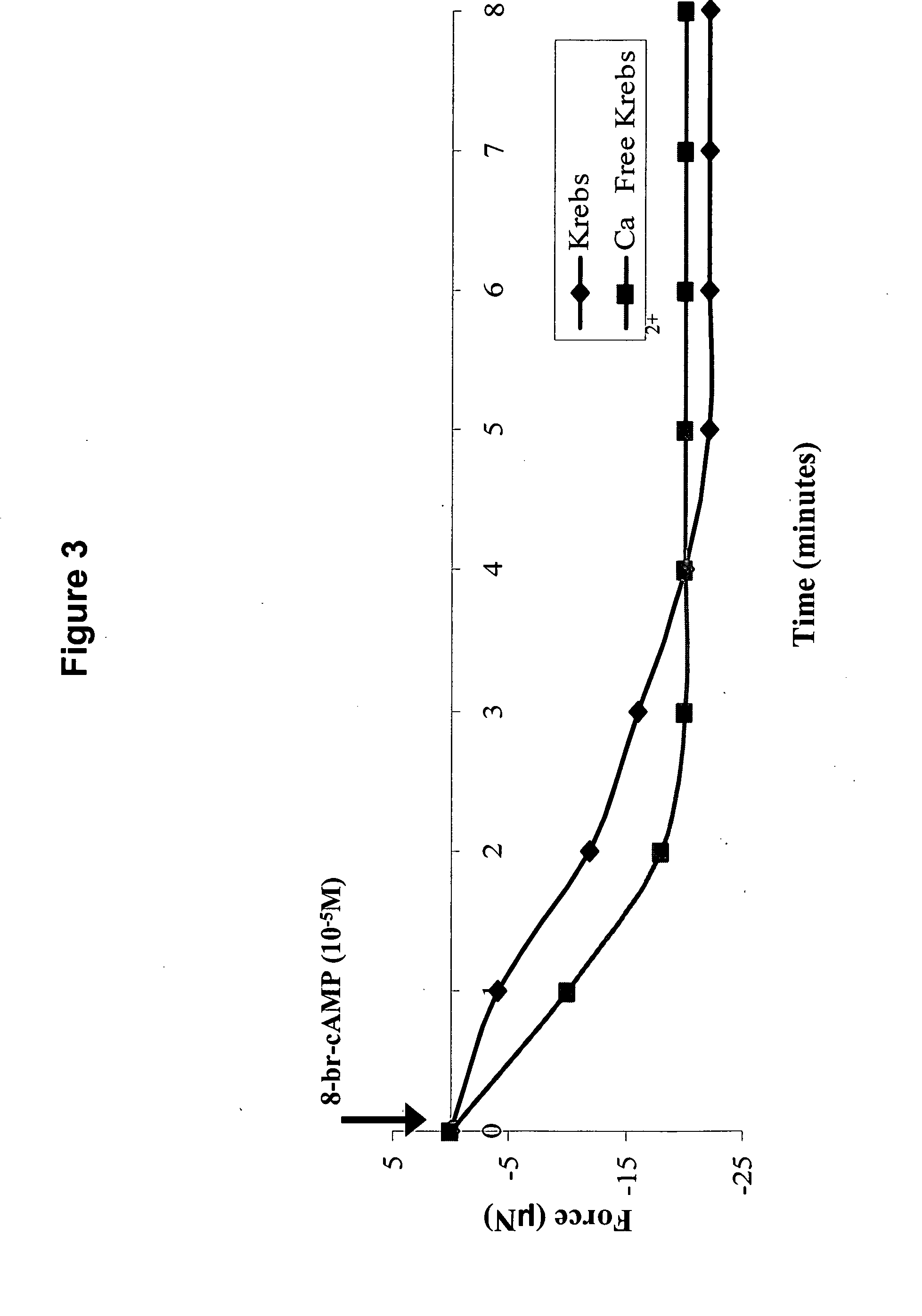
Agonist-Induced Contraction and Relaxation of Bioengineered Sphincter Rings

[0112] Excitability and contractility of bioengineered IAS rings was measured using techniques adapted from previous work (Dennis et al., In Vitro Cellular &Developmental Biology Animal 36: 327-335, 2000. Dennis et al., American Journal of Physiology Cell Physiology 280: C288-C295, 2001). The diameter of the sphincter rings was measured and used to calculate cross-sectional area (CSA). The bioengineered sphincters were separated from their molds using forceps (FIG. 1 Panel E) and the minimum ring diameter was measured using a calibrated eyepiece and a 5× or 10× objective lens on an inverted microscope (Zeiss Axiovert 25, Thornwood, N.Y.). The cross section was circular, and therefore the CSA was calculated using the measured diameter.

[0113] To measure the passive baseline force, the differentiation media in the plate containing the bioengineered sphincter was replaced with 37° C. Krebs solution (NaCl (119 m...


PUM

PropertyMeasurementUnit
diameteraaaaaaaaaa
volumeaaaaaaaaaa
thickaaaaaaaaaa

Abstract

This invention relates to a three dimensional bioengineered sphincter such as a bioengineered internal anal sphincter (IAS) and three-dimensional physiological models of smooth muscle cells. The invention also related to methods for generating a bioengineered sphincter and smooth muscle rings in culture. In addition, the invention provides for methods of using a bioengineered sphincter or bioengineered smooth muscle ring to identify agents that modify contractile force and relaxation in smooth muscle cells.

Description

RELATED APPLICATIONS [0001] The present application claims priority benefit of U.S. Provisional Application No. 60 / 630,816 filed Nov. 24, 2004 which is herein incorporated by reference in its entirety.[0002] Scientific work relating to the invention was supported by Grant No. DK-042876 from the United States National Institute of Diabetes and Digestive and Kidney Diseases. The United States government may have certain rights in the invention.FIELD OF THE INVENTION [0003] This invention relates to a three dimensional bioengineered smooth muscle cell ring and a three dimensional bioengineered sphincter such as a bioengineered internal anal sphincter (IAS). The invention also related to methods for generating a bioengineered sphincter in culture and methods of using a bioengineered sphincter to identify agents that modify contractile force and relaxation in smooth muscle cells. BACKGROUND [0004] Sphincter malfunction results from sluggish contractile response of the skeletal and smooth...

Claims


Application Information

Patent Timeline
Patent Type & Authority Applications(United States)
IPC IPC(8): A61K35/37A61K35/34A61K35/38
CPCA61F2/0004A61F2/08A61K38/1709C12N5/0661C12N2503/04C12N2533/56A61K35/34A61K35/38G01N33/5088
Inventor BITAR, KHALIL N.HECKER, LOUISEBAAR, KEITHSOMARA, SITA
Owner RGT UNIV OF MICHIGAN
Why Eureka
Try Eureka