Cryopreservation of human blastocyst-derived stem cells by use of a closed straw vitrification method
a technology of blastocyst and cell culture, which is applied in the field of improved methods for vitrification of biological cells, can solve the problems of difficult application of cryopreservation using conventional methods to complex and sensitive biological materials, poor survival of undifferentiated human embryonic stem cells, and the majority of cell differentiation or death
- Summary
- Abstract
- Description
- Claims
- Application Information
AI Technical Summary
Benefits of technology
Problems solved by technology
Method used
Image
Examples
example 7
[0124] Comparison Between Using Different Concentrations of Trehalose in the Vitrification Medium
[0125] Human BS cells (cell line SA121) were vitrified and thawed following the procedure described in Example 1 & 2 with the exception that trehalose was used in two different concentrations (0.3M and 0.5M) in the second vitrification solution (solution B). When 0.3 M trehalose was used in solution B, solution C contained 0.2 M trehalose and solution D contained 0.1 M trehalose. When 0.5 M trehalose was used in solution B, solution C contained 0.4 M trehalose and solution D contained 0.2 M trehalose. Forty-eight hours after seeding in culture dishes on top of mouse embryonic feeder cells the hBS cell colonies were evaluated and counted. The thawing recovery was calculated as the ratio between the number of viable thawed colonies (displaying appropriate hBS cell morphology) and the number of hBS cell pieces originally vitrified. Two separate experiments using two different human BS cell...
example 8
[0126] Extensive Evaluation of Vitrification and Devitrification Procedure Using Closed Straws
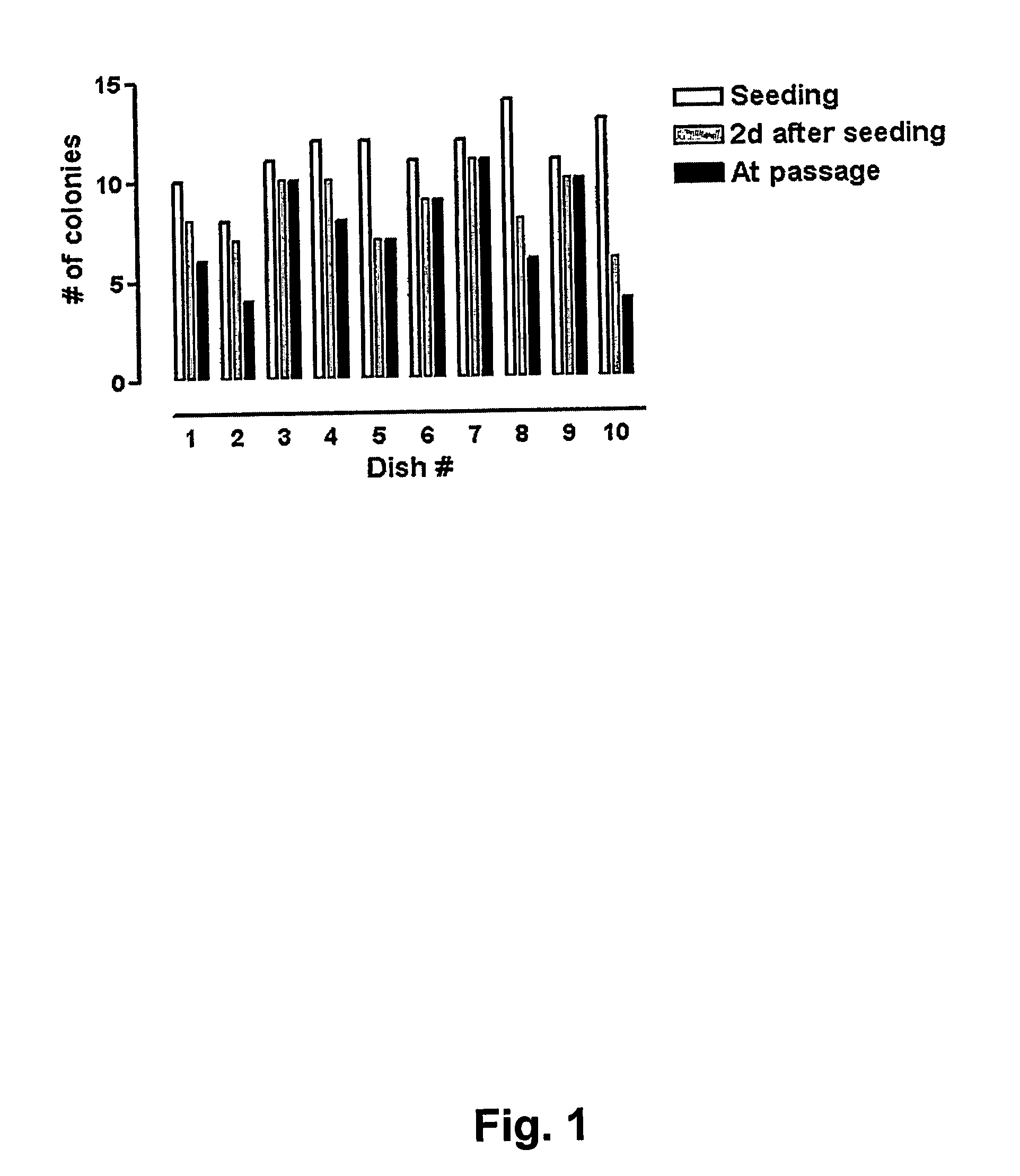
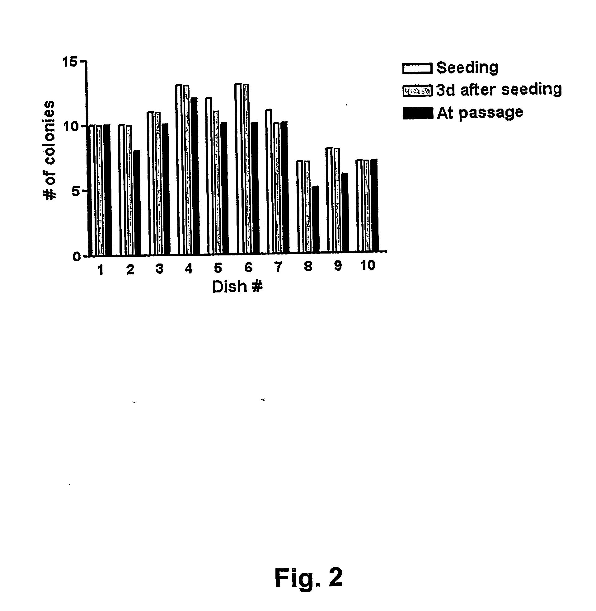
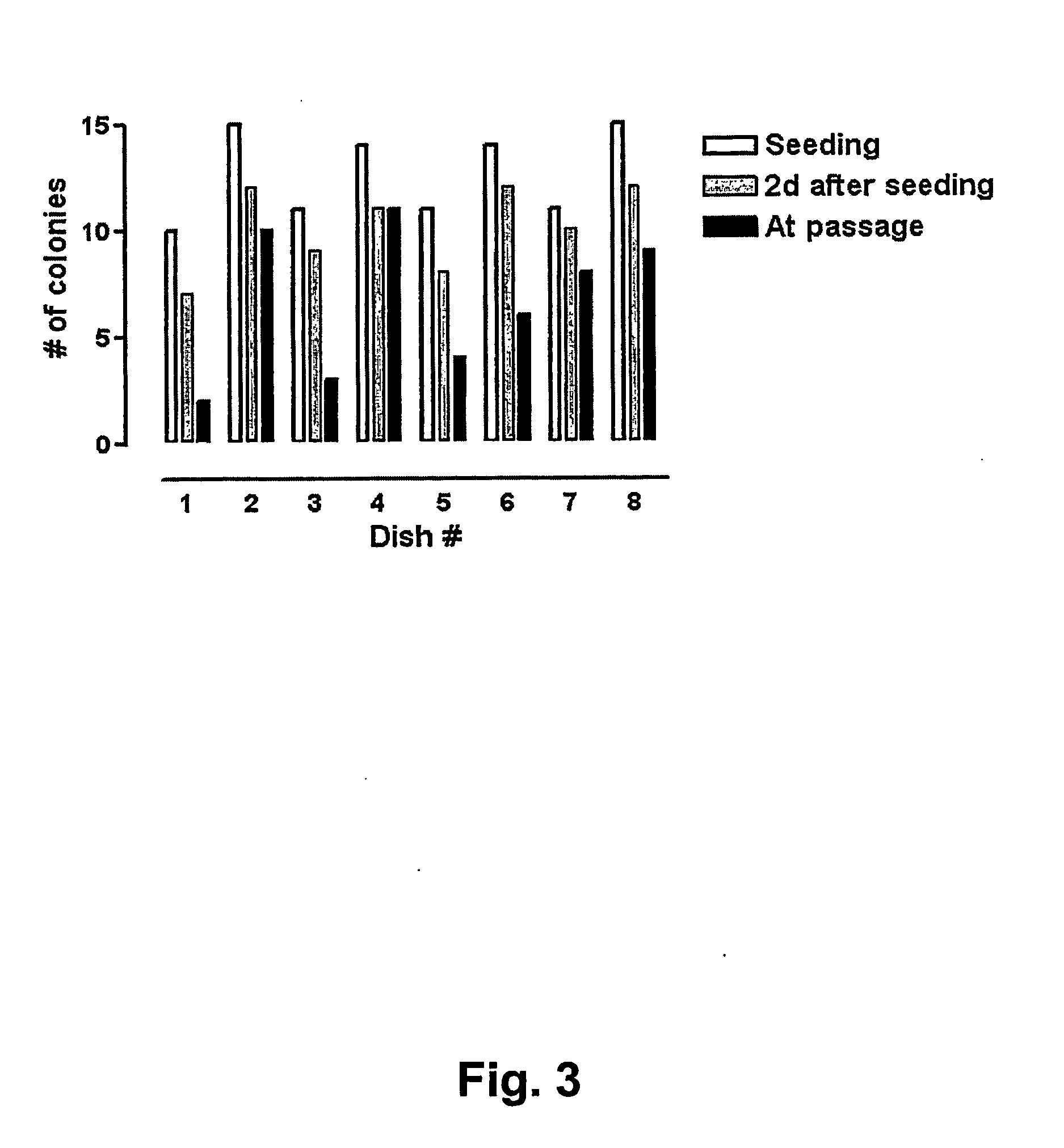
[0127] In order to evaluate the quality of the vitrification process large quantities of human BS cells were vitrified (as described in Example 1 above) at three different occasions using three different human BS cell lines (SA001, SA002, and AS034). At each occasion >100 straws were vitrified from each cell line. Eight to 10 straws each from of these large batches were randomly selected, devitrified (as described in Example 2 above) and seeded in separated dishes on top of mouse embryonic feeder cells. The number of hBS cell clumps that were seeded and that attached, proliferated, and displayed appropriate morphology was determined in each dish. The results are presented in FIGS. 1, 2 and 3 and show that every straw gave rise to viable hBS cell colonies that subsequently were passaged according to standard procedures and characterized.
example 9
[0128] Typical Morphology of Human BS Cell Before and After Vitrification and Thawing
[0129] Typical morphology of the human BS colonies (cell line SA001) before vitrification is shown in FIG. 4. After devitrification and seeding, viable colonies proliferated and displayed morphology characteristic for undifferentiated human BS cells (FIG. 5). Subsequently, these cells were propagated and passaged according to standard procedures and representative illustrations of the human BS cell colonies are shown in FIG. 6. Similar results were obtained for human BS cell line SA002 and AS034 (data not shown).
PUM
 Abstract
Description
Claims
Application Information
 - R&D
- Intellectual Property
- Life Sciences
- Materials
- Tech Scout
- Unparalleled Data Quality
- Higher Quality Content
- 60% Fewer Hallucinations
Browse by: Latest US Patents, China's latest patents, Technical Efficacy Thesaurus, Application Domain, Technology Topic, Popular Technical Reports.
© 2025 PatSnap. All rights reserved.Legal|Privacy policy|Modern Slavery Act Transparency Statement|Sitemap|About US| Contact US: help@patsnap.com


