

Reconstituted human breast tumor model
a human breast tumor and reconstructed technology, applied in the field of molecular biology and oncology, can solve the problems of significant differences in the properties and behavior of xenografted cells, human cells maintained in culture as distinct cell lines, etc., and achieve the effect of propagate the tumor
- Summary
- Abstract
- Description
- Claims
- Application Information
AI Technical Summary
Benefits of technology
Problems solved by technology
Method used
Image

Examples
example 1
Construction of Breast Tumor Model
[0046] Human Tissues and Cell Lines
[0047] Fresh human breast tissue from reduction mammoplasty was provided by Dr. Andrea Richardson at the Brigham and Women's Hospital, Boston, Mass., in compliance with institutional guidelines and RB approval. The fresh tissue was digested overnight at 37° C. using 2.8 mg / ml collagenase and 0.6 mg / ml hyaluronidase. The following morning, the digested human epithelial organoids (clusters of epithelial cells) and primary fibroblasts were collected by centrifugation. The mixture of epithelial organoids and primary fibroblasts were frozen in standard freezing medium and stored in liquid nitrogen.The immortalized human mammary fibroblast cell lines were provided by Charlotte Kuperwasser at the Whitehead Instituted.
[0048] Vector Constructs
[0049] Lentivirus vectors were used for transduction of the human mammary epithelial cells. The lentivirus backbone used in constructing all of the following lentivirus vectors was...
example 2
Efficiency of Lentivirus Infection and p53 shRNA Knockdown
[0067] To determine the efficiency of p53 shRNA in knocking down the expression of p53 and to determine the efficiency of gene transfer through lentiviral system, 293T cells and HMEC organoids were infected with lentivirus expressing pLenti-U6-p53shRNA+CMV-KRAS+SV40-GFP. The sample processing and lentiviral preparation and infection were as described in Example 1. Three days after infection, the cells were observed under UV light for estimation of infection rate and were collected for analyzing p53 and KRAS expression by real-time RT-PCR. Almost all of 293T cells were infected with the lentivirus. Since the expression level of p53 in infected 293T cells was about 3.6% of mock infected cells, we concluded that about 97.4% knockdown of p53 transcription was achieved in 293T cells through lentiviral infection. As for HMEC organoids, about 50% of the cells were infected with the lentivirus. And since the expression level of p53 ...
example 3
Human Breast Pre-Malignant Lesions and CIS Developed from HMECs Transduced with p53 shRNA and erbB2 or KRAS
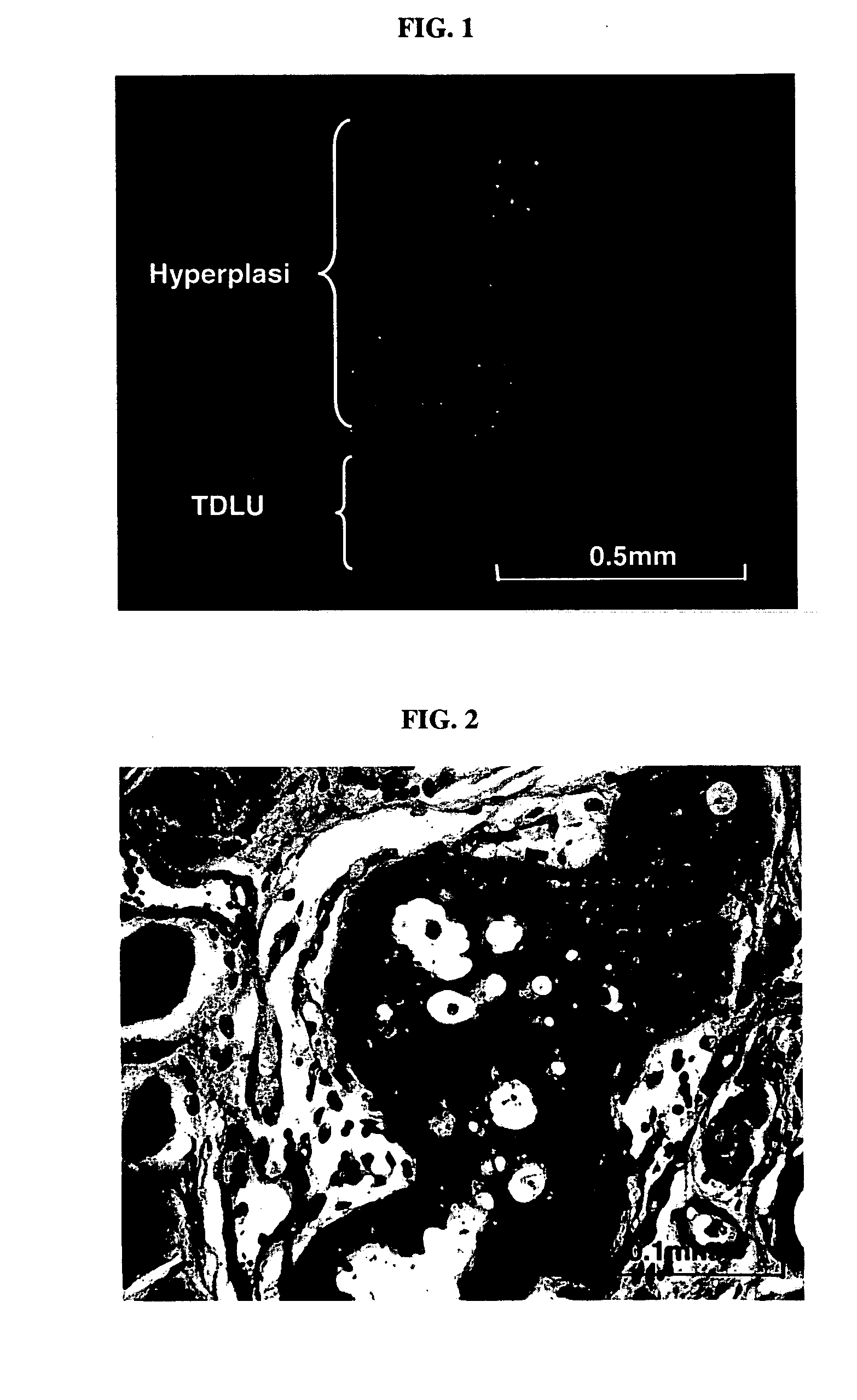
[0068] Employing the lentiviral system, we decided to determine if human breast cancer could be generated from normal primary HMECs transduced with a defined set of genetic elements. The procedures for sample processing, cell culture, lentiviral construct generation, virus production, and mouse surgery were as described in Example 1. As a first step towards generating human breast tumor model in mouse, we transduced human mammary epithelial cells with p53 shRNA plus either erbB2V659E or KRASG12V, using lentiviral construct pLenti-CMV-KRAS+SV40-GFP or pLenti-CMV-erBb2. The infected HMEC organoids were either mixed with human breast fibroblasts prior to injection or injected alone into fat pads that had been injected with immortalized human fibroblasts two weeks previously. In all combinations, normal, e.g. terminal ductual lobular unit, and hyperplastic human breast structures ...
PUM
 Abstract
Description
Claims
Application Information