

Method and apparatus for direct spectrophotometric measurements in unaltered whole blood
Patent Information
- Authority / Receiving Office
- US · United States
- Patent Type
- Applications(United States)
- Current Assignee / Owner
- SHEPHERD A P
- Publication Date
- 2006-09-14
- Estimated Expiration
- Not applicable · inactive patent
Smart Images
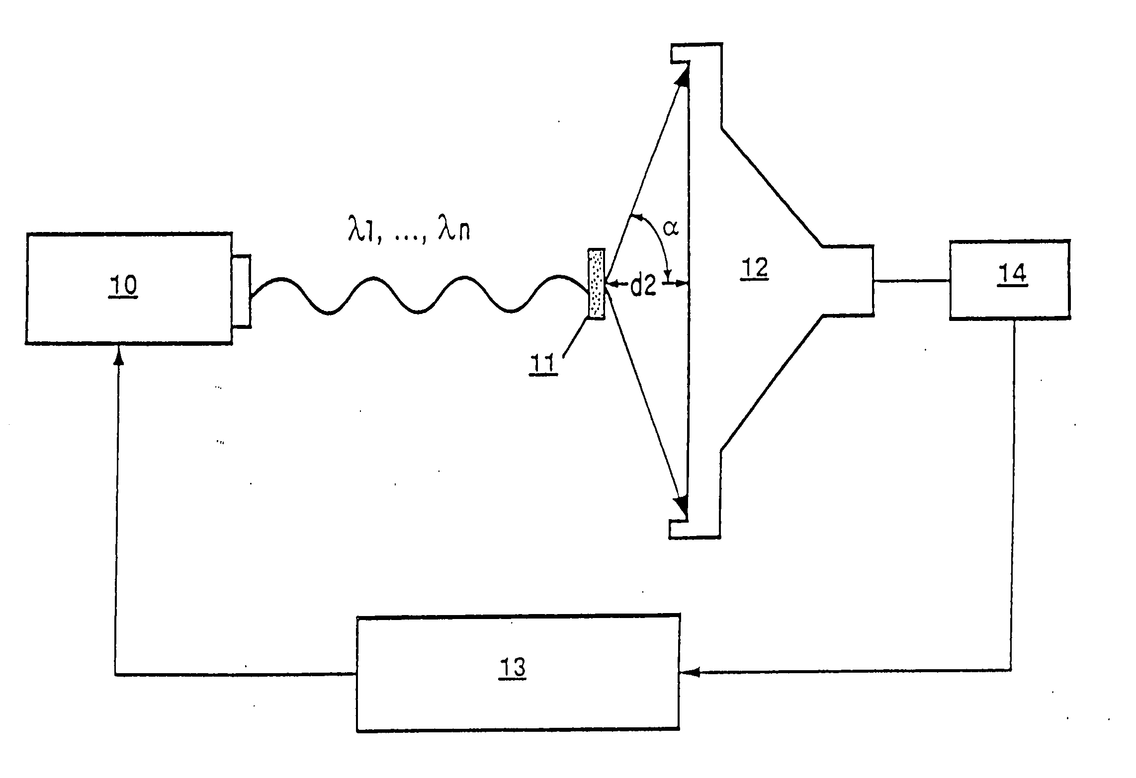
Figure 1 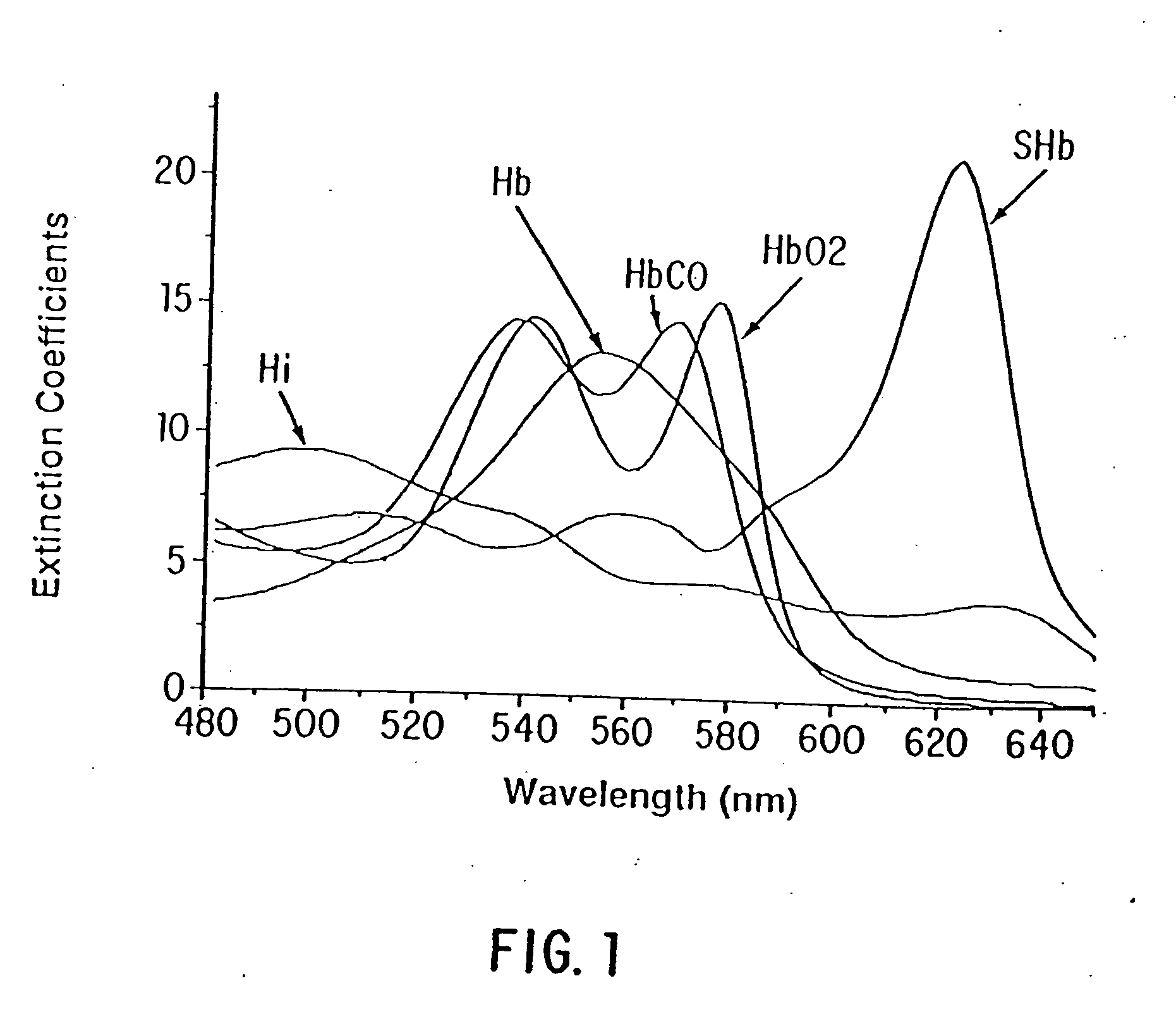
Figure 2 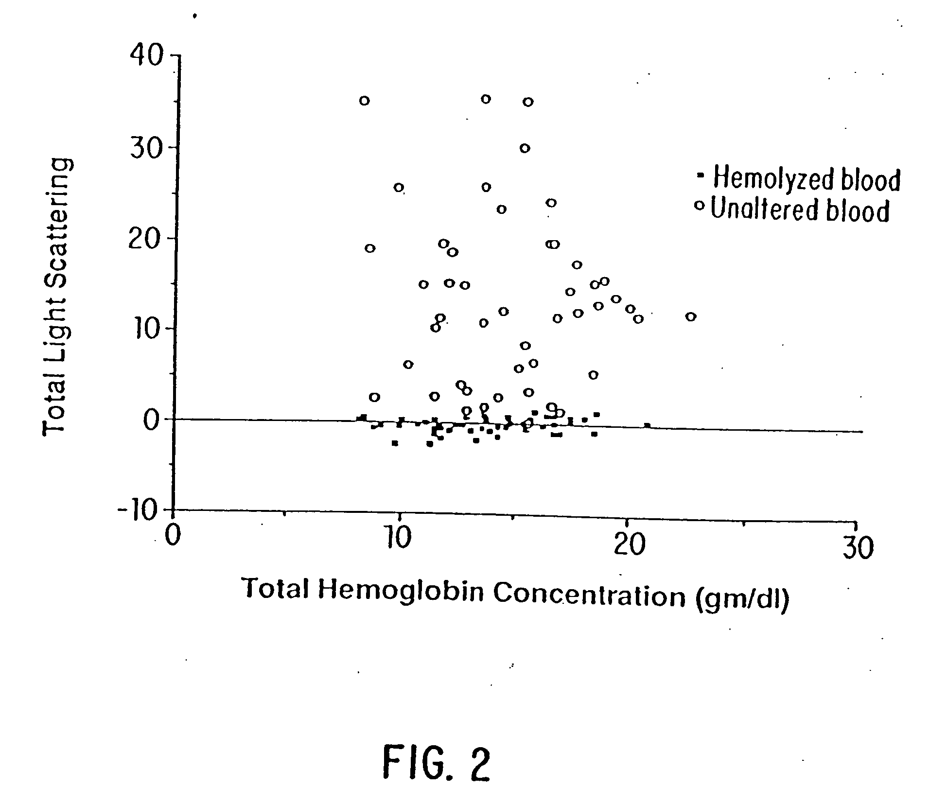
Figure 3
Abstract
Description
[0001] A portion of the disclosure of this patent document contains material which is subject to copyright protection. The copyright owner has no objection to the facsimile reproduction by anyone of the patent document or the patent disclosure, as it appears in the Patent and Trademark Office patent file or records, but otherwise reserves all copyright rights whatsoever. FIELD OF THE INVENTION
[0002] This invention relates to a method and apparatus to assess the optical transmittance of a sample of unaltered whole blood at multiple wavelengths to attain an accurate measurement of its total hemoglobin concentration, the concentration of bilirubin, and the concentrations of oxy-, deoxy-, carboxy-, met-, and sulfhemoglobin. BACKGROUND
[0003] To the best of the Applicants' knowledge, no prior art whether patented or not has ever successfully exploited the optical transmittance of unaltered whole blood to achieve an accurate measurement of the total hemoglobin concentration (THb) in a bl...