

Derivation of embryonic stem cells
a technology stem cells, which is applied in the field of derivation of embryonic stem cells, can solve the problems of a relatively low success rate in the culturing of embryos to blastocysts
- Summary
- Abstract
- Description
- Claims
- Application Information
AI Technical Summary
Benefits of technology
Problems solved by technology
Method used
Image

Examples
example 1
Generation of ES Cell Lines
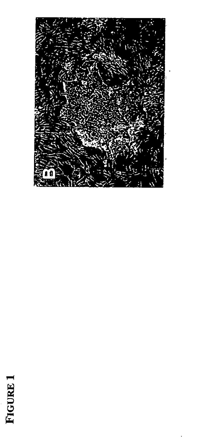
[0056] Single blastomeres were isolated from 8-cell stage 129 / Sv-ROSA26:LacZ mouse embryos either by biopsy through a hole in the zona pellucida drilled using Piezo-pulse or by disaggregating of zona-denuded embryos in Ca++ / Mg++ free PBS for 10 minutes. The biopsied (7-cell) embryos were transferred to the oviducts of 1.5 days post coitum (d.p.c.) synchronized surrogate mothers, and each separated blastomere aggregated with a small clump (approximately 100 cells) of green fluorescent protein (GFP)-positive 129Sv / CD-1 mouse ES (mES) cells in a 300 um depression created by pressing an aggregation needle into the bottom of a plastic tissue culture plate. After incubation for 24-48h a growing “bud” of GFP-negative cells was observed on the sides of the majority (60%) of GFP-mES clusters (See FIG. 4 A, B). The cell clumps were plated onto mitomicin C-treated mouse embryonic fibroblasts (MEF) and cultured in knockout DMEM (15% FCS, penicillin / streptomycin, Glut...
example 2
Differentiation of ES Cells
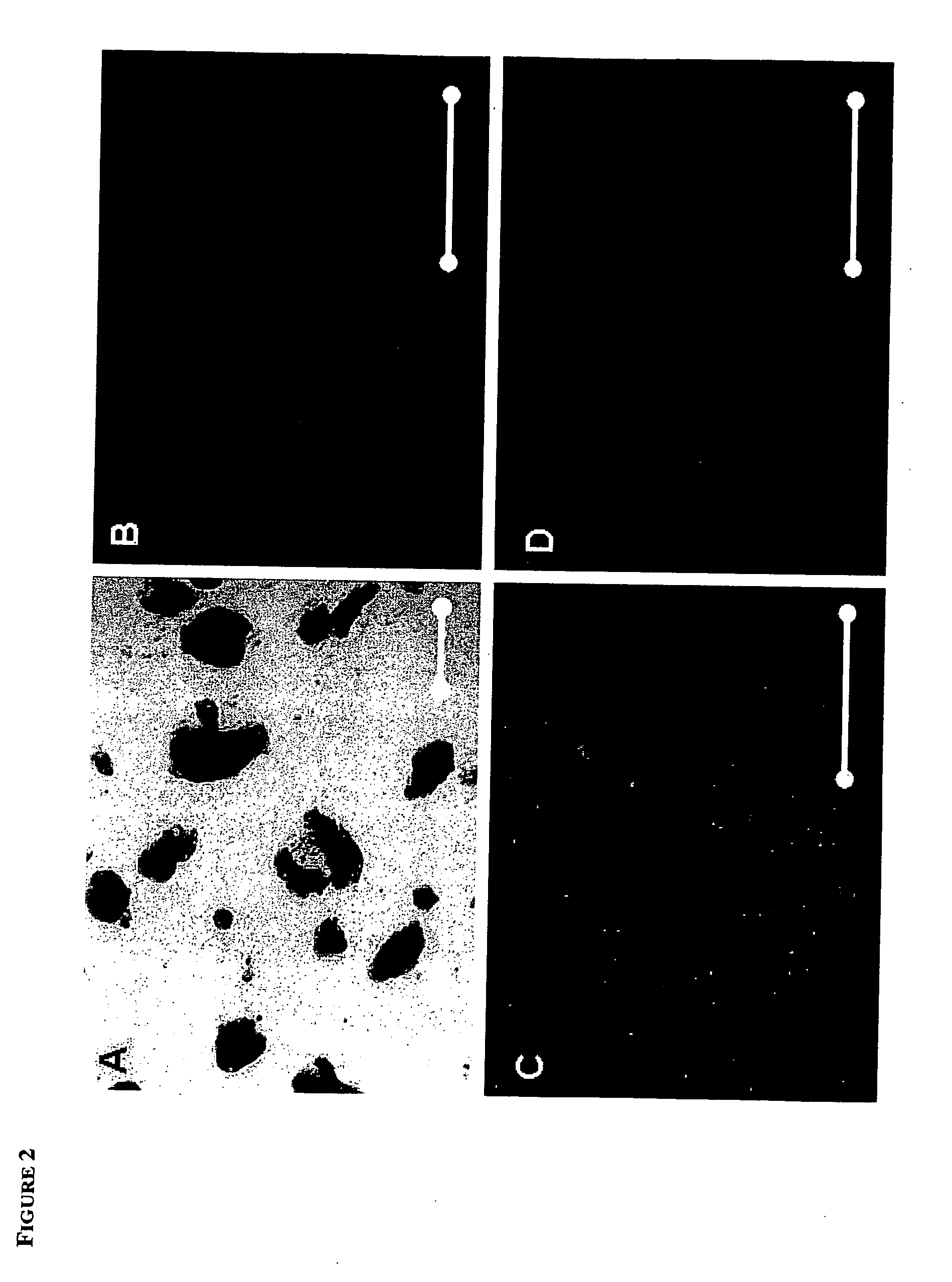
[0060] When the ES-like cell cultures were allowed to overgrow, they spontaneously differentiated into cells of all three germ layers, as evidenced by immunostaining with antibodies to muscle actin (mesoderm), βIII tubulin (ectoderm), and α-feto protein (primitive endoderm)(FIG. 3 B-D and FIG. 8 A-C). Beating heart muscle, extraembryonic endoderm and multiple neuronal cell types were also routinely observed in differentiating cultures. To further demonstrate the pluripotency of the derived ES cells, ES cell lines were either injected into CD-1 mouse blastocysts or aggregated with 8-cell stage morulae as described previously (Hogan et al., supra) and transferred to recipient females. X-Gal (5-bromo—4-chloro—3-indolyl-beta-D-galactopyranoside) staining of the resulting chimeric fetuses showed that the ES cell lines contributed to all organs (FIG. 3 A), such as, heart, kidney, liver, lung, intestine, brain, blood, skin and genital ridge. Twenty-four of the f...
example 3
Generation of TS Cell Lines
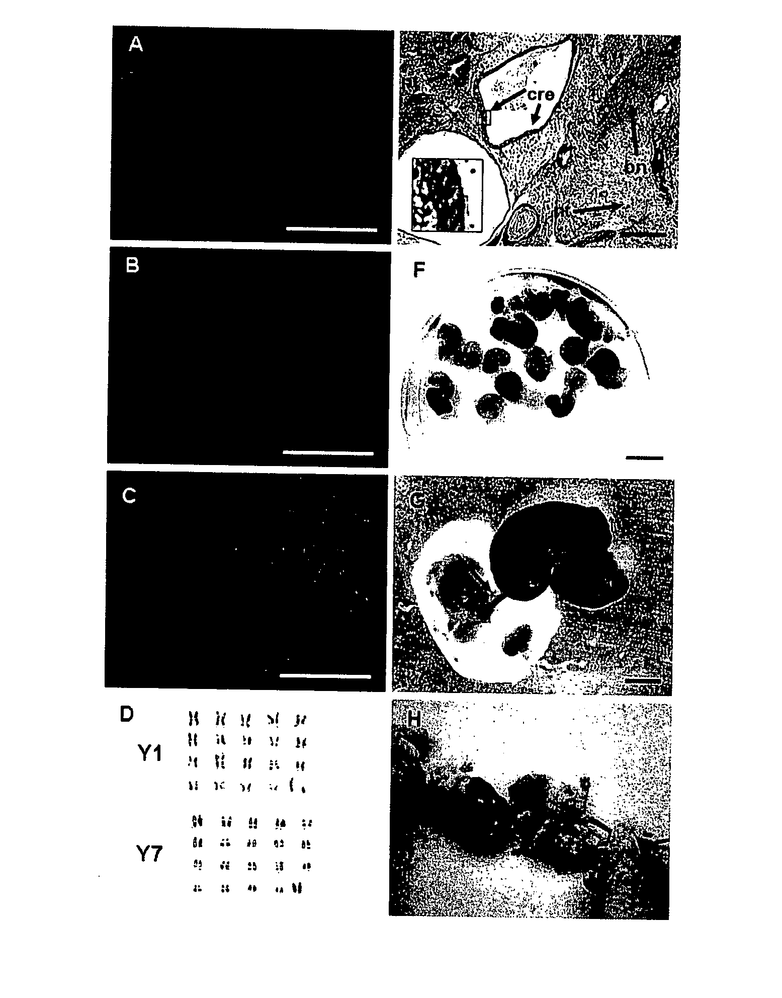
[0064] Blastomere outgrowths that morphologically resembled trophoblast and extraembryonic endoderm but not ES cells were further cultured in the mES cell medium with 50 ng / ml FGF-4 produced trophoblast stem (TS)-like cells that were maintained under these conditions and passaged with trypsin. Seven putative TS lines were established, which maintained normal karyotype and expressed markers of TS cells (FIG. 6 B, D, F, H, J). These cells were negative for Oct-4 (FIG. 6 H) and for α-feto protein. Putative TS cells contributed to the extraembryonic lineage in chimeric fetuses generated by aggregation with the LacZ+ TS-cells (FIG. 7). RT-PCR analysis confirmed that these cells expressed Cdx-2, but not Oct-4 (FIG. 5 C and F). Nanog and Rex-1 were expressed in both the putative TS and ES cell lines (FIG. 5 D and E).
[0065] Briefly, RT-PCR analysis was performed as follows: Total RNA was isolated from ES and TS cells using an RNAeasy Mini Kit (Qiagen), and 1 μg ...
PUM
| Property | Measurement | Unit |
|---|---|---|
| Composition | aaaaa | aaaaa |
Abstract
Description
Claims
Application Information