Medical hyperspectral imaging for evaluation of tissue and tumor

a hyperspectral imaging and tumor technology, applied in the field of hyperspectral imaging analysis, can solve the problems of not translating into, contributing to significant local morbidity, cost of care and anxiety,

Active Publication Date: 2006-11-02
HYPERMED IMAGING
View PDF40 Cites 158 Cited by
  • Summary
  • Abstract
  • Description
  • Claims
  • Application Information

AI Technical Summary

Benefits of technology

[0012] One embodiment of the invention is directed to a medical instrument comprising a first-stage optic responsive to illumination of a tissue, a spectral separator, one or more polarizers, an imaging sensor, a diagnostic processor, a filter control interface, and a general-purpose operating module. Preferably, the spectral separator is optically responsive to the first-stage optic and has a control input, the polarizer compiles a plurality of light beams into a plane of polarization before entering the imaging sensor, the imaging sensor is optically responsive to the spectral separator and has an image data output, the diagnostic processor comprises an image acquisition interface with an input responsive to the imaging sensor and one or more diagnostic protocol modules wherein each diagnostic protocol module contains a set of instructions for operating the spectral separator and for operating the filter control interface, the filter control interface comprises a control output provided to the control input of the spectral separator, which directs the spectral separator independently of the illumination to receive one or more wavelengths of the illumination to provide multispectral or hyperspectral information as determined by the set of instructions provided by the one or more diagnostic protocol module, and the general-purpose operating module performs filtering and acquiring steps one or more times depending on the set of instructions provided by the one or more diagnostic protocol modules.
[0013] The instrument may also comprise a second-stage optic responsive to illumination of the tissue. Preferably, the one or more wavelengths of illumination is one or a combination of V, visible, NIR, and IR. In preferred embodiments, the multispectral or hyperspectral information determines one or more of presence of cancer for screening or diagnosis, presence of residual cancer in a surgical excision bed, and cancer progression, preferably wherein the presence of cancer is breast, lymphoma or any cancer readily visualized by hyperspectral imaging. Preferred embodiments include the multispectral or hyperspectral information applied laparoscopically, thoracoscopically, cystoscopically, hysteroscopically, bronchoscopically, or mediastinoscopically to assess presence of tumor, adequacy of surgical resection or nodal or intracavitary spread.

Problems solved by technology

Early studies suggested that LR does not translate into death from disease.4 However, recent data showing lower LR rates and survival benefit by adding radiation therapy to mastectomy for patients with higher stage cancers indicate the potential importance of freedom from local recurrence.5 In addition, LR contributes to significant local morbidity, usually requiring a mastectomy.
Finally LR contributes to the cost of care and anxiety for the patient.
Despite these issues, 20-60% of patients undergoing BCT are found to have positive margins requiring additional surgical procedures, either re-excisional lumpectomies, or mastectomy.6 These additional procedures result in increased cost, increased anxiety for the patient, and, importantly, a delay in initiation of important systemic chemotherapy or radiation therapy.
At operation, the surgeon attempts to completely resect the cancer with negative microscopic margins, but faces several difficulties.
However, the microscopic extent of disease is difficult to gauge.
Frozen section analysis of breast biopsy margins is difficult and unreliable, because the fat content of breast tissue results in difficulty in sectioning frozen specimens.
This is difficult, and impractical in most institutions, but also does not provide real time information while the patient is in the operating room.
The former method does not allow re-resection of a specific positive margin and results in resection of a larger volume of breast tissue since the cavity side with a positive margin is not known.
With both approaches, the ink may creep into crevices, resulting in falsely positive margins.
With the multi-colored approach, inks may run together, resulting in confusion as to the location of a specific positive margin.
Many surgeons perform wide excisions, potentially resulting in significant breast deformity that is added to by the breast shrinkage associated with radiation therapy.
This delay results in patient anxiety and longer time to completion of the patient's surgical treatment.
The accuracy of sentinel node evaluation by frozen section is problematic,6 with a significant false negative risk, when compared to the final report.
Patients already treated for lymphoma are at the greatest risk.

Method used


Image

  • Medical hyperspectral imaging for evaluation of tissue and tumor
  • Medical hyperspectral imaging for evaluation of tissue and tumor
  • Medical hyperspectral imaging for evaluation of tissue and tumor

Examples


Embodiment Construction

Hyperspectral Imaging System

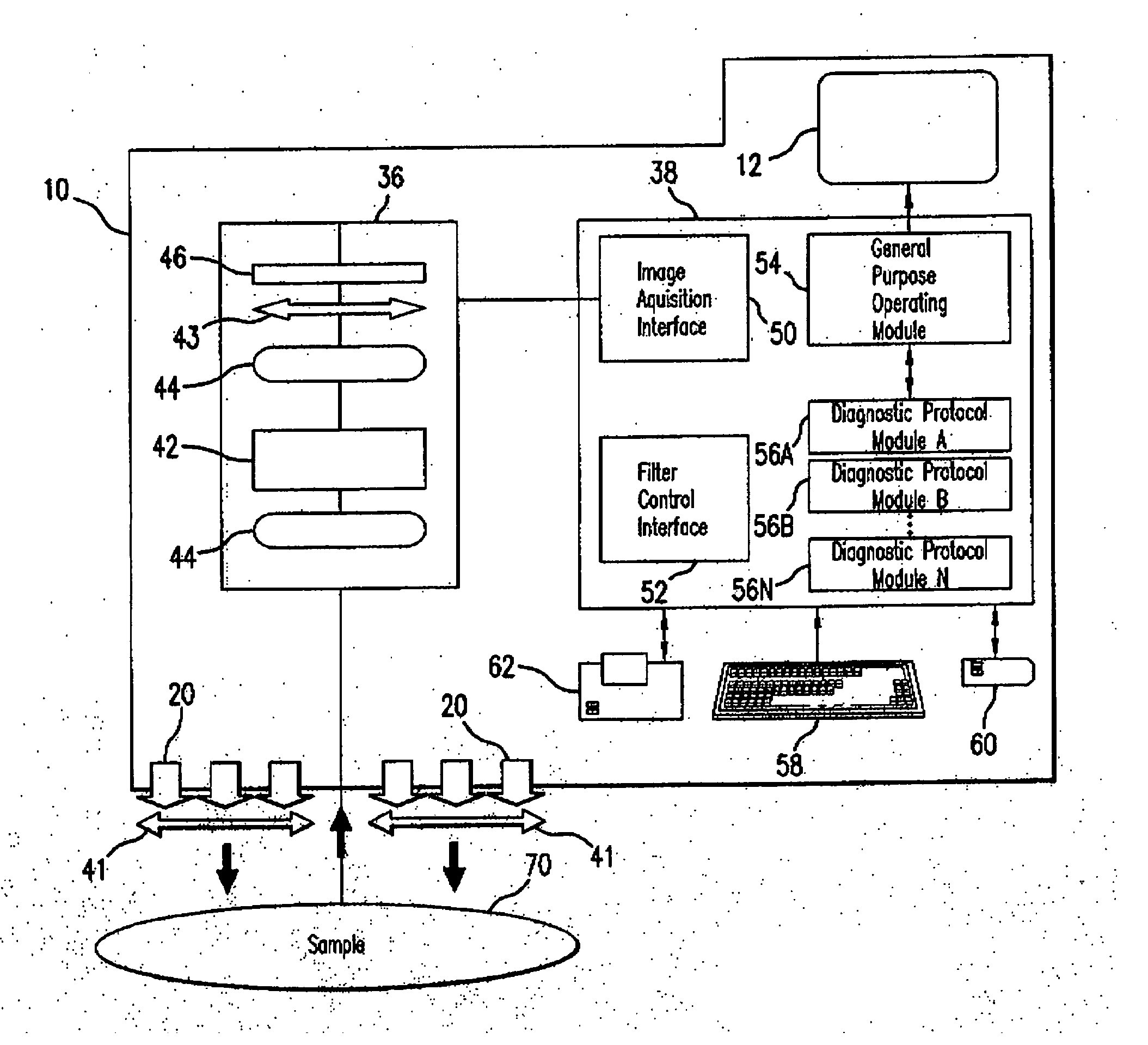
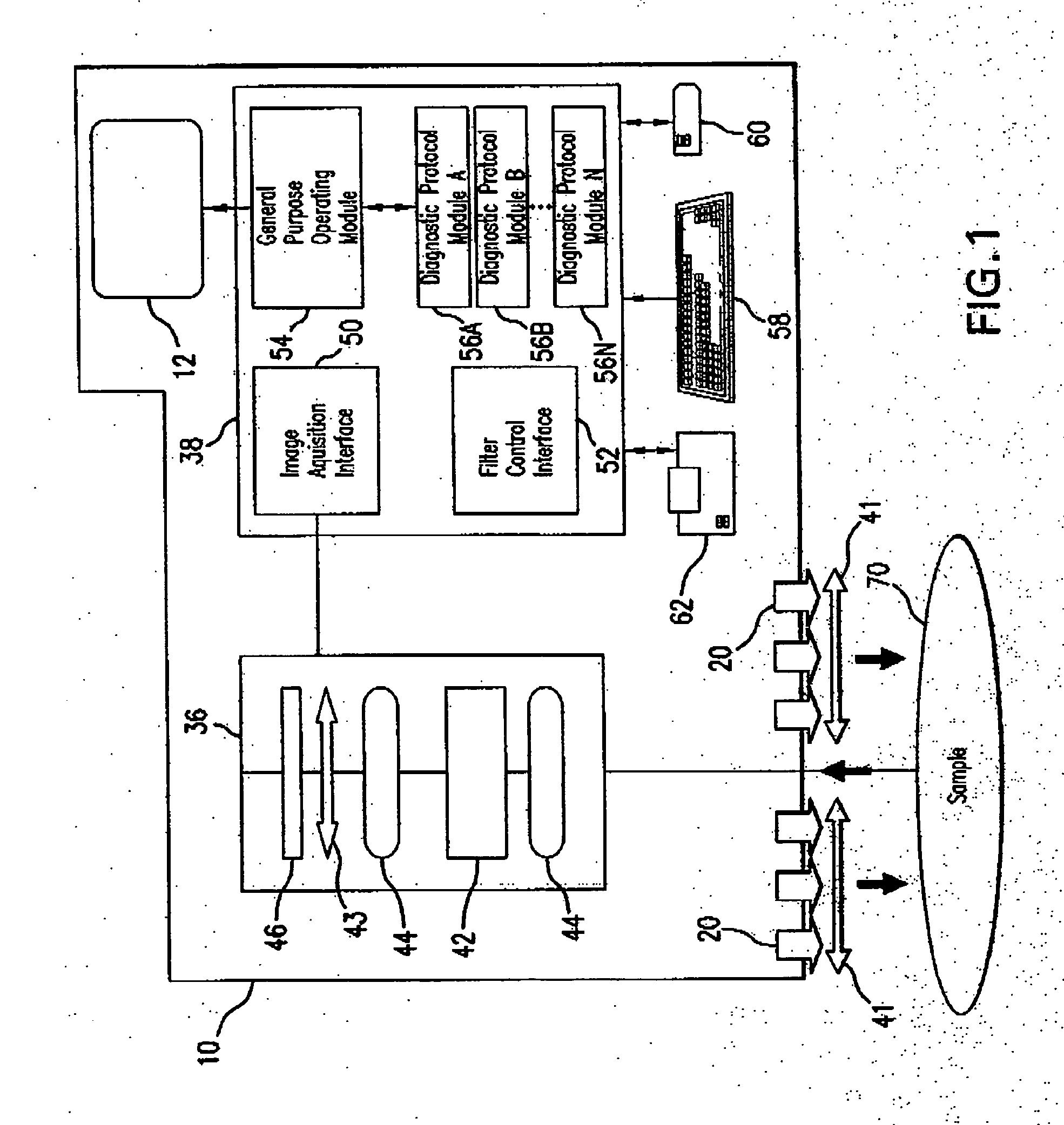
[0031] Hyperspectral imaging (HSI) is a novel method of “imaging spectroscopy” that generates a map of a region of interest based on local chemical composition. HSI has been used in non-medical applications including satellite investigation to indicate areas of chemical weapons production and to assess the condition of agricultural fields. HSI has recently been applied to the investigation of physiologic and pathologic changes in living tissue in animal and human studies to provide information as to the health or disease of tissue that is otherwise unavailable. MHSI has been shown to accurately predict viability and survival of tissue deprived of adequate perfusion, and to differentiate diseased tissue (e.g. tumor) and growth due to cancerous angiogenesis in a rat model system of breast cancer.

[0032] HSI is a remote sensing technology in which a 2-dimensional image is created having spectral data inherent in each pixel. These stacks of images comprise ...


PUM


Abstract

Apparatus and methods for hyperspectral imaging analysis that assists in real and near-real time assessment of biological tissue condition, viability, and type, and monitoring the above over time. Embodiments of the invention are particularly useful in surgery, clinical procedures, tissue assessment, diagnostic procedures, health monitoring, and medical evaluations, especially in the detection and treatment of cancer.

Description

REFERENCE TO RELATED APPLICATIONS [0001] The present application claims priority to U.S. Provisional Patent Applications Ser. No. 60 / 631,135 entitled Hyperspectral Imaging in Medical Applications, filed Nov. 29, 2004, Ser. No. 60 / 667,678 entitled Hyperspectral Imaging in Breast Cancer, filed on Apr. 4, 2005, and Serial Number 60 / ______ entitled Hyperspectral Analysis for the Detection of Lymphoma, filed on Nov. 2, 2005, which are hereby incorporated by reference.FIELD OF THE INVENTION [0002] The invention is directed to a hyperspectral imaging analysis that assists in real and near-real time assessment of biological tissue condition, viability, and type, and monitoring the above over time. Embodiments of the invention are particularly useful in surgery, clinical procedures, tissue assessment, diagnostic procedures, health monitoring, and medical evaluations. BACKGROUND OF THE INVENTION [0003] In 2005, 212,000 new cases of breast cancer are expected, and approximately 40,000 women wi...

Claims


Application Information

Patent Timeline
Patent Type & Authority Applications(United States)
IPC IPC(8): A61B5/05
CPCA61B5/0059G01N21/359A61B5/416A61B5/418G01J3/02G01J3/0224G01J3/0264G01J3/10G01J3/2823G01N21/21G01N21/31G01N21/35G01N2021/3137G01N2021/3155G01N2021/317A61B5/0091A61B5/02007A61B5/4842A61B5/7425A61B5/415A61B5/742
Inventor PANASYUK, SVETLANA V.FREEMAN, JENNY E.PANASYUK, ALEXANDER A.
Owner HYPERMED IMAGING
Why Eureka
Try Eureka