Method for the characterization of the three-dimensional structure of proteins employing mass spectrometric analysis and computational feedback modeling

a three-dimensional structure and protein technology, applied in the field of three-dimensional surface structure characterization of proteins and protein complexes, can solve the problems of reducing sensitivity, reducing the sensitivity of ions, and expensive operation of strong countercurrent gas flow

Inactive Publication Date: 2006-12-07
HV OPS H NU
View PDF37 Cites 7 Cited by
  • Summary
  • Abstract
  • Description
  • Claims
  • Application Information

AI Technical Summary

Benefits of technology

[0015] More than one molecule may be bound to the protein surface during an experimental determination of (KB). This would allow multiple binding constants (KB) to be determined during one

Problems solved by technology

The use of such a strong countercurrent gas flow is expensive and difficult to operate because the gas flow rate and the temperature need to be controlled precisely and be optimized for each analyte and solvent system.
If proper gas flow and temperature conditions are not attained, it can re

Method used


Image

  • Method for the characterization of the three-dimensional structure of proteins employing mass spectrometric analysis and computational feedback modeling
  • Method for the characterization of the three-dimensional structure of proteins employing mass spectrometric analysis and computational feedback modeling
  • Method for the characterization of the three-dimensional structure of proteins employing mass spectrometric analysis and computational feedback modeling

Examples


example one

[0070] At least three kinds of quadrupole mass spectrometers have been used with this invention to characterize the ES probe: two single quadrupoles and one custom, double quadrupole instrument. However, any type of mass spectrometer system could accommodate this ES probe interface.

[0071] In this experiment, the following polypeptides were used to optimize the ES probe of this invention: angiotensin III (Sigma #A-0903, 30 pmol / μL, bradykinin (Sigma #B-3259, 47 pmol / μL), renin substrate (Sigma #R-8380, 56 pmol / μL, melittin (Sigma #M-2272, 50 pmol / μL), and glucagon (FIG. 6, Sigma #G-1774, 50 pmol / μL). These polypeptides were prepared with equal parts of methanol and 1% acetic acid / water. Cytochrome c (FIG. 7, horse heart, Sigma #C-2506, 67 pmol / μL) was prepared with 2% acetic acid / water and methanol.

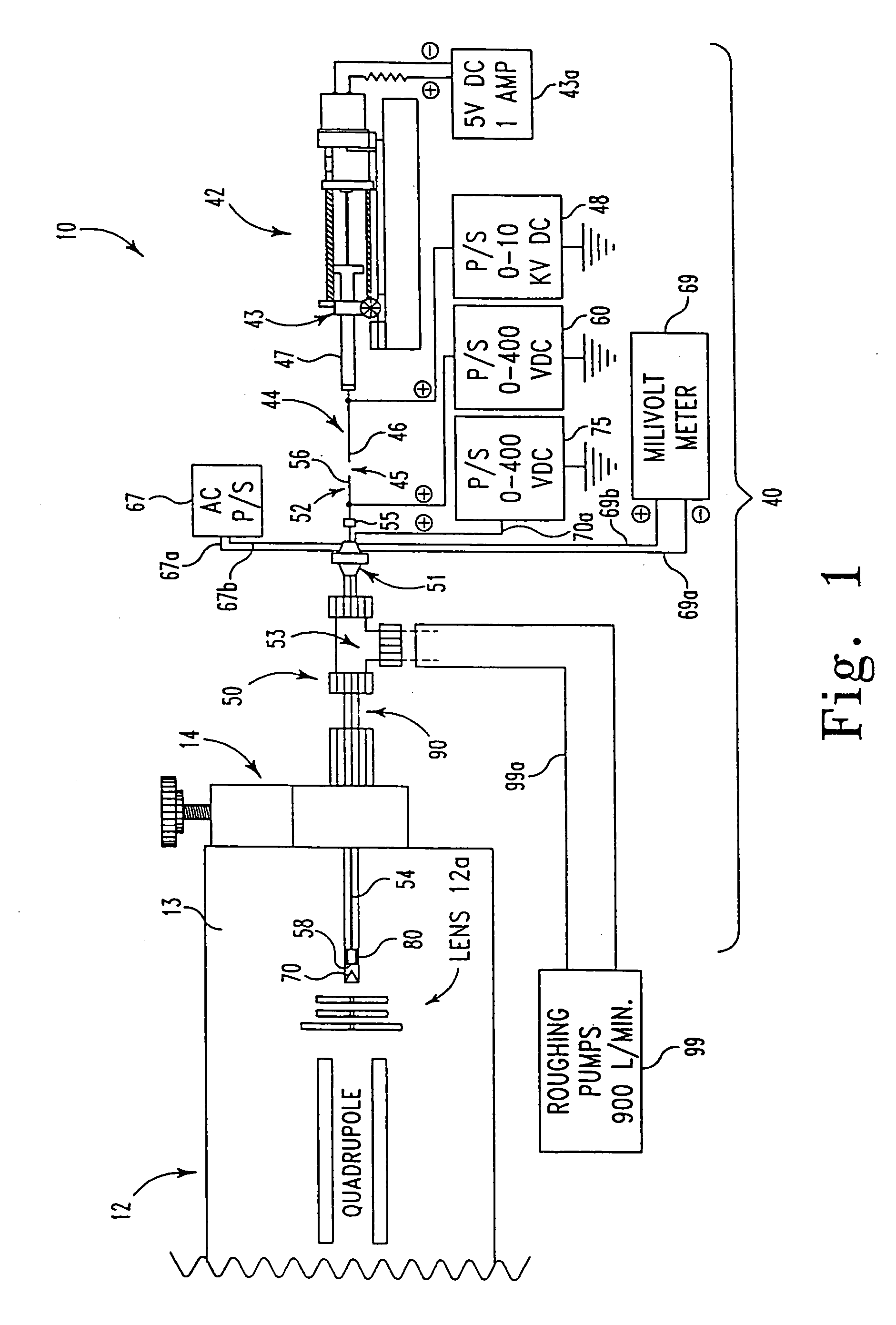
[0072] The ES spectrum of glucagon depicted in FIG. 6 was characterized using a standard EI / CI lens assembly on an Extrel dual quadrupole mass spectrometer (50 in 50:50 MeOH:H2O, 1% acet...

example two

[0074] A study was conducted of the non-covalent interactions of three crown ethers, dicyclohexano-18-crown-6 (#1), 18-crown-6 (#2), and dibenzo-18-crown-6 (#3) (FIGS. 17A-17C) with three types of cytochrome c; horse tuna and yeast, utilizing the ES probe 50 and method of this invention. Each of the three different types of cytochrom c displayed different degrees of binding for each of the three crown ethers; however, the binding of the crown ethers was found to increase in the order given above with dicyclohexano-18-crown-6 binding the most tightly and dibenzo-18-crown-6 binding the least tightly.

[0075] More particularly, the experiments showing binding of crowns to cytochrome c were done by adding 1, 2, and 3 mol ratios to a 70 pmol / μL mixture of the three different cytochrome c's. The solutions were prepared with equal parts of methanol / water with 1% acetic acid. Typical sample conditions were, 95° C., 2 μL / min, 4000 VDC on the syringe, 170 VDC on the capillary tube, and 60 VDC ...


PUM


Abstract

A method for characterizing the three-dimensional surface structure of molecules, particularly proteins and protein complexes, employing mass spectrometric analysis, an electrospray ionization (ES) source, a novel data interpretation process that utilizes comparisons of particular binding constants (KB) and heats of formation (ΔHf), and computational feedback modeling.

Description

REFERENCE TO RELATED APPLICATIONS [0001] This application is a continuation-in-part application of U.S. Ser. No. 09 / 287,307, now U.S. Pat. No. 7,047,171, which is a continuation-in-part application of U.S. Ser. No. 08 / 569,358, now abandoned, which is a continuation-in-part application of U.S. Ser. No. 08 / 147,688, now U.S. Pat. No. 5,504,327.BACKGROUND OF THE INVENTION [0002] 1. Field of Invention [0003] The present invention relates to methods for characterization of the structure of molecules and, more particularly, to a method for characterizing the three-dimensional surface structure of proteins and protein complexes employing electrospray mass spectrometric analysis (ES-MS) techniques and computational feedback modeling. [0004] 2. Description of the Prior Art [0005] The employment of mass spectrometry for identification of chemical structures, molecular weights, determination of mixtures, and quantitative elemental analysis, based on the application of the mass spectrometer, is ...

Claims


Application Information

Patent Timeline
IPC IPC(8): G06G7/48
CPCG01N33/6848Y10T436/25Y10T436/24H01J49/0431
Inventor SPROCH, NORMAN K.KRUGER, TERRY L.KRUGER, KARYN
Owner HV OPS H NU
Why Eureka
Try Eureka