
Electrophysiology catheter and system for gentle and firm wall contact

a technology of electrodes and walls, applied in the field of electrophysiology medical procedures in the intracardiac cavity, can solve the problems of reducing the effectiveness of mapping and ablation, difficulty in maintaining the desired contact with the moving surface of the heart, and local anomalies, and achieves the effect of satisfying and sa

Inactive Publication Date: 2006-12-14
STEREOTAXIS
View PDF10 Cites 102 Cited by
  • Summary
  • Abstract
  • Description
  • Claims
  • Application Information

AI Technical Summary

Benefits of technology

"The present invention provides improved control of medical devices in contact with anatomical surfaces, particularly moving surfaces such as the heart. The invention uses flexible or stiffer devices that maintain a firm but gentle contact with the surface through the use of feedback control systems. The devices can be telescoped from a guide sheath and provide satisfactory and safe contact for cardiac mapping, pacing, and ablation. The invention also allows for control of contact pressure between predetermined minimum and maximum values. Overall, the invention provides a safer and more effective means of controlling medical device contact with anatomical surfaces."

Problems solved by technology

It is difficult to maintain the desired contact with the moving surface of the heart during the entire cardiac cycle.
This tends to locally distend the tissue during part of the cycle, and cause relatively wide variance in the contact force between the device and the tissue, potentially reducing the effectiveness of mapping and ablation.
This distention may also create a local anomaly of the electrical activity that the physician is attempting to map.

Method used


Image

  • Electrophysiology catheter and system for gentle and firm wall contact
  • Electrophysiology catheter and system for gentle and firm wall contact
  • Electrophysiology catheter and system for gentle and firm wall contact

Examples


second embodiment

[0019] Alternatively, in a second embodiment, the catheter actuated by a remote navigation system can be advanced (possibly by using a joystick or other control), or magnetic field or other control variable applied, until distal catheter shaft prolapse is visible on an X-ray image or an ultrasound image. This prolapse of the catheter can be continually monitored by the user during the diagnostic process, or during the therapy delivery portion of the procedure (such as RF ablation).

third embodiment

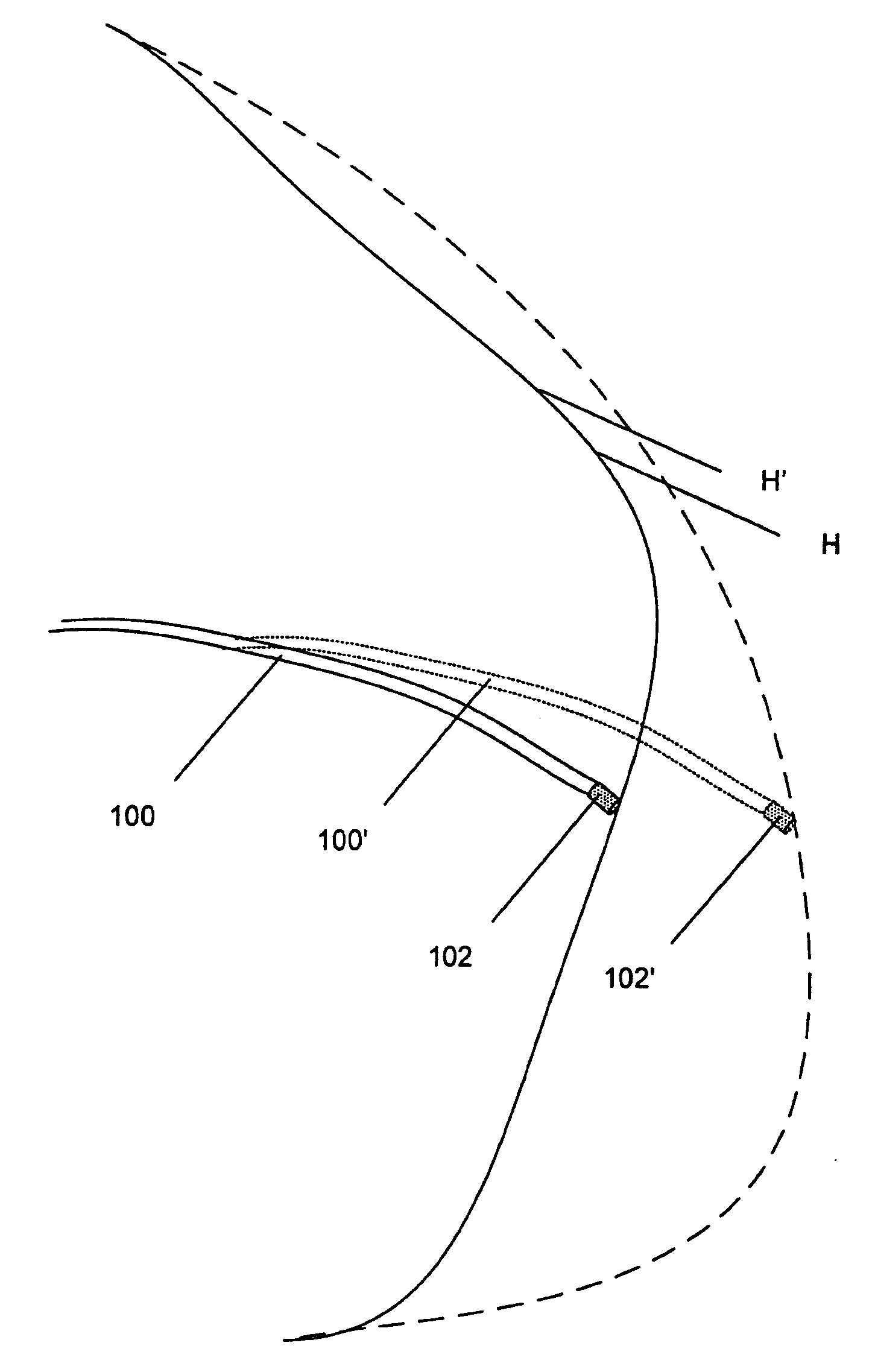
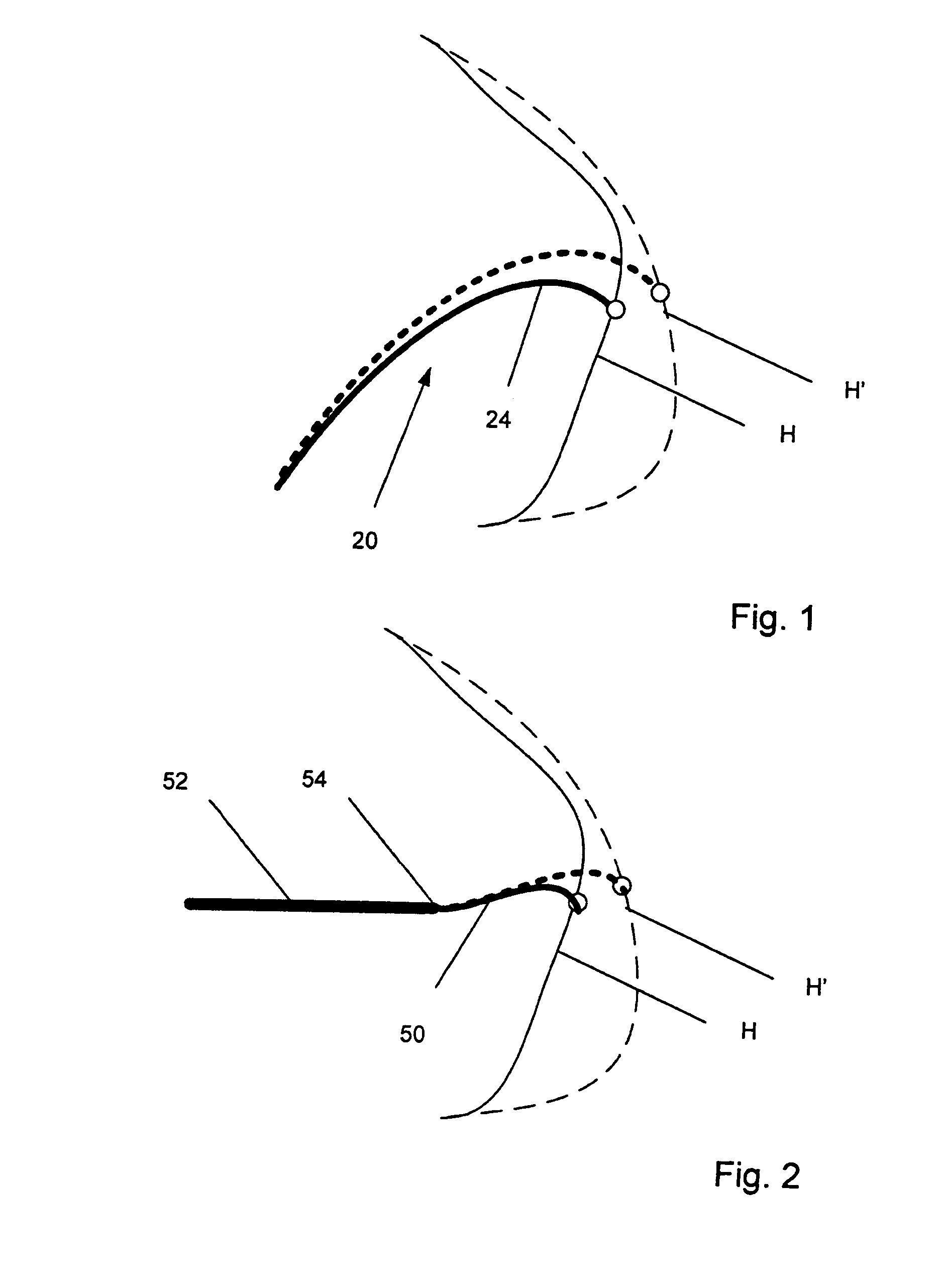
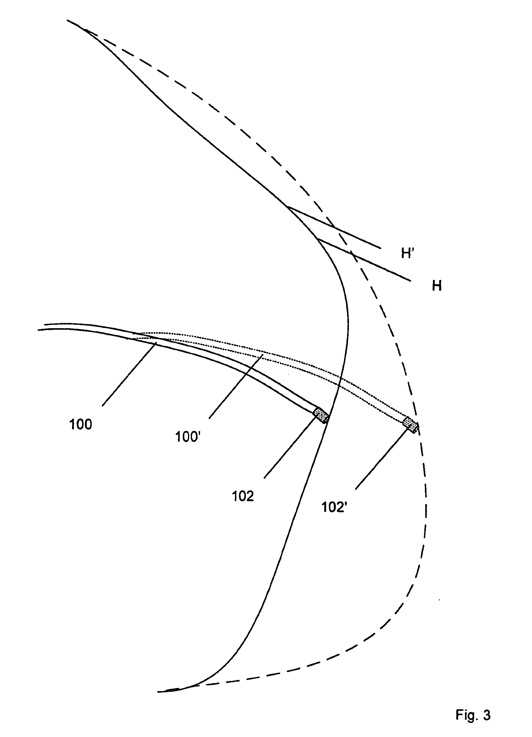
[0020] In a third embodiment shown in FIG. 2, the flexible catheter 50 is disposed inside a guide sheath 52. The guide sheath 52 is navigated to a position adjacent to and opposed to the anatomical surface of interest. This can be conveniently done with a remote navigation system, such as a magnetic navigation system or a mechanical navigation system that orients the distal end of the guide sheath. Once the distal end 54 of the guide sheath 52 is positioned, the catheter 50 is advanced until it contacts the anatomical surface and buckles. More specifically, the catheter 50 is advanced until it remains buckled during the entire cycle of movement. This is illustrated in FIG. 2 which shows that when the heart is contracted, the catheter 50 (shown in solid lines) contacts the wall of the heart H (shown in solid lines, and when the heart is expanded, the catheter indicated as 50′ (shown by the dashed lines) contacts the wall of the heart indicated as H′ (shown in dashed lines).

[0021] By ...

fourth embodiment

[0022] Alternatively, in a fourth embodiment, a guide sheath actuated by the remote navigation system can be advanced (possibly by using a joystick or other control), or magnetic field or other applied control variable, until distal catheter shaft prolapse is visible on an X-ray image or an Ultrasound image. This prolapse of the catheter can be continually monitored by the user during the diagnostic process, or during the therapy delivery portion of the procedure (such as RF ablation).

[0023] Examples of a guide sheaths are disclosed in U.S. Pat. No. 6,527,782, issued Mar. 4, 2003, for “Guide for Medical Devices”, incorporated herein by reference. In one preferred embodiment the guide sheath can be actuated mechanically with pull-wire cables, as also described therein. The wires can be driven with computer-controlled servo motors or other mechanical means. The soft catheter passes through the sheath and the length of catheter that extends from the distal end of the sheath can itself ...


PUM


Abstract

A method of applying an electrode on the end of a flexible medical device to the surface of a body structure, the method including navigating the distal end of the device to the surface by orienting the distal end and advancing the device until the tip of the device contacts the surface and the portion of the device proximal to the end prolapses. Alternatively the pressure can be monitored with a pressure sensor, and used as an input in a feed back control to maintain contact pressure within a pre-determined range.

Description

CROSS-REFERENCE TO RELATED APPLICATIONS [0001] This application is a continuation of U.S. patent application Ser. No. 11 / 446,522, Files Jun. 2, 2006, which claims the benefit of U.S. Provisional Patent Application Ser. No. 60 / 686,786, filed Jun. 2, 2005, the entire disclosures of which is incorporated herein by reference.BACKGROUND OF THE INVENTION [0002] In intracardiac electrophysiology medical procedures, catheters have been routinely used for many years to map cardiac electrical abnormalities (arrhythmias) for diagnostic purposes, and to deliver therapy by Radio Frequency (RF) ablation of diseased tissue or abnormal electrical nodes. Usually, such catheters have been navigated within the anatomy by deflecting them with a manually operated handle, and torquing or twisting them by hand. Typically, the handle is connected to mechanical pull wires that deflect or manipulate the distal portion of the device through suitably applied tension or compression. [0003] For certain cardiac m...

Claims


Application Information

Patent Timeline
Patent Type & Authority Applications(United States)
IPC IPC(8): A61B19/00
CPCA61B18/1492A61B19/52A61B19/5244A61B2017/00053A61B2017/00243A61B2019/5276A61B2019/2253A61B2019/464A61B2019/465A61B2019/481A61B2019/5238A61B2018/00839A61B34/73A61B90/36A61B34/20A61B2090/064A61B2090/376A61B2090/065A61B2090/08021A61B2090/378
Inventor VISWANATHAN, RAJU R.
Owner STEREOTAXIS
Learn More