
Method for modulating gene expression in epithelial cells
- Summary
- Abstract
- Description
- Claims
- Application Information
AI Technical Summary
Benefits of technology
Problems solved by technology
Method used
Image

Examples
example 1
GM-284 Promotes Wound Healing
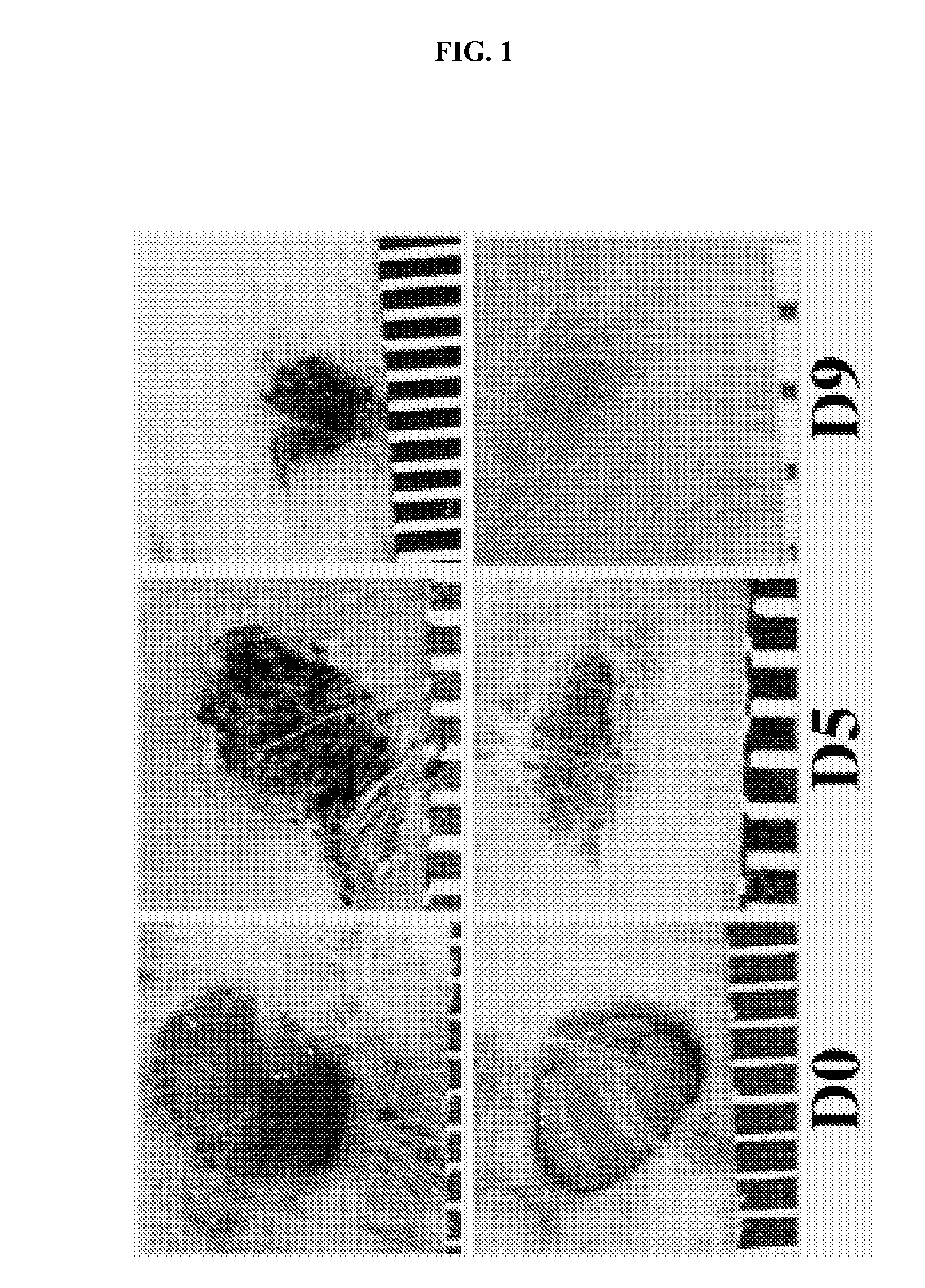
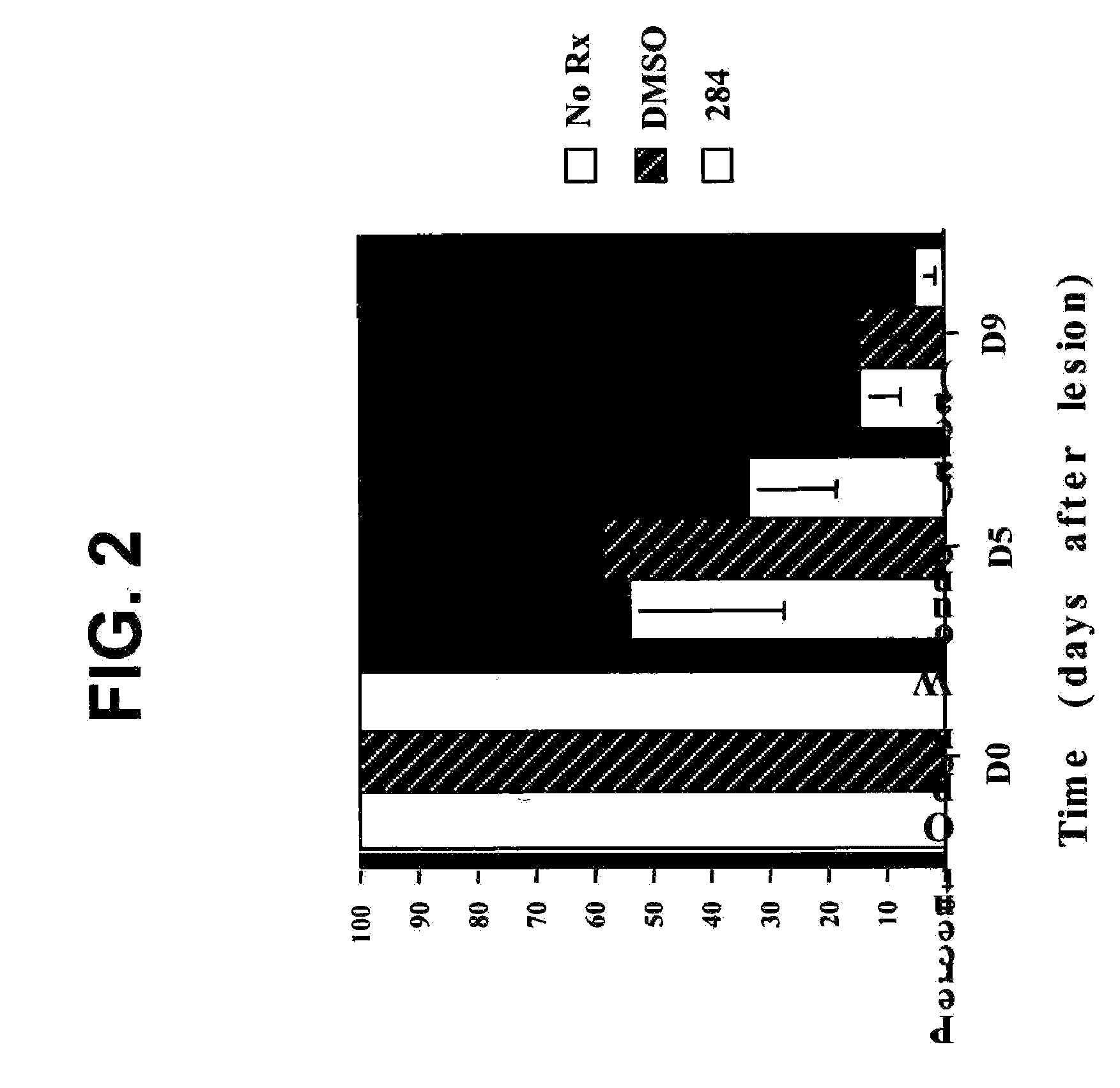
[0077] In recent studies, the inventor demonstrated myelin and axonal hypertrophy following mechanical transection of the sciatic nerve. These studies required surgical exposure of the right lower quadrant of all study animals. Following exposure of the sciatic nerve from the sciatic notch to the knee, and manipulation of the nerve, the muscle and overlaying skin were closed with suture material. Each animal was subsequently treated with a daily gavage of either GM-284 (5 mg / kg), FK506 (5 mg / kg), or methylcellulose vehicle alone. During the post-operative course, it was noted by an investigator who was blinded to the treatment groups that the surgical wounds of one group of animals appeared to be qualitatively improved over those of the other two groups, with less erythemia and more rapid closure. Following unblinding of the investigator, it was learned that the group demonstrating improved healing had received treatment with GM-284.
[0078] To determine...
example 2
GM-284 Doubles the Keratinocyte Proliferative Index
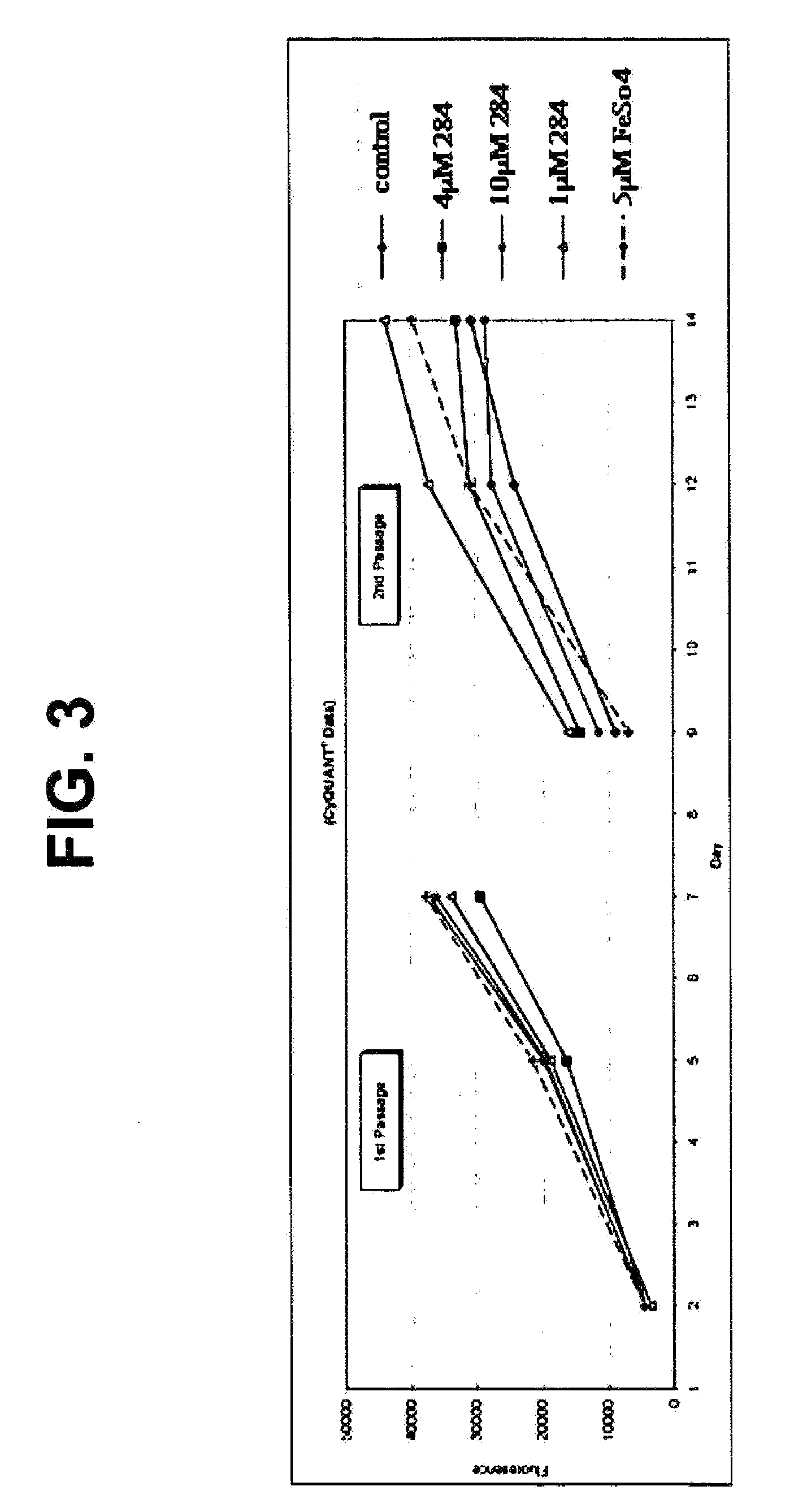
[0081] As detailed above, keratinocyte proliferation is influenced by a number of factors, including innervation (Hsieh and Lin, Modulation of keratinocyte proliferation by skin innervation. J. Invest. Dermatol., 113:579-86, 1999) and growth-factor treatment (Castagnino et al., Neu differentiation factor / heregulin induction by hepatocyte and keratinocyte growth factors. Oncogene, 19:640-48, 2000; and reviewed by Werner and Smola, Paracrine regulation of keratinocyte proliferation and differentiation. Trends Cell Biol., 11:143-46, 2001). Thus, by culturing human-foreskin keratinocytes with increasing concentrations of GM-284, the inventor investigated the possibility that GM-284 was able to influence keratinocyte growth. As shown in FIG. 3, treatment with 1 μM of GM-284 increased the rate of keratinocyte proliferation by more than 150% after 14 days of treatment. Not surprisingly, a comparison of the growth rates of keratinocytes gr...
example 3
Topical Treatment with GM-284 Increases Epidermal Thickness In Vivo
[0082] The increased rate of wound closure in the presence of GM-284, taken with the accelerated keratinocyte proliferation, suggested to the inventor the possibility that GM-284 treatment might alter epidermal thickness in vivo. To examine this possibility, 5-mm, full-skin thickness biopsies were taken from the hindquarters of both male and female ICR retired breeders. The wounds were treated daily, for 8 days, with topical administration of either the DMSO-based vehicle, or FK506 or GM-284 in the vehicle. Thereafter, the original biopsy site was rebiopsied. Comparison of epidermal thickness of the FK506- and vehicle-treated animals showed no observable differences (cf. FIG. 4, panels a and b). In contrast, the epidermis of the GM-284-treated animals was much thicker, and appeared to be much more cellular, than the epidermis of animals under the other two conditions (FIG. 4, panel c). Without being bound by theory,...
PUM
| Property | Measurement | Unit |
|---|---|---|
| Cell angle | aaaaa | aaaaa |
| Gene expression profile | aaaaa | aaaaa |
Abstract
Description
Claims
Application Information