Method and apparatus for x-ray imaging of a patient during a shockwave treatment

a technology for x-ray imaging and shockwave treatment, which is applied in the field of method and an apparatus for x-ray imaging of a patient, can solve the problems that the exposure to x-ray radiation is not risk-free for the patient or the personnel, and achieve the effects of less power, less radiation, and less radiation

Inactive Publication Date: 2007-02-01
SIEMENS AG
View PDF5 Cites 10 Cited by
  • Summary
  • Abstract
  • Description
  • Claims
  • Application Information

AI Technical Summary

Benefits of technology

[0043] With the memory it is possible to provide the time-invariant information of the image data set for the later evaluation with regard to the information, or to store said time-invariant information up to the second point in time, or the acquisition of the x-ray image and the representation of the information. The x-ray system for the x-ray image can be made smaller, with less power and at a lower cost than the system for acquisition of the image data set since—as noted above—the x-ray image is to be acquired with a lower dose or smaller image field. A weaker-power x-ray system in

Problems solved by technology

Due to the danger of radiation injury, exposure to x-ray radiation is not

Method used


Image

  • Method and apparatus for x-ray imaging of a patient during a shockwave treatment
  • Method and apparatus for x-ray imaging of a patient during a shockwave treatment

Examples


Embodiment Construction

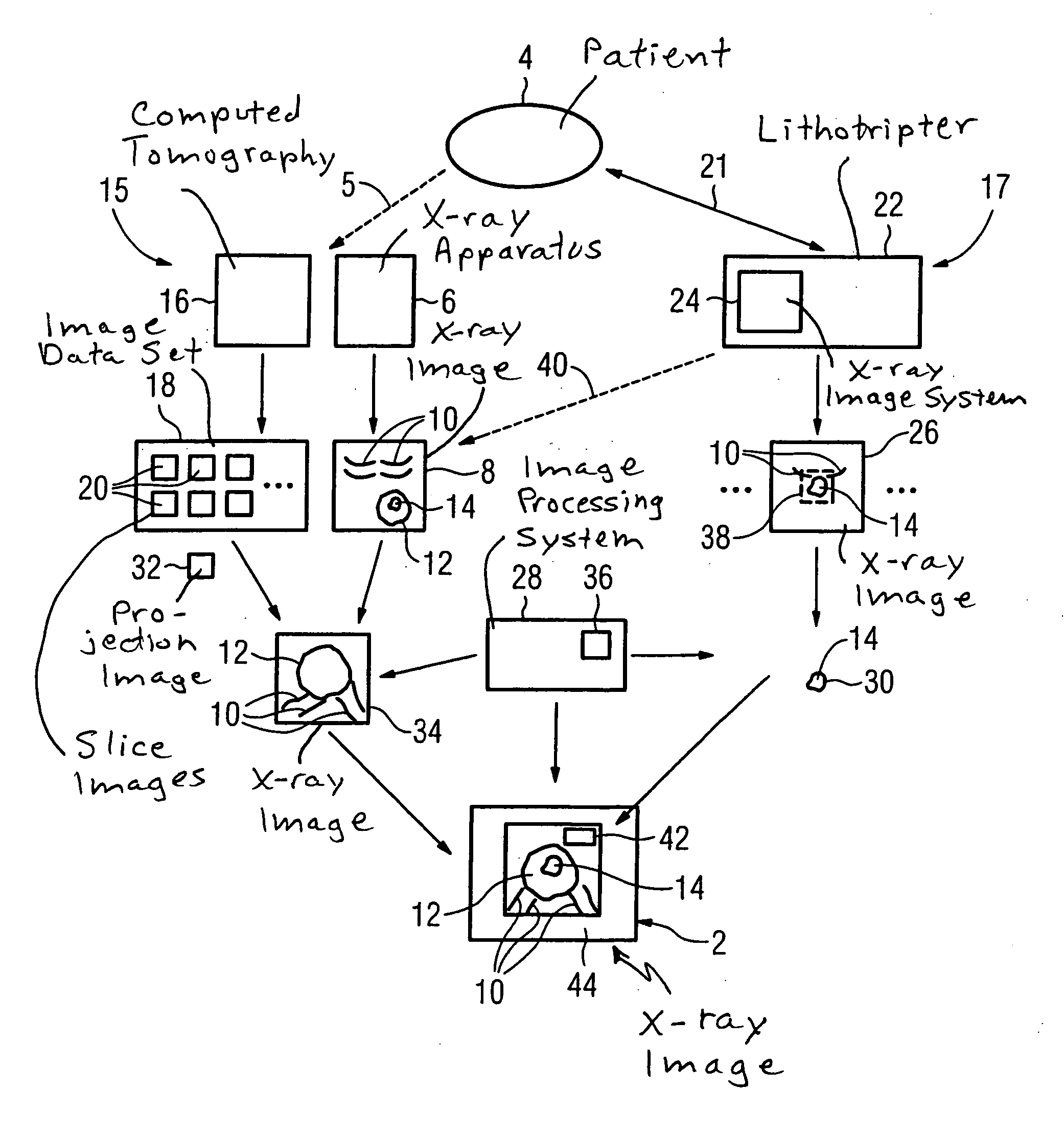
[0051] The FIGURE shows an exemplary workflow scenario for generation of an x-ray image 2 of a patient 4 during a kidney stone lithotripsy. The case history is that a patient 4 seeks out a doctor (not shown) and complains of abdominal pains. With an x-ray apparatus 6, the doctor (not shown) immediately produces an x-ray image 8 of the patient 4 by exposure of the patient with a standard dose of x-rays. In addition to the ribs 10 of the patient, his kidneys 12 with a kidney stone 14 located therein are visible on the x-ray image 8.

[0052] In order to confirm the initial diagnosis of the kidney stone 14 in the patient 4, the doctor arranges a further examination of the patient 4 at a later point in time 15. This corresponds to the inventive first point in time. By a computed tomography 16, a 3D image data set 18 containing a number of slice exposures 20 of the patient is hereby produced (likewise indicated by the arrow 5). The evaluation of the 3D image data set 18 confirms the initia...


PUM


Abstract

In a method for x-ray imaging given a patient containing a subject to be represented during a shockwave treatment, an image data set containing the subject and a marker is generated at a first point in time; an x-ray image showing essentially only the subject (14) and the marker is acquired at a second point in time, the x-ray image is correctly spatially associated with the image data set using the marker, the x-ray image is displayed together with information extracted from the image data set during the shockwave treatment. An apparatus for x-ray imaging a patient containing a subject to be represented during a shockwave treatment has a memory for an image data set generated at a first point in time and containing the subject and a marker, an x-ray system for acquisition of an x-ray image showing essentially only the subject and the marker at a second point in time; an evaluation unit for spatially-accurate association of the x-ray image with the image data set using the marker and for extraction of information from the image data set, and a display unit for display of the x-ray image together with the information during the shockwave treatment.

Description

BACKGROUND OF THE INVENTION [0001] 1. Field of the Invention [0002] The present invention concerns a method and an apparatus for x-ray imaging OF a patient containing a subject to be represented during a shockwave treatment. [0003] 2. Description of the Prior Art [0004] Imaging methods are used today in many fields of medicine. One of the most widespread methods is x-ray imaging, in which a living person or animal as a patient is irradiated with ionizing x-ray radiation. The goal is the representation or imaging of a subject of interest located in the patient. This can, for example, an internal organ, a foreign body or the like. [0005] In addition to the many known advantages of x-ray imaging there are certain disadvantages associated therewith. Due to the danger of radiation injury, exposure to x-ray radiation is not risk-free for the patient or for the personnel conducting the x-ray exam. The x-ray dose radiated on a living patient is therefore legally limited to maximum values fo...

Claims


Application Information

Patent Timeline
IPC IPC(8): A61N5/10
CPCA61B6/12A61B17/2256A61B19/54G06T2207/30004G06T7/0028G06T7/0038A61B2019/5238A61B2090/376A61B90/39G06T7/33G06T7/38
Inventor BUCHHOLTZ, GERHARDFEHRE, JENSGRANZ, BERNDHOHEISEL, MARTINKRUFT, WERNERLANSKI, MARKUSMAHLER, MATTHIASMEINERT, CHRISTIANMERTELMEIER, THOMASNANKE, RALFRATTNER, MANFRED
Owner SIEMENS AG
Why Eureka
Try Eureka