

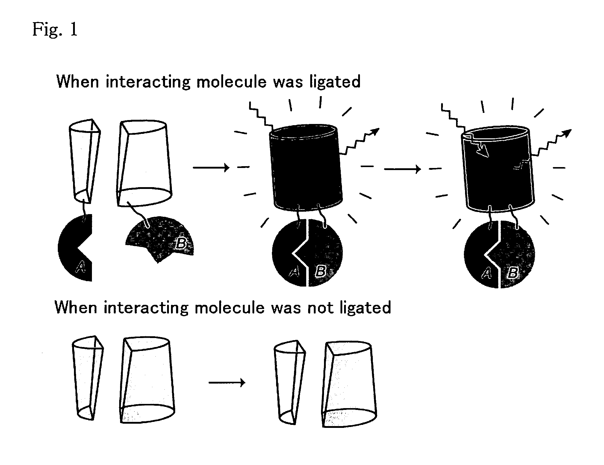
Method for analysis of protein interaction using fluorescent protein
a protein and fluorescent protein technology, applied in the field of protein interaction analysis using fluorescent proteins, can solve problems such as inability to monitor
- Summary
- Abstract
- Description
- Claims
- Application Information
AI Technical Summary
Benefits of technology
Problems solved by technology
Method used
Image

Examples
example 1
Preparation of mKO Mutant by Point Mutagenesis in which Multimer Formation is Blocked
[0066] The multimer formation boundary was predicted from the amino acid sequence of KO-1, and amino acids in the multimer formation boundary were substituted so that KO-1 was monomerized but the fluorescence characteristic was maintained. The introduction of the point mutation was carried out in an E. coli expression vector with KO-1 inserted thereinto (pRSET B) (the expression vector containing DNA encoding KO-1 described in International Publication WO03 / 54191) using primers for point mutagenesis. In particular, a multiplicity of point mutagenesis primers were annealed at the same time with one chain of the template plasmid and extended with polymerase. The DNA fragments extended by each primer were ligated in the same reaction mixture using DNA ligase. In this procedure, the DNA produced was complementary to the template except where the mutation was introduced. Since the ends of the DNA must b...
example 2
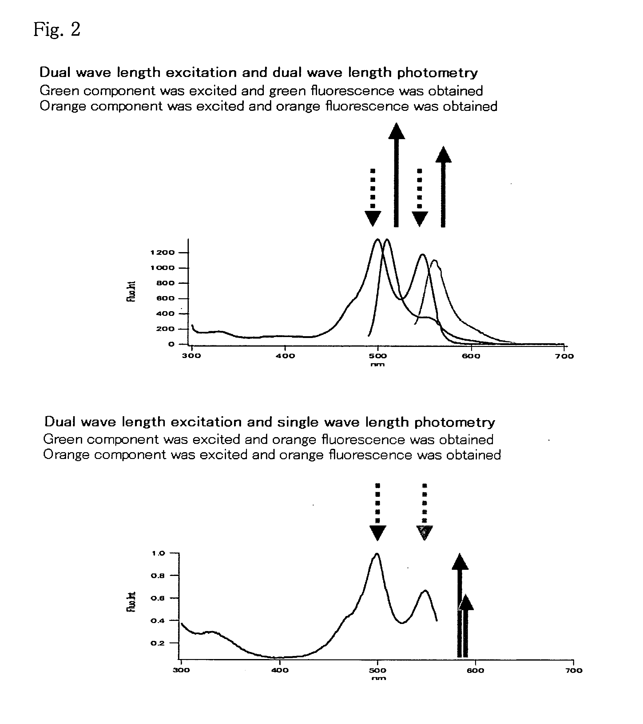
Analysis of Fluorescence Characteristic
[0083] Fluorescent and absorption spectra of mKO protein purified in Example 1 were measured as follows, and quantum yield and molar absorption coefficient were calculated.
[0084] Absorption spectra were measured using the 20 μM fluorescent protein, 50 mM HEPES pH 7.5 solution. The molar absorption coefficient was calculated from the peak values of the spectra. The absorption peak was observed at 548 nm for mKO. The fluorescent protein was diluted with the aforementioned buffer so that the absorbance at 500 nm was 0.0025, and the fluorescence spectra with excitation by 500 nm and excitation spectra by fluorescence at 590 nm were measured. DsRed (CLONTECH) was similarly diluted so that the absorbance at 500 nm was 0.0025, and the fluorescent spectra were measured and the quantum yield of mKO was obtained assuming the quantum yield of DsRed was 0.29.
[0085] The results are shown in Table 1. The data of KO protein (dimer protein) described in Int...
example 3
Measurement of Molecular Weight by Ultracentrifugation Analysis
[0086] mKO protein solution was prepared in 150 mM KCl, 50 mM HEPES-KOH, pH 7.4, and subjected to ultracentrifugation analysis to determine the molecular weight of mKO. The solution was centrifuged using an ultracentrifuge XL-1 (Beckman Coulter) at 25,000 rpm for 22 hours, and absorbance at 540 nm, which was close to the absorption peak (548 nm) of mKO, was measured. The molecular weight of mKO was calculated to be 28 kDa from the result of the measurement. This value is almost the same with 26 kDa which is predicted from the amino acid sequence, confirming that mKO exists as monomer.
PUM
 Abstract
Description
Claims
Application Information