Diagnosis device for radiographic and nuclear medical examinations

a radiographic and nuclear medical examination technology, applied in the direction of radiographic control devices, instruments, tomography, etc., can solve the problems of limiting the accuracy of detection, unable to provide good anatomical imaging, and unable to accurately locate tumors or metastases in the body of patients, etc., to achieve good anatomical imaging, good accessibility, and high registration precision

Inactive Publication Date: 2007-05-10
SIEMENS HEATHCARE GMBH
View PDF12 Cites 16 Cited by
  • Summary
  • Abstract
  • Description
  • Claims
  • Application Information

AI Technical Summary

Benefits of technology

[0046] The advantages achieved with the invention thus particularly consist in the good anatomical imaging by means of the radiographic examination, combined with the functional imaging with the display of the metabolism by m

Problems solved by technology

As is known, the SPECT technique does not provide good anatomical imaging, thereby rendering the localization of detected tumors or metastases in the body of the patient problematic for instance.
The challenge which then presents itself is that of correct registration, n

Method used


Image

  • Diagnosis device for radiographic and nuclear medical examinations
  • Diagnosis device for radiographic and nuclear medical examinations
  • Diagnosis device for radiographic and nuclear medical examinations

Examples


Embodiment Construction

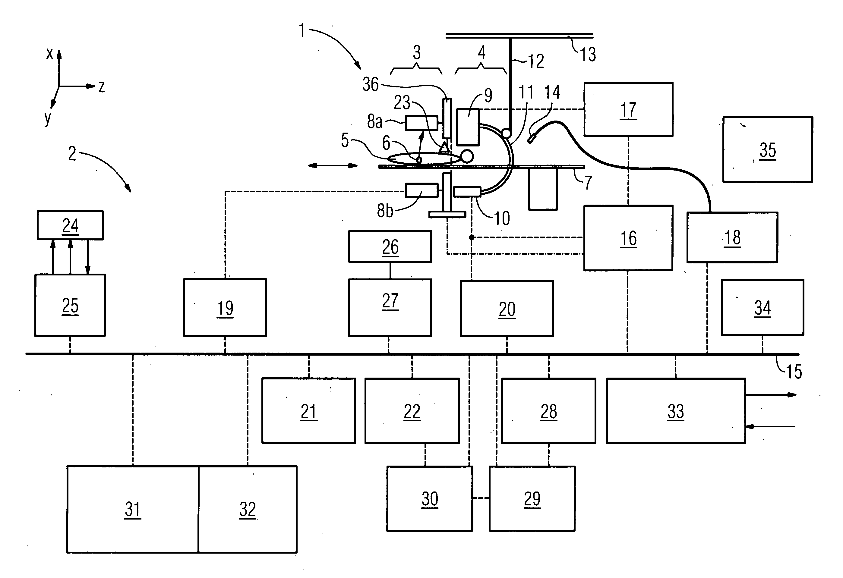
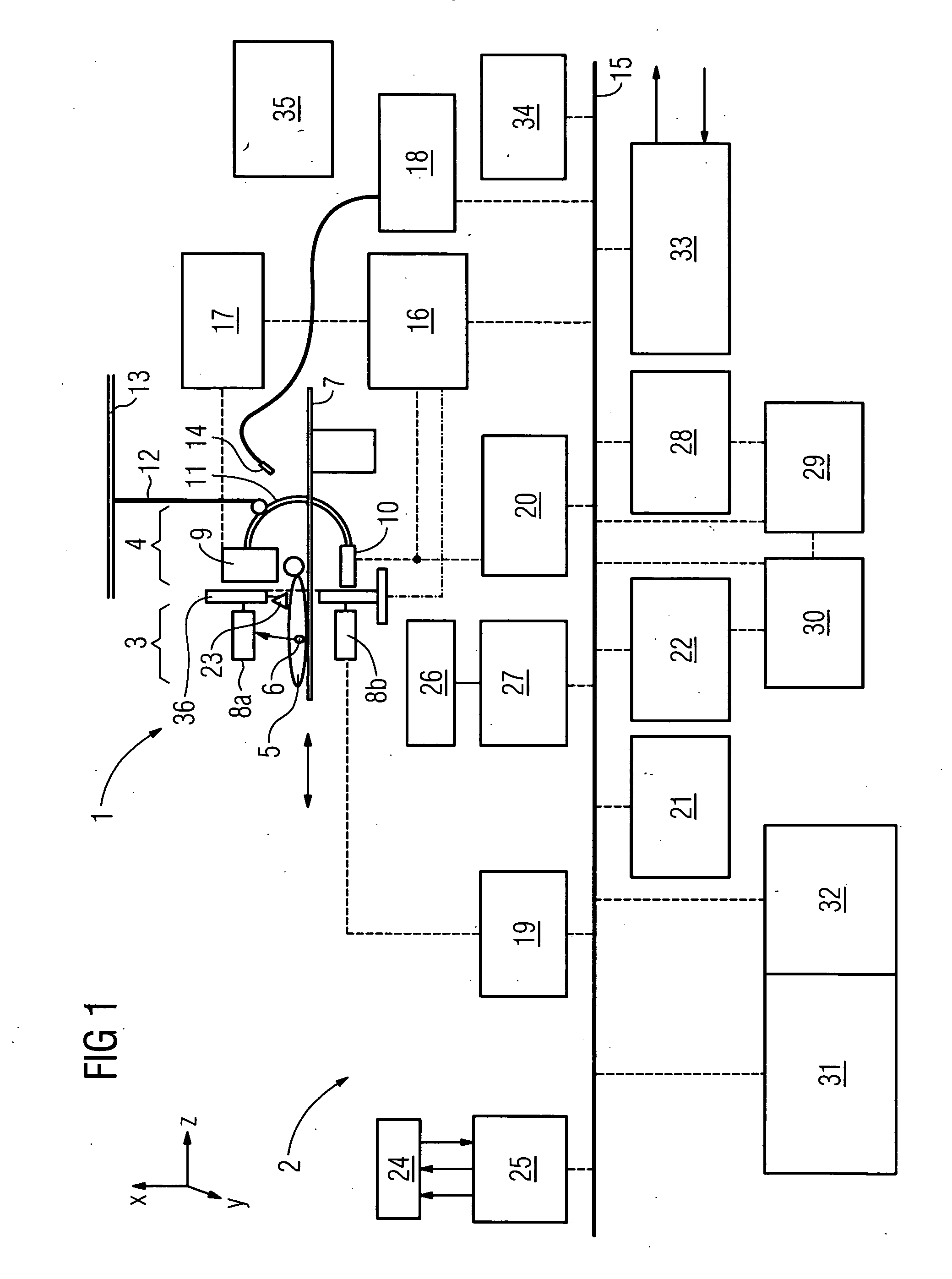
[0055]FIG. 1 shows a schematic representation of a first exemplary embodiment of an inventive diagnosis device 1 with the block diagram structure of an evaluation and / control device 2.

[0056] The diagnosis device 1 comprises a first measurement area 3 for recording SPE or SPECT images and a second measurement area 4 for recording x-ray images. A patient 5 administered with radiopharmacs containing radioactive materials prior to the examination, which accumulate in a tumor as tracer 6, is moved by means of a patient bed in the form of support 7, which can be moved in the Z-direction, for an examination, firstly through the first measurement area 3 and then through or into the second measurement area 4.

[0057] The first measurement area 3 comprises an SPE or SPECT measuring head, which is designed in FIG. 1 as two opposing detector arrays 8a, b which are aligned to one another. The detector arrays 8a, b each comprise a collimator (not shown in further detail), which operates as a dire...


PUM


Abstract

Diagnosis device for combined and/or combinable radiographic and nuclear medical examinations Digital imaging methods have now become common practice in medical diagnostics. Methods of this type have been used for some time, e.g. in computer tomography, for magnetic resonance examinations, ultrasound examinations and for nuclear medical methods.
The object underlying the invention is to propose a diagnosis device for increasing accessibility to a patient. To this end, a diagnosis device (1) is proposed for combined and/or combinable radiographic and nuclear medical examinations with an x-ray source (9), with an examination room for accommodating a patient (5), with a gamma radiation source (6) arranged in the body, with the diagnosis device (1) being designed in order to carry out the radiographic examination by evaluating the measurement of the x-rays, and in order to carry out a single photon emission (SPE) examination as a nuclear medical examination by evaluating the gamma radiation, with a detector system (8a,b, 10) which has a detector surface for simultaneously measuring the x-ray and gamma radiation without changing the patient's position and/or which is designed to record a two-dimensional locally-resolved and object-imaging individual x-ray projection image.

Description

CROSS REFERENCE TO RELATED APPLICATIONS [0001] This application claims priority of German application No. 10 2005 053 994.7 filed Nov. 10, 2005, which is incorporated by reference herein in its entirety. FIELD OF THE INVENTION [0002] The invention relates to a diagnosis device for combined or combinable radiographic and nuclear medical examinations having an x-ray source, an examination room for accommodating a patient with a gamma radiation source arranged in the body, with the diagnosis device being designed in order to carry out the radiographic examination by evaluating the measurement of the x-rays and to carry out a single-photon-emission (SPE) examination as a nuclear medical examination by evaluating the gamma radiation. BACKGROUND OF THE INVENTION [0003] Digital imaging methods have now become common practice in medical diagnostics. Methods of this type have been used for some time, e.g. in computer tomography, for magnetic resonance examinations, ultrasound examinations an...

Claims


Application Information

Patent Timeline
IPC IPC(8): G01T1/24
CPCG01T1/1615A61B6/037G01T1/1648G01T1/1642A61B6/4464
Inventor MASCHKE, MICHAEL
Owner SIEMENS HEATHCARE GMBH
Why Eureka
Try Eureka