
Reinforced small intestinal submucosa

a submucosa and small intestinal technology, applied in the field of bioprosthetics, to achieve the effect of improving mechanical and handling properties

Inactive Publication Date: 2007-06-07
DEPUY SYNTHES PROD INC
View PDF99 Cites 35 Cited by
  • Summary
  • Abstract
  • Description
  • Claims
  • Application Information

AI Technical Summary

Benefits of technology

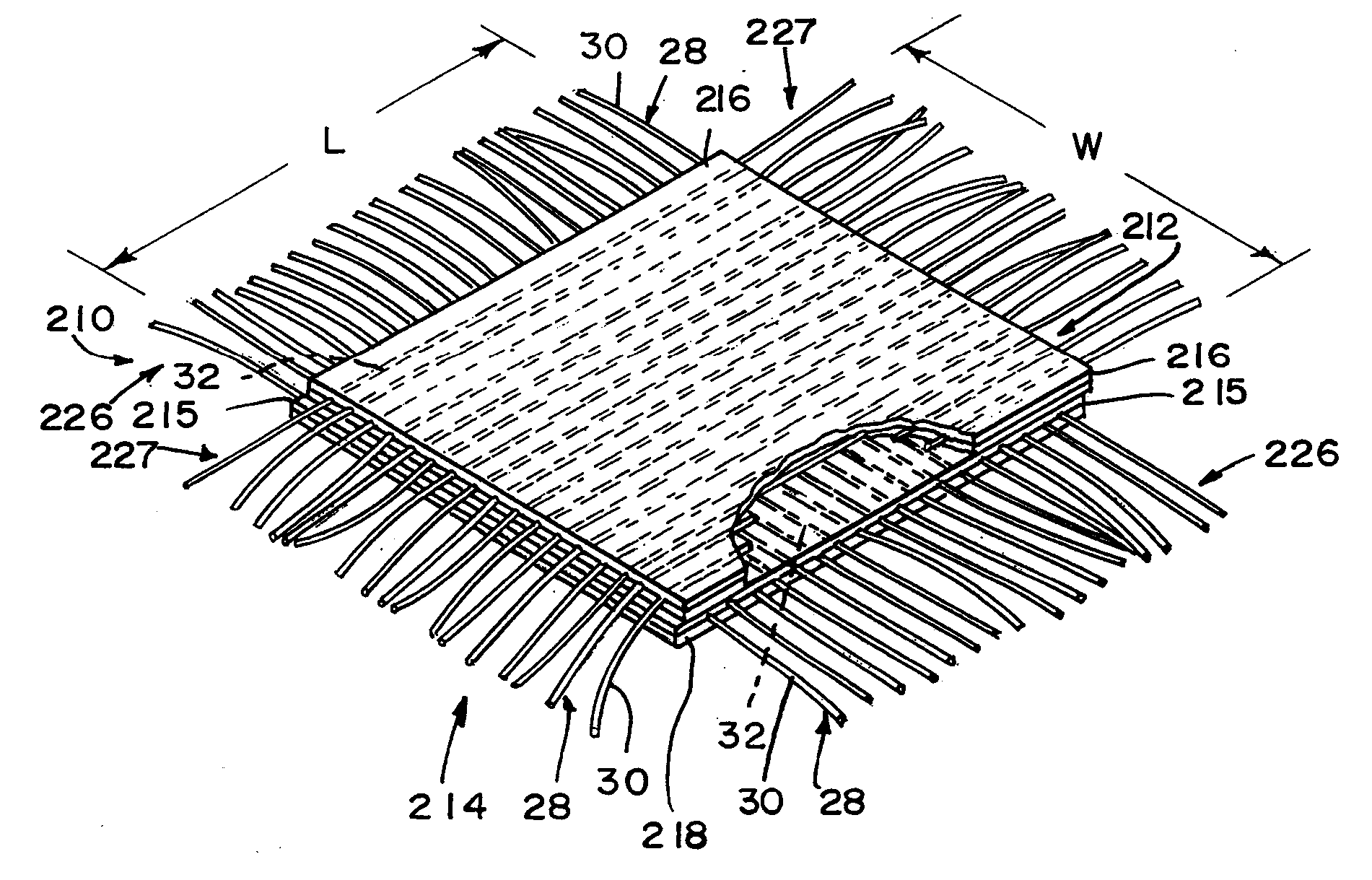
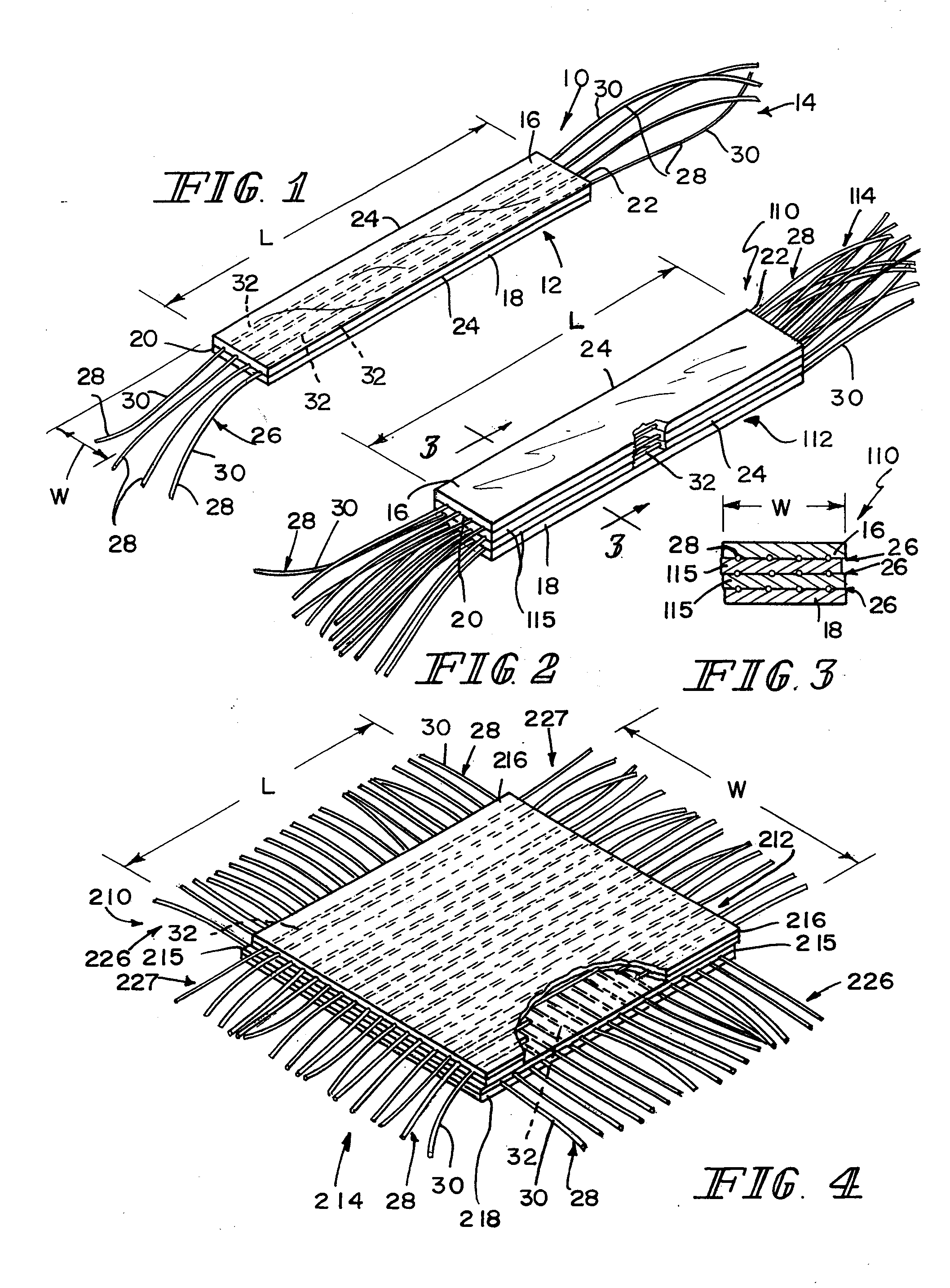
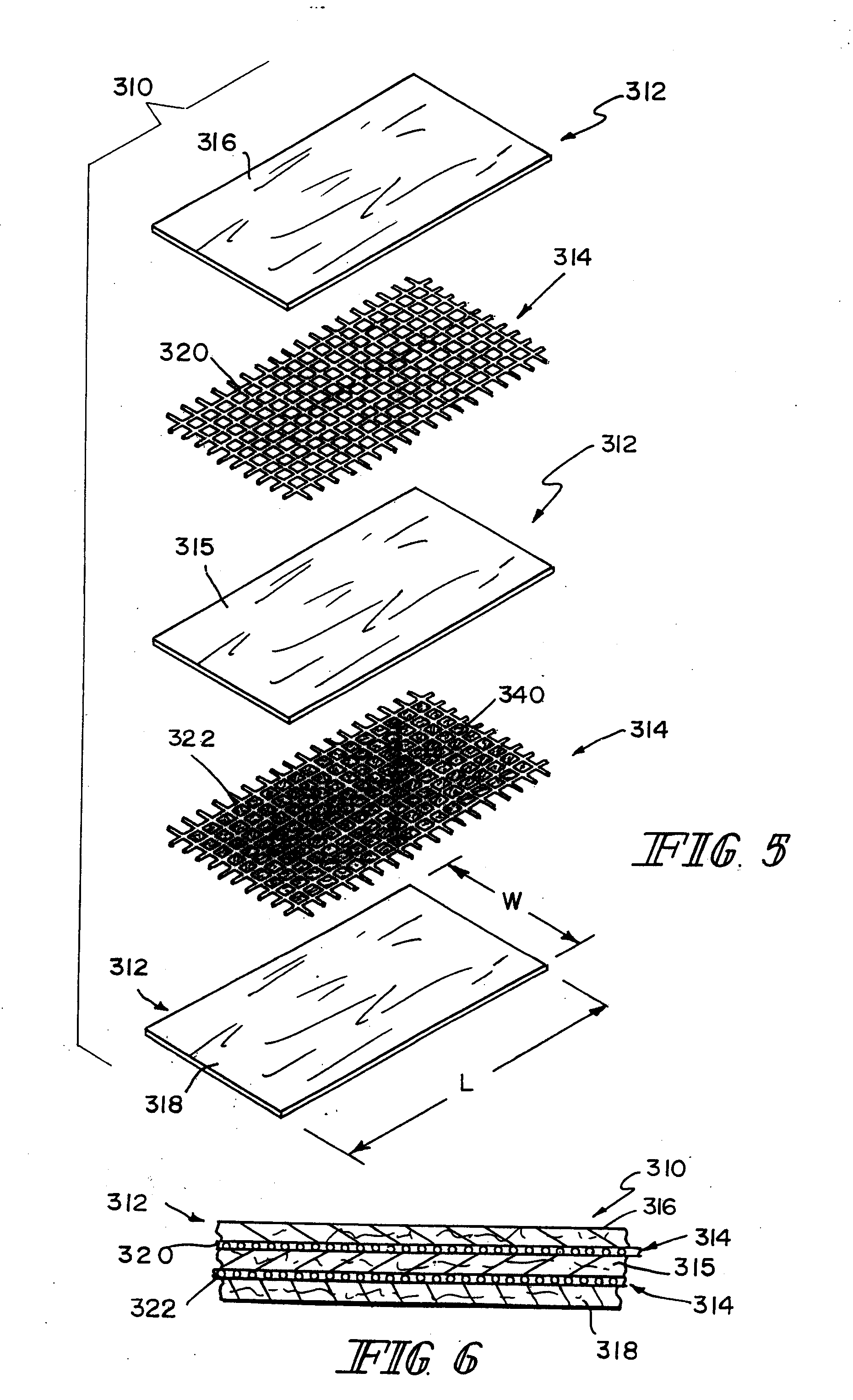
The present invention provides a bioprosthetic device for soft tissue attachment, reinforcement, and reconstruction. The device includes a small intestine submucosa (SIS) or other naturally occurring extracellular matrix (ECM) that is formed to include a tissue layer of SIS and a synthetic portion coupled to the SIS tissue layer. The synthetic portion includes a row of fibers positioned to lie between the top and bottom tissue layers of SIS. The device can be used to reinforce, reconstruct, or attach soft tissue. The invention also provides a method for making the bioprosthetic device by cleaning, delaminating, and / or comminuting the ECM, or even cross-linking the collagen fibers within the ECM. The reinforcing component of the device can be made of any absorbable or non-absorbable biocompatible material, including textiles with woven, knitted, warped knitted, non-woven, and braided structures.

Problems solved by technology

However, it is not within the definition of a naturally occurring ECM to extract and purify the natural fibers and refabricate a matrix material from purified natural fibers.

Method used


Image

  • Reinforced small intestinal submucosa
  • Reinforced small intestinal submucosa
  • Reinforced small intestinal submucosa

Examples


example 2

[0039] A soaking test was performed to test resistance to delamination. Implants made as specified in Example 1 (both reinforced and non-reinforced) were cut into several strips 1 cm wide by 5 cm long, using a #10 scalpel blade. The strips were immersed in RO water, at room temperature for 1, 2, 5, 10, 20, 30, or 60 minutes. Delamination was detected at the edges of the implants by direct visual observation. All implants showed obvious signs of delamination at 1 hour. In non-reinforced implants, delamination was first visually observed between 40-60 minutes, whereas in the reinforced samples delamination was apparent between 20-30 minutes.

example 3

[0040] This example illustrates the enhanced mechanical properties of a construct reinforced with absorbable mesh. Preparation of three-dimensional elastomeric tissue implants with and without a reinforcement in the form of a biodegradable mesh are described. While a foam is used for the elastomeric tissue in this example, it is expected that similar results will be achieved with an ECM and a biodegradable mesh.

[0041] A solution of the polymer to be lyophilized to form the foam component was prepared in a four step process. A 95 / 5 weight ratio solution of 1,4-dioxane / (40 / 60 PCL / PLA) was made and poured into a flask. The flask was placed in a water bath, stirring at 70° C. for 5 hrs. The solution was filtered using an extraction thimble, extra coarse porosity, type ASTM 170-220 (EC) and stored in flasks.

[0042] Reinforcing mesh materials formed of a 90 / 10 copolymer of polyglycolic / polylactic acid (PGA / PLA) knitted (Code VKM-M) and woven (Code VWM-M), both sold under the tradename VI...

example 4

[0045] Lyophilized 40 / 60 polycaprolactone / polylactic acid, (PCL / PLA) foam, as well as the same foam reinforced with an embedded VICRYL knitted mesh, were fabricated as described in Example 3. These reinforced implants were tested for suture pull-out strength and compared to non-reinforced foam prepared following the procedure of Example 3.

[0046] For the suture pull-out strength test, the dimensions of the specimens were approximately 5 cm×9 cm. Specimens were tested for pull-out strength in the wale direction of the mesh (knitting machine axis). A size 0 polypropylene monofilament suture (Code 8834H), sold under the tradename PROLENE (by Ethicon, Inc., Somerville, N.J.) was passed through the mesh 6.25 mm from the edge of the specimens. The ends of the suture were clamped into the upper jaw and the mesh or the reinforced foam was clamped into the lower jaw of an Instron model 4501 (Canton, Mass.). The Instron machine, with a 20 lb load cell, was activated using a cross-head speed o...


PUM

PropertyMeasurementUnit
bed temperatureaaaaaaaaaa
angleaaaaaaaaaa
sizeaaaaaaaaaa

Abstract

Bioprosthetic devices for soft tissue attachment, reinforcement, or construction are provided. The devices comprise a sheet of naturally occurring extracellular matrix and a sheet of synthetic mesh coupled to the naturally occurring extracellular matrix portion.

Description

[0001] This application is a continuation of U.S. patent application No. 10 / 656,345, filed Sep. 5, 2003, now U.S. Pat. No. ______, which is a continuation of U.S. patent application Ser. No. 09 / 918,116, filed Jul. 30, 2001, now U.S. Pat. No. 6,638,312, which claims priority under 35 U.S.C. 119(e) to U.S. Provisional Application Ser. No. 60 / 223,399, filed Aug. 4, 2000, each of which is expressly incorporated by reference herein.FIELD OF THE INVENTION [0002] The present invention relates to bioprosthetics and particularly to the use of bioprosthetics for the repair and replacement of connective tissue. More particularly, the present invention relates to the use of a composite bioprosthetic device made up of a synthetic portion and heterologous animal tissue. BACKGROUND AND SUMMARY OF THE INVENTION [0003] Currently there are multiple patents and publications that describe in detail the characteristics and properties of small intestine submucosa (SIS). See, for example, U.S. Pat. Nos. 5...

Claims


Application Information

Patent Timeline
Patent Type & Authority Applications(United States)
IPC IPC(8): A61F2/02A61F2/08A61F2/00A61B17/00A61F2/28A61F2/30A61F2/38A61F2/44A61L27/00A61L27/18A61L27/36A61L27/40
CPCA61F2/0063A61F2/08A61L27/18A61L27/3629A61L27/3633A61L27/3641A61L27/3683A61L27/40C08L67/04
Inventor PLOUHAR, PAMELA L.MALAVIYA, PRASANNAFERGUSON, JOE W.MELICAN, MORA C.
Owner DEPUY SYNTHES PROD INC
Learn More