Tool for quantitative real-time analysis of viral gene expression dynamics in single living cells
a technology of viral gene expression and real-time analysis, applied in the field of quantitative real-time analysis of viral gene expression dynamics in single living cells, can solve the problems of inability to visualise virus at or beyond the point of pre-integration complex formation, lack of sensitivity and duration of recording of fluorescence based methods following hiv gene expression, and limited study to steady-state (end-point) measurement of viral infection levels
- Summary
- Abstract
- Description
- Claims
- Application Information
AI Technical Summary
Problems solved by technology
Method used
Image
Examples
example 1
Detection of HIV-1 Infection in Living Primary Target Cells
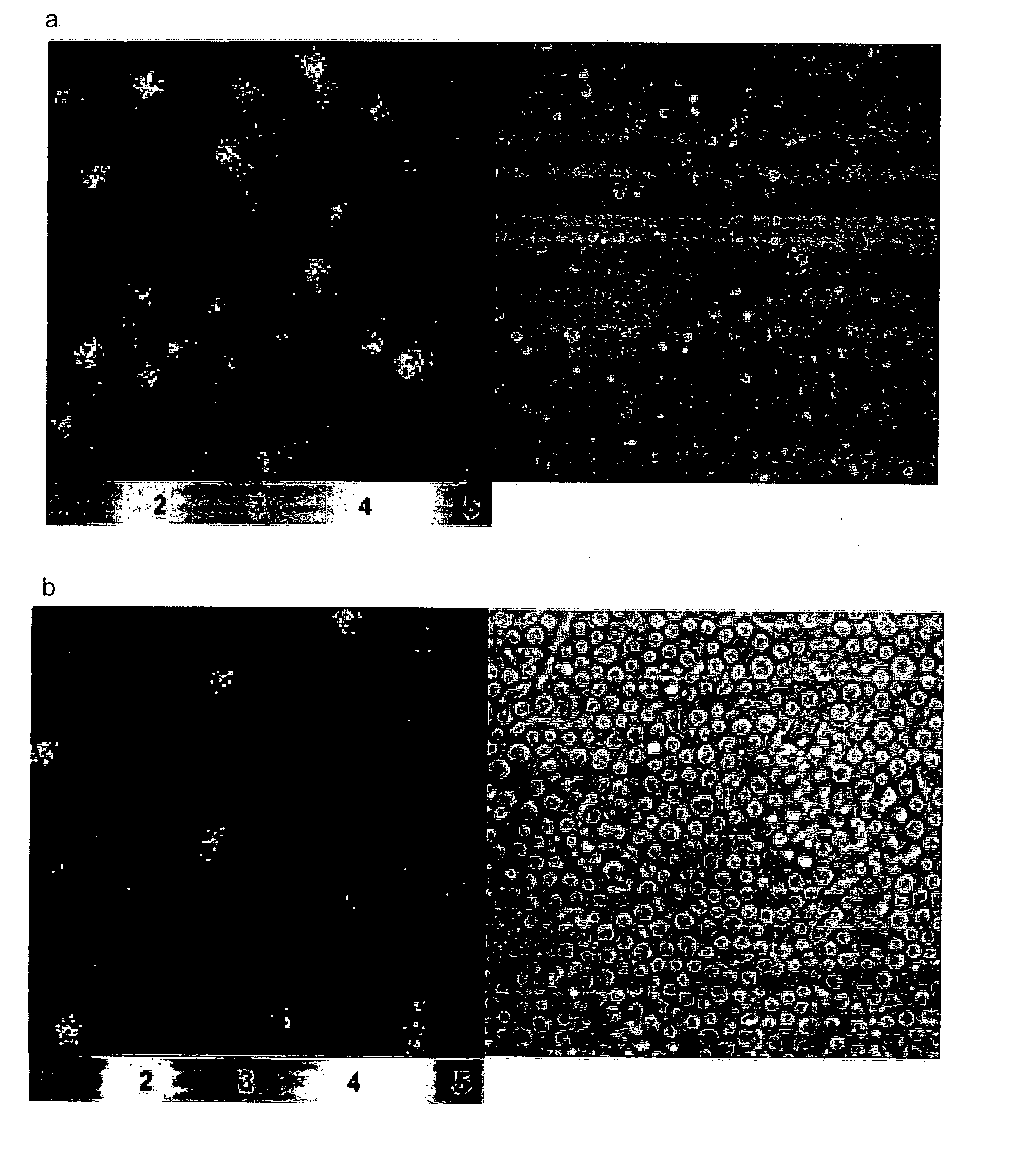
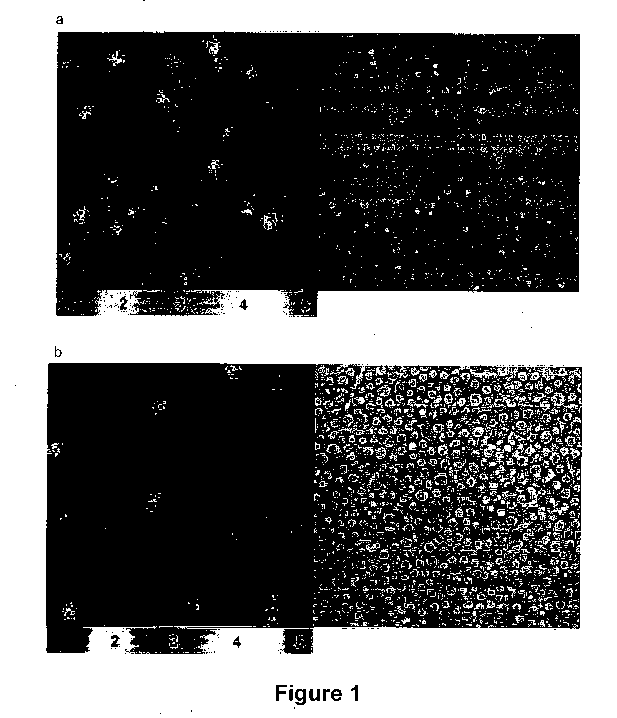
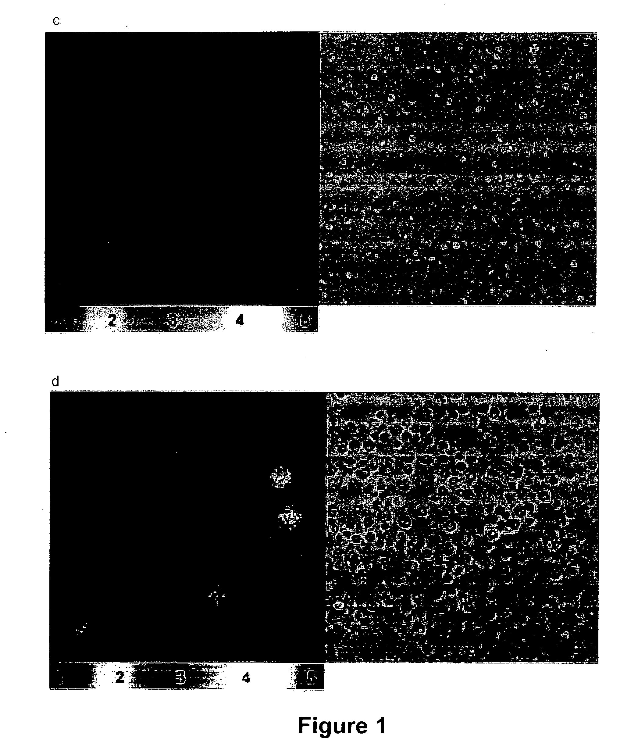
[0085] We tested the performance of the bioluminescence system in single round infections of two major natural HIV-1 target cells: human monocyte derived macrophages (MDM) (FIG. 1a) and activated CD4 T lymphocytes from peripheral blood (FIG. 1b). Target cells were infected with NL4-3-luc HIV-1 particles, engineered such that part of the viral nef gene is replaced by the luciferase reporter gene (Connor et al. (1995) Virology 206:935-44), pseudotyped with the Vesicular Stomatitis Virus protein G (VSV-G) that uses a membrane phospholipid as a receptor for entry and yields a high efficiency of infection compared with HIV-1 envelopes (Aiken (1997) J. Virol. 71:5871-7). Cells were checked for bioluminescence emission 5 and 3 days after infection respectively, times that yielded for each cell system maximal luciferase activity in cell lysates (not shown). Emission of bioluminescence was readily detected in the infected cells upon...
example 2
Quantifying HIV-1 Infection at the Single Cell Level
[0086] In order to establish whether our method for detection of bioluminescent signals could be used as a way to quantify single-cell infection levels, we examined the effect of the diketo acid L-731,988, an integrase inhibitor which prevents HIV-1 integration by blocking strand transfer (Hazuda et al. (2000) Science 287:646-50). MDM cells were incubated in the presence of increasing concentrations of L-731,988 prior to, and during infection with the HIV-1 VSV-G pseudotype. Five days after infection we measured infection levels both using a luminometer in cell-lysates (Connor et al. (1995) Virology 206:935-44), and in intact single cells using our bioluminescence imaging system. As expected, the luciferase activity in cell lysates treated with the integrase inhibitor was reduced in a dose-dependent manner indicative of inhibition of HIV-1 replication in MDM cells (FIG. 2a). In parallel, the single-cell bioluminescence imaging ass...
example 3
Monitoring HIV-1 Gene Expression Dynamics in Single Living Primary Cells
[0087] In situ, under normal conditions the vast majority of CD4 T lymphocytes in the peripheral blood and lymphoid tissues are in a resting (non-activated) state, and represent a latent reservoir for HIV-1 (Pierson et al. (2000) Annu Rev Immunol 18:665-708). Along these lines, activated CD4 T lymphocytes support rapid HIV-1 replication, whereas resting CD4 T cells are incapable of sustaining infection due to several blocks during early steps of viral replication (Chiu et al. (2005) Nature 435:108-14, Ganesh et al. (2003) Nature 426:853-7). Indeed, in resting CD4 T cells HIV-1 DNA is predominantly unintegrated, but cellular activation (eg: mitogenic stimulation) can overcome this, allowing virus integration and replication (Pierson et al. (2002) J. Virol. 76:8518-31). Based upon our results showing that bioluminescence signals in single cells are a direct indicator of viral gene expression efficiency therein, w...
PUM
| Property | Measurement | Unit |
|---|---|---|
| concentration | aaaaa | aaaaa |
| temperature | aaaaa | aaaaa |
| time | aaaaa | aaaaa |
Abstract
Description
Claims
Application Information
 - R&D
- Intellectual Property
- Life Sciences
- Materials
- Tech Scout
- Unparalleled Data Quality
- Higher Quality Content
- 60% Fewer Hallucinations
Browse by: Latest US Patents, China's latest patents, Technical Efficacy Thesaurus, Application Domain, Technology Topic, Popular Technical Reports.
© 2025 PatSnap. All rights reserved.Legal|Privacy policy|Modern Slavery Act Transparency Statement|Sitemap|About US| Contact US: help@patsnap.com


