Medical tracking system using a gamma camera

a tracking system and camera technology, applied in the field of tracking of objects, can solve the problems of insufficient depth and resolution of images, inability to locate certain abnormalities, and inability to accurately locate diagnostic tools and subsequent images, so as to save operating room time, achieve image much faster, and penetrate through objects.

Inactive Publication Date: 2007-07-19
BRAINLAB
View PDF19 Cites 113 Cited by
  • Summary
  • Abstract
  • Description
  • Claims
  • Application Information

AI Technical Summary

Benefits of technology

[0008] gamma radiation is able to travel long distances and easily penetrate through objects.
[0009] On this basis, a gamma camera can provide functional real time intraoperative imaging. Advantageously, the gamma camera allows a surgeon to see anatomical structures, soft tissue and abnormalities as they appear within the body at a particular moment in time. Additionally, the real time imaging can display a surgical instrument within a region

Problems solved by technology

Some current forms of intraoperative imaging include CT and MRI; however, these diagnostic tools and subsequent images are neither real time nor precise enough to locate certain abnorma

Method used


Image

  • Medical tracking system using a gamma camera
  • Medical tracking system using a gamma camera

Examples


Embodiment Construction

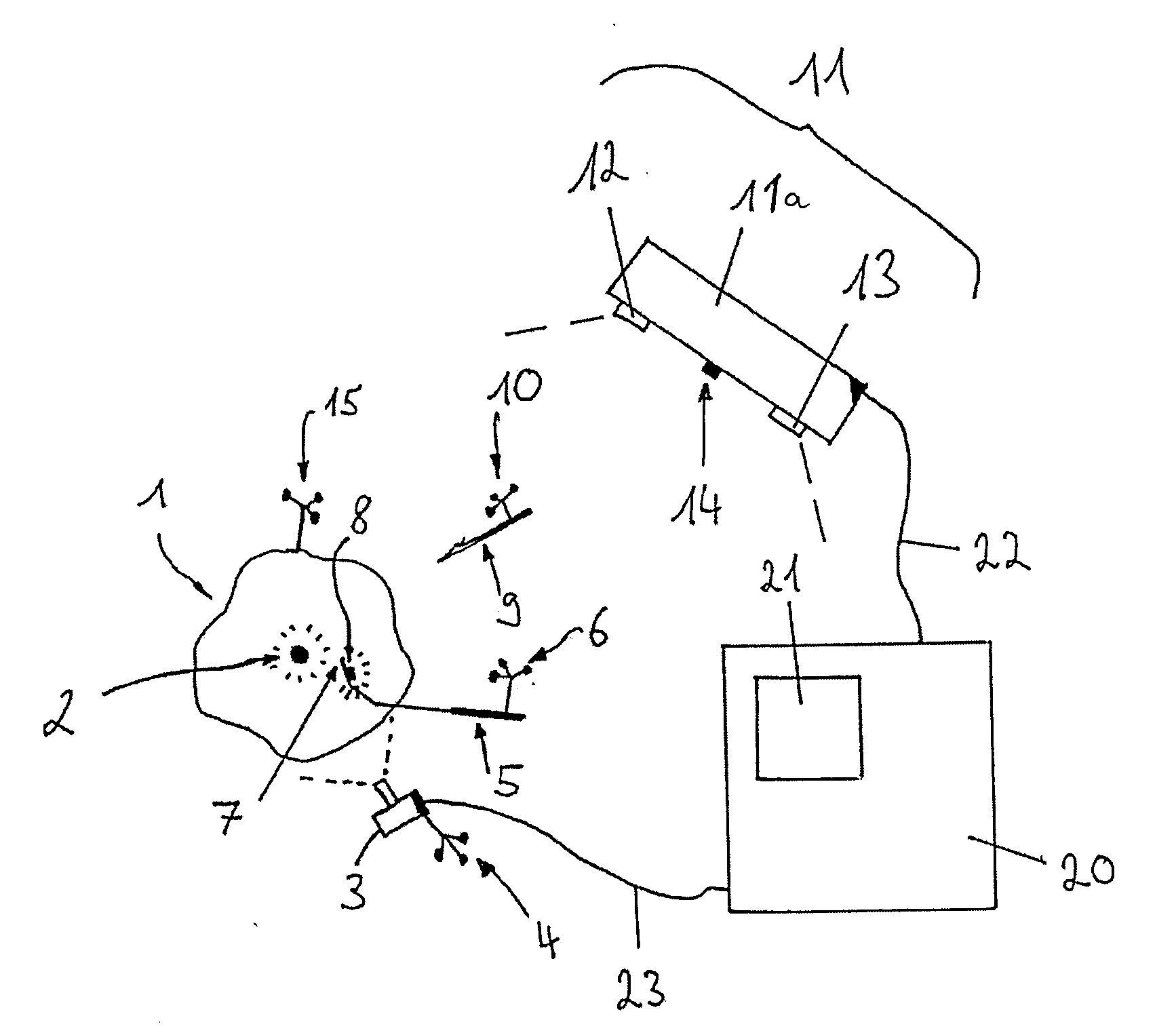
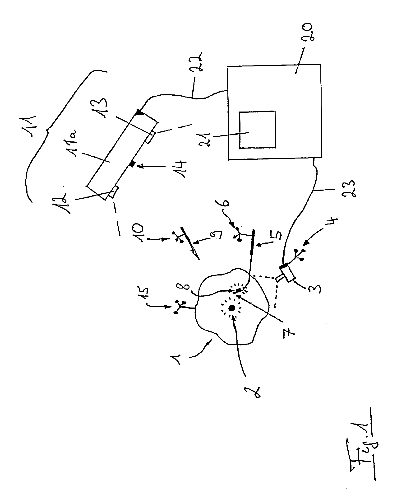
[0040]FIG. 1 is a schematic diagram showing a patient's body part 1 including a gamma radiant tracer material 2 within a region of interest, wherein a gamma camera 3 captures data from the patient's body part 1. First, second, third and fourth tracking arrays (e.g., arrays of reflective spheres) 4, 6, 10 and 15 are coupled to the gamma camera 3, a first instrument 5, a second instrument 9, and the patient's body part 1, respectively. The first instrument 5 includes a flexible tip section 7 having a tracer material 8 on the tip section 7. The second instrument 9 is a rigid instrument such as a scalpel, for example. An infrared tracking system 11 includes first and second stereoscopic infrared tracking cameras 12 and 13, each coupled to a camera holder 11a. An infrared light source 14 is coupled to the camera holder 11a. A navigation system 20, which includes a display 21, is operatively coupled to the infrared tracking system 11 and gamma camera 3 via lines 22 and 23, respectively. G...


PUM


Abstract

A medical tracking system includes a localization system operative to spatially localize medical instruments; a computer operative to assign absolute and/or relative positions of said instruments in a coordinate system; and at least one localizable gamma camera operative to detect gamma radiation emitted from a tracer material.

Description

RELATED APPLICATION DATA [0001] This application claims priority of U.S. Provisional Application No. 60 / 754,512 filed on Dec. 28, 2005, which is incorporated herein by reference in its entirety.FIELD OF THE INVENTION [0002] The present invention relates to the tracking of objects, such as medical instruments and / or bodies, in a medical environment. BACKGROUND OF THE INVENTION [0003] An exemplary medical navigation and tracking system is disclosed in DE 196 39 615 C2. Various forms of intraoperative imaging coupled with navigation are becoming standardized in today's medical environment. This trend will continue to grow in various fields, including tumor / tissue resection, patient positioning, and confirmation of planned trajectories. Some current forms of intraoperative imaging include CT and MRI; however, these diagnostic tools and subsequent images are neither real time nor precise enough to locate certain abnormalities, particularly in deep tissue areas. Although ultrasound provid...

Claims


Application Information

Patent Timeline
IPC IPC(8): A61B5/05A61B6/00
CPCA61B6/12A61B19/52A61B19/5212A61B19/5244A61B19/54A61B2019/5227A61B6/4258A61B2019/5255A61B2019/5289A61B2019/542A61B2019/547A61B2019/5483A61B2019/5231A61B34/20A61B90/36A61B90/361A61B90/39A61B2034/2055A61B2090/364A61B2090/371A61B2090/373A61B2090/392A61B2090/397A61B2090/3983
Inventor KEGLOVICH, MIKEPEDAIN, CHRISTOPH
Owner BRAINLAB
Why Eureka
Try Eureka