
Methods and Apparatus for Treating Uterine Fibroids

a technology for uterine fibroids and surgical instruments, applied in the field of uterine fibroids, can solve the problems of urinary symptoms, hysterectomy requires major invasive surgery, can involve excessive blood loss, prolong convalescence, and economic costs, and achieves the effect of safe and reliable, minimal patient trauma and procedure tim

Inactive Publication Date: 2007-09-27
AMS RES CORP
View PDF4 Cites 8 Cited by
  • Summary
  • Abstract
  • Description
  • Claims
  • Application Information

AI Technical Summary

Benefits of technology

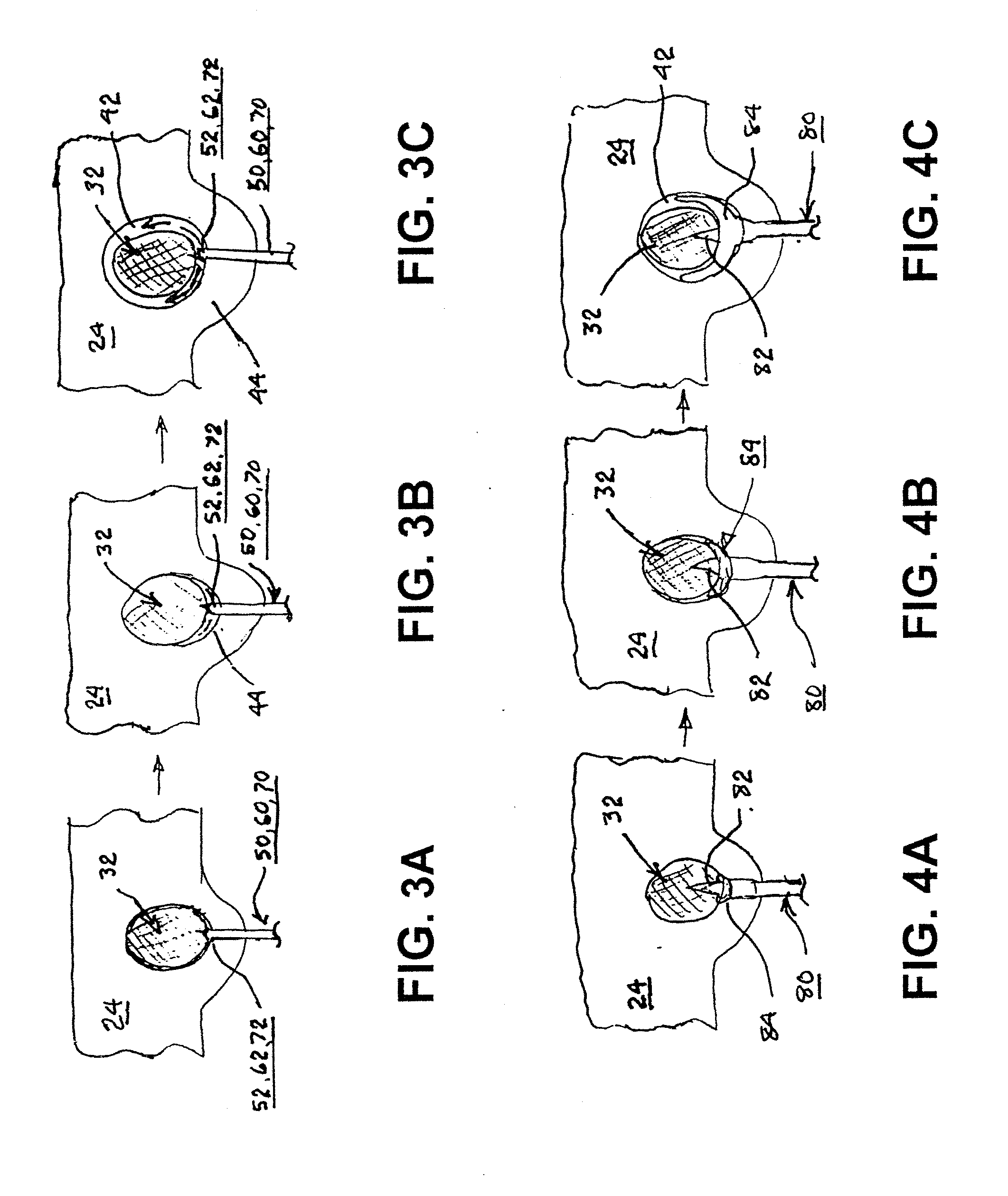
[0017]A further refinement of the instrument for detaching the fibroid comprises a fibroid mass penetrating and gripping tip that enables twisting or rotation of the fibroid mass by rotation of the instrument proximal end (or a shaft extending through the instrument body to the gripping end) sufficiently to detach the blood vessels traversing the avascular space from the fibroid mass.
[0019]Advantageously, the minimally invasive instruments and procedures minimize patient trauma and procedure time while ensuring safe and reliable

Problems solved by technology

Fibroids that project to the outside of the uterus (subserosal fibroids) can press on the bladder or ureters, causing urinary symptoms.
Subserosal fibroids that bulge from the posterior of the uterus can press either on the rectum, causing constipation, or on spinal nerves, causing backache.
Thus, in addition to the loss of reproductive capability, a hysterectomy requires major invasive surgery that can involve excessive blood loss, prolonged convalescence, attendant pain and discomfort, and economic costs.
However, the surgical exposure of the uterus through the abdominal wall in a myomectomy procedure can involve risks and complications, and fibroids may recur over time ultimately necessitating a repeated myomectomy or ultimately a hysterectomy.
This procedure involves some risk of ischemia of the normal myometrium and ovaries.
However, it would appear to be difficult to identify, isolate and penetrate such minute vessels to inject the embolyzing material therein.

Method used


Image

  • Methods and Apparatus for Treating Uterine Fibroids
  • Methods and Apparatus for Treating Uterine Fibroids
  • Methods and Apparatus for Treating Uterine Fibroids

Examples


Embodiment Construction

[0029]In the following detailed description, references are made to illustrative embodiments of methods and apparatus for carrying out the invention. It is understood that other embodiments can be utilized without departing from the scope of the invention. Preferred embodiments for minimally invasive surgical instruments and procedures for treating fibroids, particularly uterine fibroids, by isolating or excising fibroid masses are described.

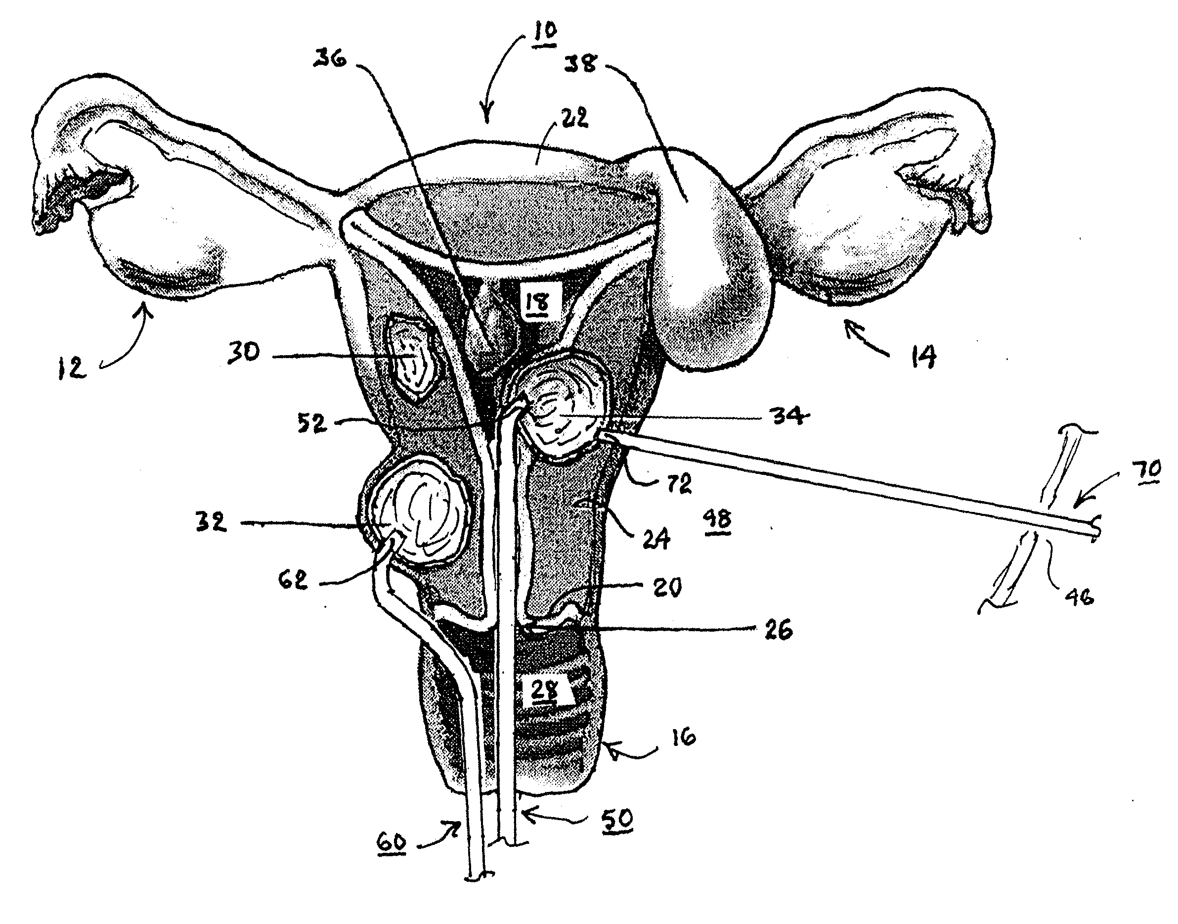
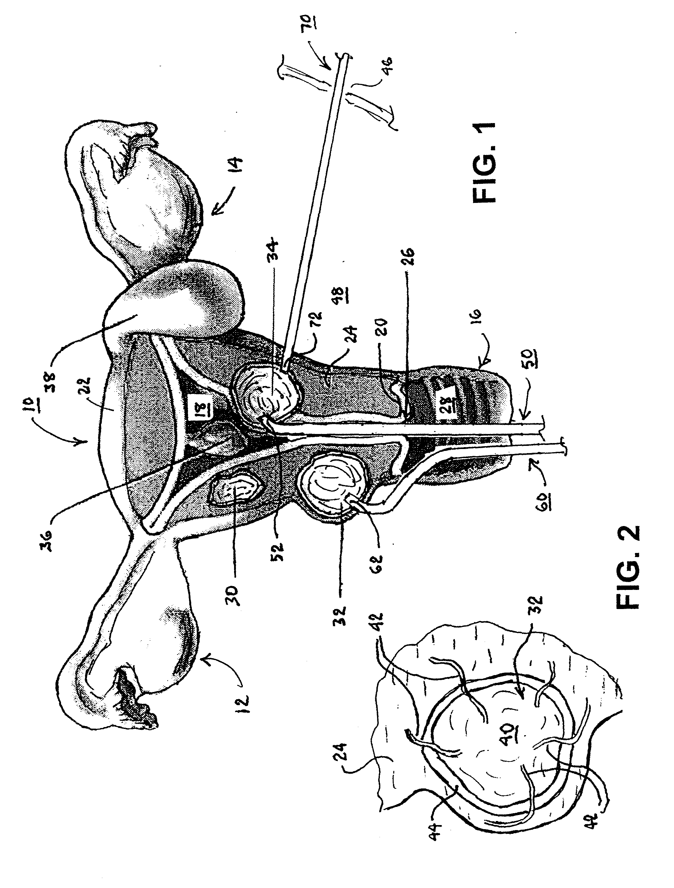
[0030]A variety of uterine fibroids are illustrated in FIG. 1 in relation to the female uterus 10, ovaries 12 and 14, and vagina 16 joined with the uterine cavity 18 at the uterine neck or cervix 20. The uterus 10 has a pear-shaped, uterine body extending between a fundus 22 extending right and left to junctions with the right and left Fallopian tubes joined with ovaries 12 and 14 and to the cervix 20 that extends to the vagina 16. The uterus 10 is formed of a smooth muscle uterine wall or myometrium 24 bounded by an outer surosa membrane and an...


PUM


Abstract

Minimally invasive surgical instruments and procedures for treating fibroids within avascular space, particularly uterine fibroids, by interrupting the blood supply to the fibroid, are disclosed. One embodiment of a dissection instrument for detaching the fibroid comprises fluid delivery into the avascular space to expand the space and detach the fibroid mass from blood vessels traversing the avascular space through hydro-dissection. The dissection instrument may comprise an inflatable balloon that is shaped to be inflated in the avascular space to expand and dissect the space, at least partially surrounding the fibroid mass, and detach the blood vessels traversing the avascular space. The dissection instrument may comprises a fibroid mass penetrating and gripping tip that enables twisting or rotation of the fibroid mass sufficiently to detach the blood vessels. The detached fibroid mass may be left in place or removed.

Description

RELATED APPLICATION[0001]This application claims priority to U.S. Provisional Application Ser. No. 60 / 784,304 filed Mar. 21, 2006, the entire content of which is incorporated herein by reference.TECHNICAL FIELD[0002]The present invention pertains to minimally invasive surgical instruments and procedures for treating fibroids, particularly uterine fibroids, by interrupting the blood supply to the fibroid.BACKGROUND[0003]Uterine fibroids are noncancerous growths of the uterus that are common in women and are not associated with an increased risk of uterine cancer and almost never develop into cancer. Uterine fibroids, also called fibromyomas, leiomyomas or myomas, develop from the smooth muscular tissue of the uterus (myometrium). A single cell reproduces repeatedly, eventually creating a pale, firm, rubbery tissue mass distinct from surrounding or neighboring normal myometrium. Fibroids range in size from seedlings, undetectable by the human eye, to bulky masses that can distort and ...

Claims


Application Information

Patent Timeline
IPC IPC(8): A61M29/00
CPCA61B17/22031A61B17/22032A61B17/3203A61B2017/320775A61B2017/22034A61B2017/320064A61B17/42
Inventor ARNAL, KEVIN R.ANDERSON, KIMBERLY A.
Owner AMS RES CORP
Learn More