Method, Apparatus and Computer Program Product for Automatic Segmenting of Cardiac Chambers

a technology of automatic segmentation and cardiac chamber, applied in the field of cardiac imaging, can solve the problems of affecting the reproducibility of segmentation, and sensitivity of segmentation methods to noise,

Active Publication Date: 2007-11-01
PIE MEDICAL IMAGING
View PDF4 Cites 53 Cited by
  • Summary
  • Abstract
  • Description
  • Claims
  • Application Information

AI Technical Summary

Benefits of technology

[0006] The present invention presents a more systematic approach, in that it does not rely on anatomy assumptions. First, it locates the left and right ventricles. Then the base short axis slice and the apex of the left ventricle short-axis slice are located. Next, the start conditions for the segmentation process are determined. The locations of the left ventricle, the left ventricle epicard and finally the right ventricle endocard are detected approximately. Based on these start conditions the segmentation process starts. Note that various standard acquisitions of MR cardiac imaging are used, such as the short axis (SA) and long axis (LA) orientations. Finally the papillary muscles of the left ventricle are detected. Advantageously, the methodology of the present invention is fully automatic without user interactions based on intermediate results, which can yield 100% reproducibility.
[0011] The segmentation process of the present invention corrects for data acquisition and / or subject-caused movement in the acquired MR image data set by using system coordinates from an MR scanner apparatus. In this way, accuracy is improved without disturbing the sequence of image pick-up operations. Moreover, the segmentation of the endocardial and epicardial features starts from an operator-defined seed contour and is based on a radial minimum cost with dynamic sign definition and tissue classification. Apart from such elementary user input, all further operations are machine-controlled, which yields excellent reproducibility.
[0012] In addition, the methodology of the present invention automatically detects Left Ventricle Right Ventricle (LVRV) connection position. Clinicians are recognizing this information as highly helpful.
[0013] Moreover, the methodology of the present invention estimates volume(s) of the heart which is / are derived from the spatial location of the heart's apex position on a curve that is obtained from the long axis cine sequences. In addition, such volume estimates preferably are derived from a mitral valve position obtained from long axis cine views. Such processing improves the accuracy of such volume estimate(s).

Problems solved by technology

However, this segmentation method is quite sensitive to noise and physically artifacted image data.
Various modifications thereto have been proposed, but the method remains sensitive to user-defined start conditions, which endanger the reproducibility of the segmentation.

Method used


Image

  • Method, Apparatus and Computer Program Product for Automatic Segmenting of Cardiac Chambers
  • Method, Apparatus and Computer Program Product for Automatic Segmenting of Cardiac Chambers
  • Method, Apparatus and Computer Program Product for Automatic Segmenting of Cardiac Chambers

Examples


Embodiment Construction

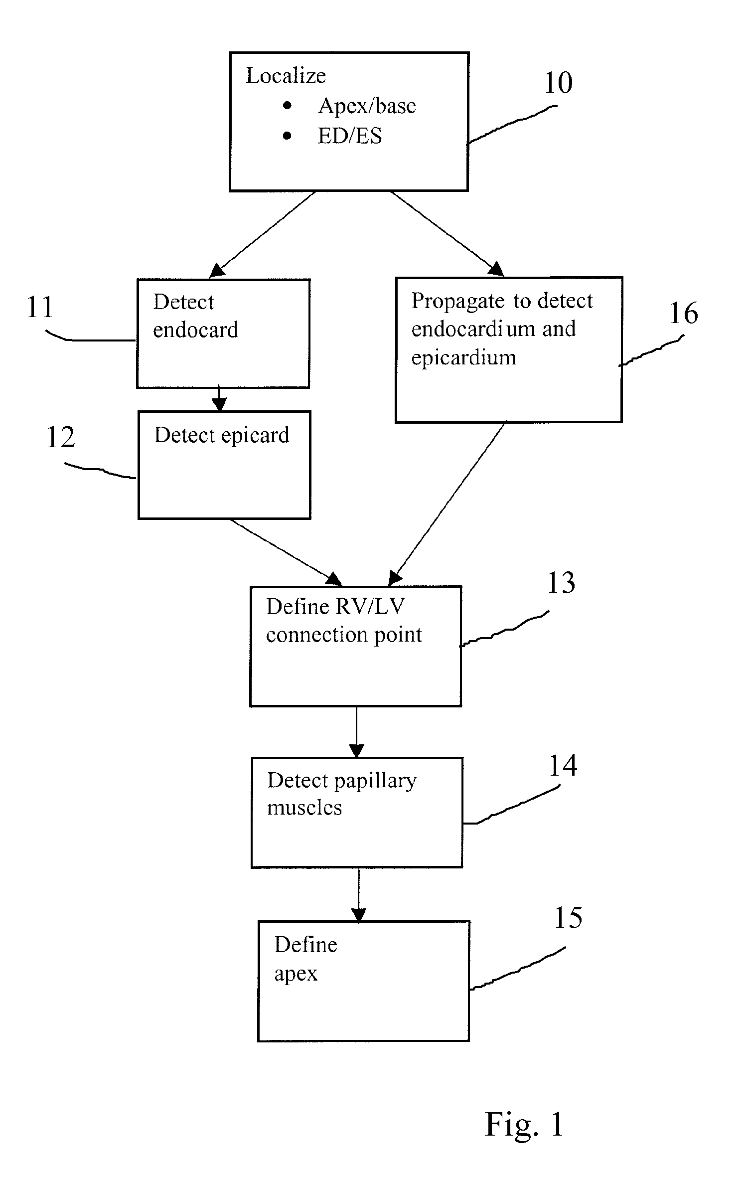
[0023] Turning now to FIG. 1, there is shown a flow chart of various processing steps in accordance with the present invention. First, in block 10 (localize Apex / Base of left ventricle, estimate epicard, find end of diastole, end of systole), the 4D MRI dataset is processed to localize the left and right ventricles therein. This dataset contains a sequence of short axis cine images that cover the heart, combined with two-chamber and four-chamber cine sequences.

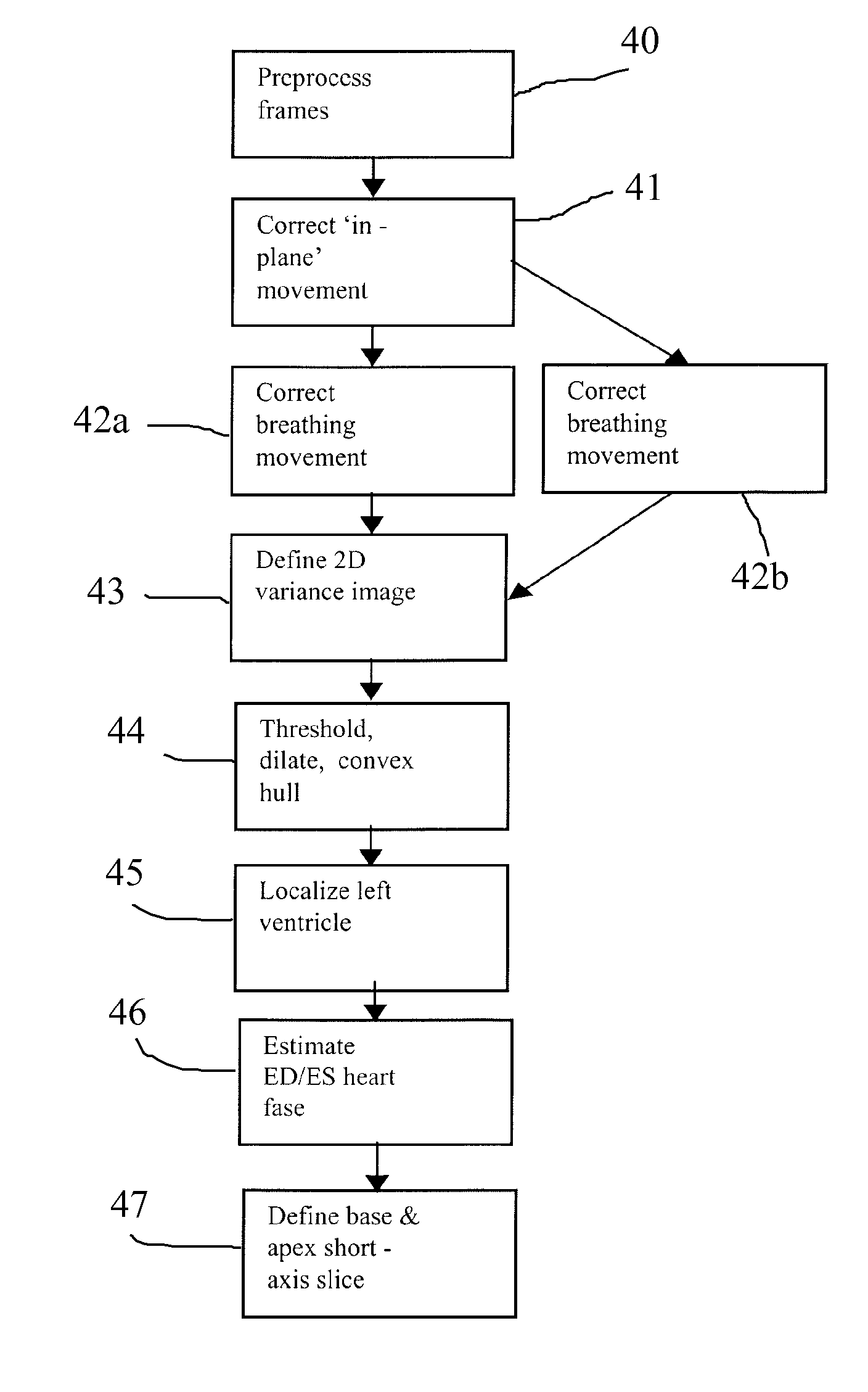
[0024] As an alternative to the above, it is possible to work with short-axis images only. Within this operation, the base and apex short axis slices are located as well as the end systole and end diastole heart phase in time. In this respect, FIG. 4 illustrates a flow diagram of the locating process for the heart's left and right ventricles, as a substitute for block 10 in FIG. 1. The remainder of FIG. 1 will be discussed later.

[0025] Turning to FIG. 4, in block 40, the images are preprocessed to eliminate high local varian...


PUM


Abstract

A method, apparatus and computer program product for machine-based segmentation of the cardiac chambers in a four-dimensional magnetic resonance image data that contains at least short-axis cine sequences. The segmenting is based on temporal variations in the data set and on features extracted from the image data. In particular, the method and apparatus process the image data to automatically segment an endocard based on fuzzy object extraction and to automatically segment an epicard through finding a radial minimum cost with dynamic sign definition and tissue classification. In addition, image data is preferably preprocessed to eliminate high local image variances and inhomogeneity. The image data is also preferably processed to identify positions of a base slice, an apex slice, an end-diastole, an end-systole and an LVRV connection.

Description

BACKGROUND OF THE INVENTION [0001] 1. Field of the Invention [0002] This invention relates broadly to cardiothoracic imaging. More particularly, this invention relates to segmenting cardiac chambers in a four-dimensional magnetic resonance image. [0003] 2. State of the Art [0004] Accurate and reproducible assessment of the left and right ventricular volumes and ejection fraction is essential for the prognosis of patients with heart disease and for evaluating therapeutic response. Magnetic resonance (MR) imaging has proven to be an accurate and reproducible imaging technique for the quantitative analysis of left and right ventricular functions. Ventricular volumes and mass as well as regional functional parameters such as wall motion and wall thickening, especially for the left ventricle, can be obtained in the true short-axis plane of the ventricle. These short-axis views have various advantages. Since wall motion and wall thickening takes place mainly perpendicularly to the viewing...

Claims


Application Information

Patent Timeline
Patent Type & Authority Applications(United States)
IPC IPC(8): G06K9/00
CPCG06T7/0081G06T7/0089G06T2207/30048G06T2207/10088G06T2207/10076G06T7/11G06T7/149
Inventor ABEN, JEAN-PAUL MARIA MICHEL
Owner PIE MEDICAL IMAGING
Why Eureka
Try Eureka