
Method and kit for detecting the early onset of renal tubular cell injury

a kidney and tubular cell technology, applied in the field of kidney tubular cell injury detection methods and kits, can solve the problems of delayed initiation, high mortality and morbidity, and high mortality

Inactive Publication Date: 2007-11-01
THE TRUSTEES OF COLUMBIA UNIV IN THE CITY OF NEW YORK +1
View PDF30 Cites 33 Cited by
  • Summary
  • Abstract
  • Description
  • Claims
  • Application Information

AI Technical Summary

Benefits of technology

The present invention relates to a method for detecting a renal tubular cell injury in a mammal. This is done by obtaining a urine sample from the mammal and contacting it with an antibody specific to a biomarker for renal tubular cell injury, such as NGAL. The antibody forms a complex with the biomarker, which is then detected. This method can be used to monitor the effectiveness of a treatment for renal tubular cell injury by obtaining a post-treatment urine sample and detecting the presence of the biomarker. The invention also provides a kit for detecting the biomarker and a method for identifying the extent of the injury based on the time of onset of the biomarker in the urine sample.

Problems solved by technology

Acute renal failure (ARF) secondary to a renal tubular cell injury, including an ischemic injury or a nephrotoxic injury remains a common and potentially devastating problem in clinical medicine and nephrology, with a persistently high rate of mortality and morbidity despite significant advances in supportive care.
While these studies have suggested possible therapeutic approaches in animal models, translational research efforts in humans have yielded disappointing results.
The reasons for this may include the multifaceted response of the kidney to ischemic injury and nephrotoxins, and a paucity of early biomarkers for ARF with a resultant delay in initiating therapy.
These indicators are not only insensitive and nonspecific, but also do not allow for early detection of the disease.
Thus, the detection of Cyr61 in the urine is problematic with respect to specificity as well as the cumbersome nature of the procedure.

Method used


Image

  • Method and kit for detecting the early onset of renal tubular cell injury
  • Method and kit for detecting the early onset of renal tubular cell injury
  • Method and kit for detecting the early onset of renal tubular cell injury

Examples


example 1

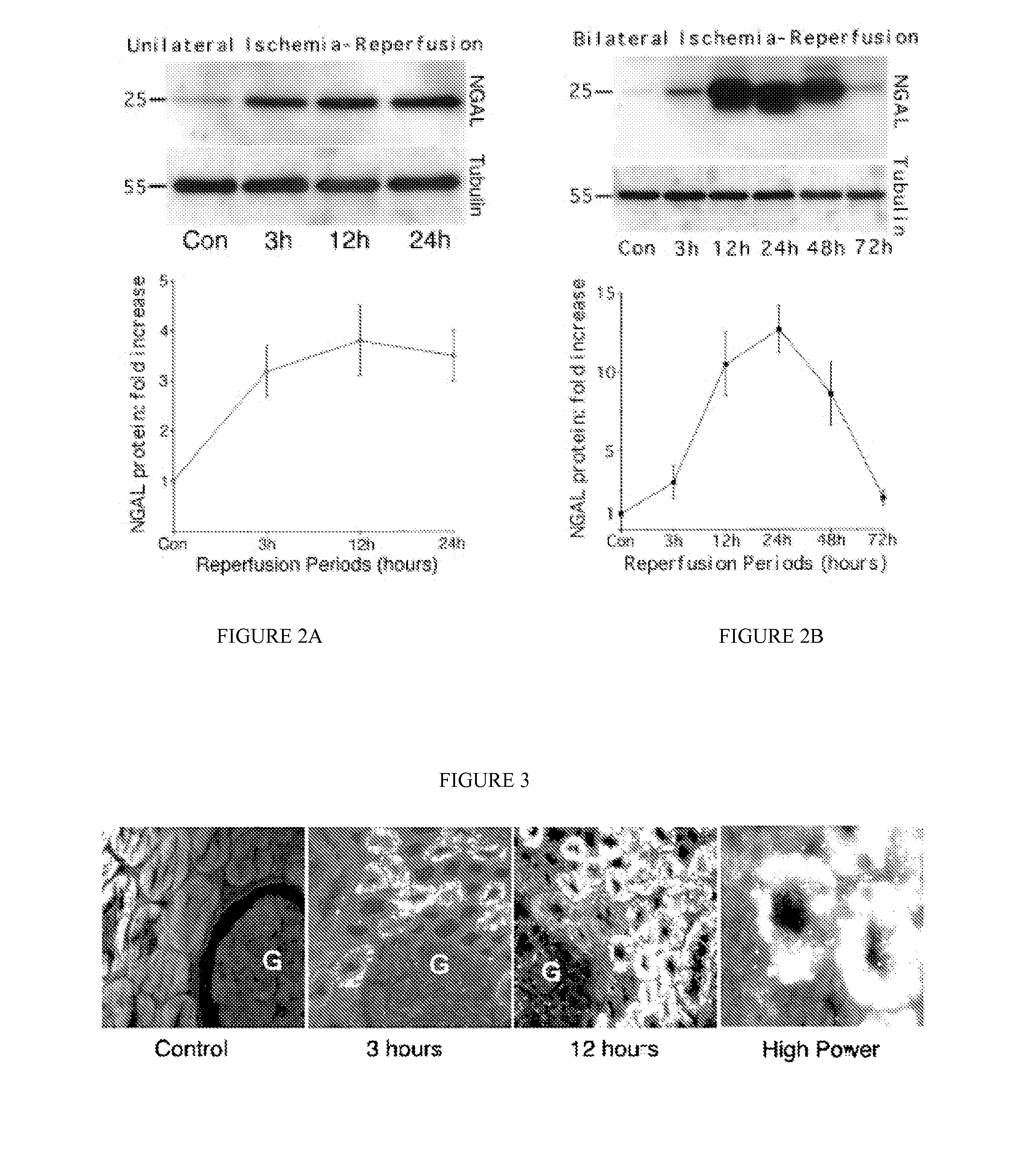
[0083] NGAL is a small protease-resistant, secreted polypeptide that is detectable in the urine. The marked upregulation of NGAL mRNA and protein levels has been shown in the early post-ischemic mouse kidney. NGAL protein expression was detected predominantly in proximal tubule cells, in a punctate cytoplasmic distribution reminiscent of a secreted protein. Indeed, NGAL was easily and rapidly detected in the urine (in the very first urine output) following ischemic injury in both mouse and rat models of ARF, at which time no leukocytic infiltration of the kidney was observed. The origin of NGAL from tubule cells was further confirmed in cultured human proximal tubule cells subjected to in vitro ischemic injury, where NGAL mRNA was markedly and promptly induced in the cells, and NGAL protein readily detectable in the culture medium within one hour of mild ATP depletion. Our results indicate that NGAL may represent a novel early urinary biomarker for ischemic renal injury.

Identifica...

example 2

NGAL Protein is Easily Detected in the Urine Immediately After Induction of ARF in Rats:

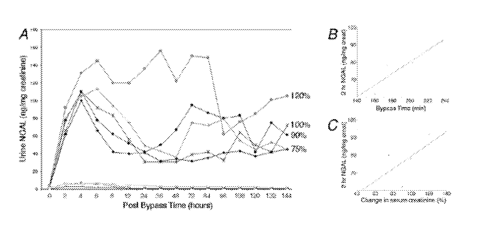
[0091] Since a debate exists regarding species differences in the responses to renal artery occlusion (10), we next examined the behavior of NGAL in a different animal model, namely a well-established rat model of renal ischemia-reperfusion injury. Using urinary creatinine concentrations to equalize for sample loading, NGAL was absent from the urine prior to rat renal ischemia. In marked contrast, NGAL was manifest as a 25 kDa immunoreactive peptide within 3 h of the injury (in the very first urine output following ischemia), as shown in FIG. 6. In comparison, the serum creatinine in this model of ischemic injury was elevated only after 24 h of reperfusion (not shown). Once again, NGAL was detectable in 1 μl of unprocessed urine and persisted for the entire duration examined (24 h of reperfusion).

example 3

NGAL mRNA is Induced in Cultured Human Proximal Tubule Cells After Early Mild Ischemia:

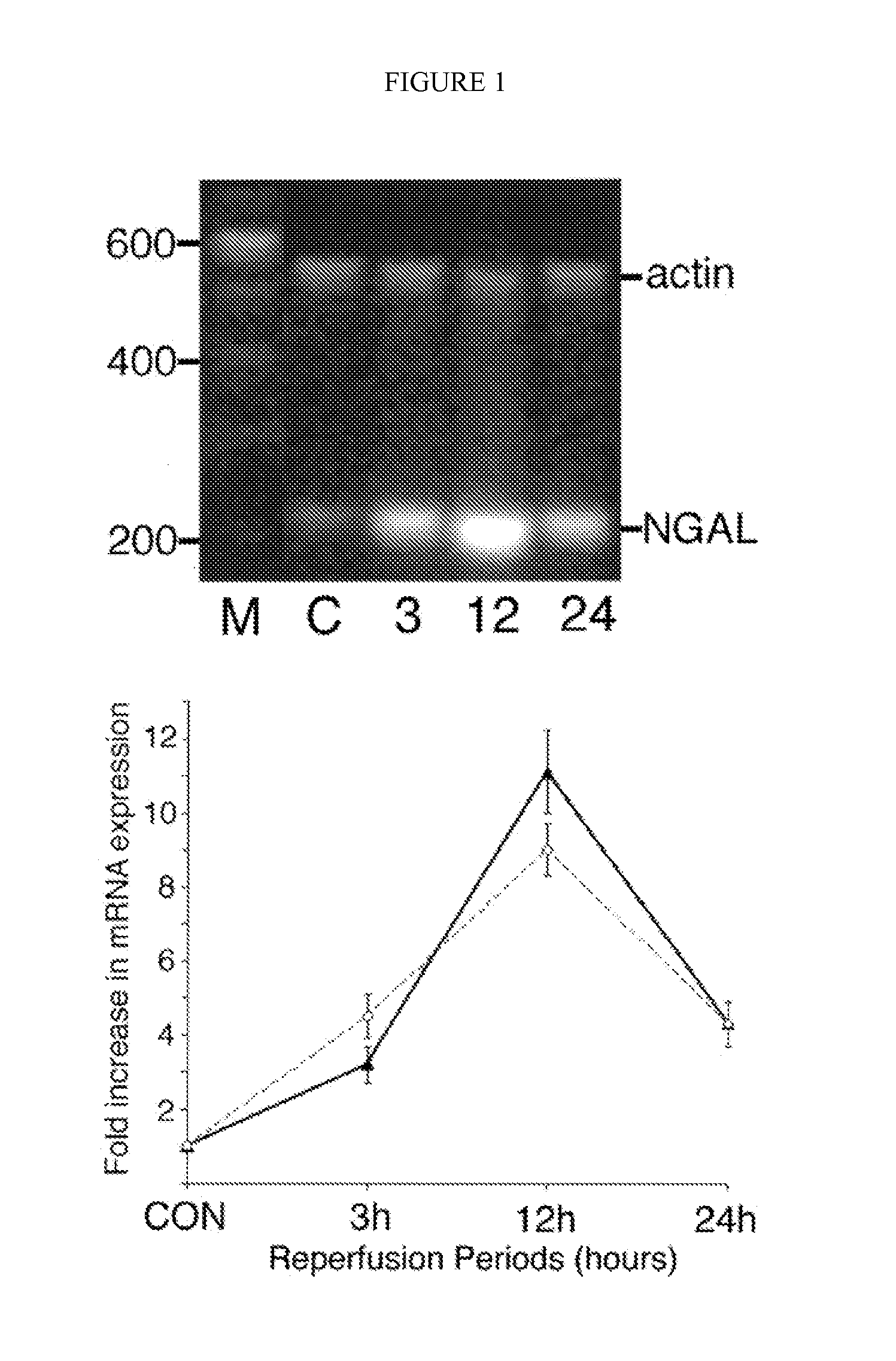
[0092] In order to confirm the origin of NGAL from ischemic proximal tubule cells, we modified previously described protocols of in vitro ischemia by ATP depletion in cultured human proximal tubule cells (RPTEC). Incubation in 1 μm antimycin resulted in a mild partial ATP depletion to about 83±3% of control within 1 h, with a more gradual decrease to about 75±3% of control by 6 h (mean ±SD from four experiments). No morphological consequences of this mild ATP depletion were discernible. NGAL mRNA was just detectable in resting cells. However, following partial ATP depletion, a rapid and duration-dependent induction of NGAL mRNA was evident by RT-PCR, as shown in FIG. 7.

NGAL Protein is Easily Detected in the Medium After Early Ischemia in vitro:

[0093] We next examined NGAL protein expression in RPTEC cells and the culture medium following mild ATP depletion. NGAL protein was detectable in cont...


PUM

PropertyMeasurementUnit
concentrationaaaaaaaaaa

Abstract

A method and kit for detecting the early onset of renal tubular cell injury, utilizing NGAL as an early urinary biomarker. NGAL is a small secreted polypeptide that is protease resistant and consequently readily detected in the urine following renal tubule cell injury. NGAL protein expression is detected predominantly in proximal tubule cells, in a punctate cytoplasmic distribution reminiscent of a secreted protein. The appearance NGAL in the urine is related to the dose and duration of renal ischemia and nephrotoxemia, and is diagnostic of renal tubule cell injury and renal failure. NGAL detection is also a useful marker for monitoring the nephrotoxic side effects of drugs or other therapeutic agents.

Description

CROSS-REFERENCE TO RELATED APPLICATIONS [0001] This application claims the benefit of co-pending U.S. Provisional Application Nos. 60 / 458,143, filed Mar. 27, 2003, and 60 / 481,596, filed Nov. 4, 2003.INTERESTS [0002] This invention was made with Government support awarded by the National Institute of Health (NIH) / National Institute of Diabetes and Digestive and Kidney Diseases, under Grant Nos. DK53289, DK52612, DK55388, and DK58872. The Government may have certain rights in this invention.BACKGROUND OF THE INVENTION [0003] Acute renal failure (ARF) secondary to a renal tubular cell injury, including an ischemic injury or a nephrotoxic injury remains a common and potentially devastating problem in clinical medicine and nephrology, with a persistently high rate of mortality and morbidity despite significant advances in supportive care. Pioneering studies over several decades have illuminated the roles of persistent vasoconstriction, tubular obstruction, cellular structural and metabol...

Claims


Application Information

Patent Timeline
Patent Type & Authority Applications(United States)
IPC IPC(8): G01N33/48A61K31/675G01NG01N33/53
CPCA61K31/675G01N33/6893C12Q1/37G01N2800/52G01N2800/56G01N2800/347C12Q1/6883C12Q2600/118C12Q2600/16G01N2333/475
Inventor DEVARAJAN, PRASADBARASCH, JONATHAN
Owner THE TRUSTEES OF COLUMBIA UNIV IN THE CITY OF NEW YORK
Learn More