Implant for repairing a cartilage defect

a cartilage defect and implant technology, applied in the field of cartilage defect repair implants, can solve the problems of cartilage defects that cannot be repaired, cartilage defects are associated with pain, cartilage defects are not easy to heal, and achieve the effect of effective and reliable treatment for cartilage defects

Inactive Publication Date: 2007-11-15
TETEC TISSUE ENG TECH
View PDF5 Cites 17 Cited by
  • Summary
  • Abstract
  • Description
  • Claims
  • Application Information

AI Technical Summary

Benefits of technology

[0016] In view of the above it is an object of the present invention to overcome the mentioned dis

Problems solved by technology

Cartilage suffers from a very limited ability for repair of joint surface damage.
It typically fails to heal on its own and cartilage defects can be associated with pain, loss of function and disability.
Nevertheless, the capability of fibrocartilage to withstand shock or shearing forces is low compared to hyaline cartilage.
Further, fibrocartilage is degenerating over the time, and resulting in the return of clinical syndromes.
These “autografts” result in the formation of a patchwork or mosaic.
However, this method is rather limited due to fact that healthy tissue is not available indefinitely and that very often the congruence of the cartilage surfaces is not satisfying.
In spite of the achieved promising results, the conventional ACT method reveals disadvantages and obstacles.
The arthrotomy—while often being enduring—is accompanied by postoperative complaints.

Method used


Image

  • Implant for repairing a cartilage defect
  • Implant for repairing a cartilage defect

Examples


example 1

Preparation of an Implant According to the Invention

[0079] An implant according to the invention can be prepared—for example—by a method disclosed in EP 1 275 405. By using this method a sponge-like layer or protein matrix can be anchored in a membrane-like layer. Briefly, a membrane-like layer was provided comprising collagen—other suitable materials are, e.g., bioresorbable polymers such as polylactide or polyglycolic acid, collagen, pericardium, composites, glycosaminoglycanes, natural tissue sources like elastin, and mixtures of two or more of these materials. The sponge-like layer was applied thereon in form of a suspension. Alternatively it can be supplied as a dispersion or paste. The suspension was comprising collagen—other materials can be used, e.g. hyaluronic acid, alginate, chitosan, gelatine, processed materials, composites, blood born components such as fibrin, and mixtures of two or more of these materials—and was introduced into the membrane-like structure by means ...

example 2

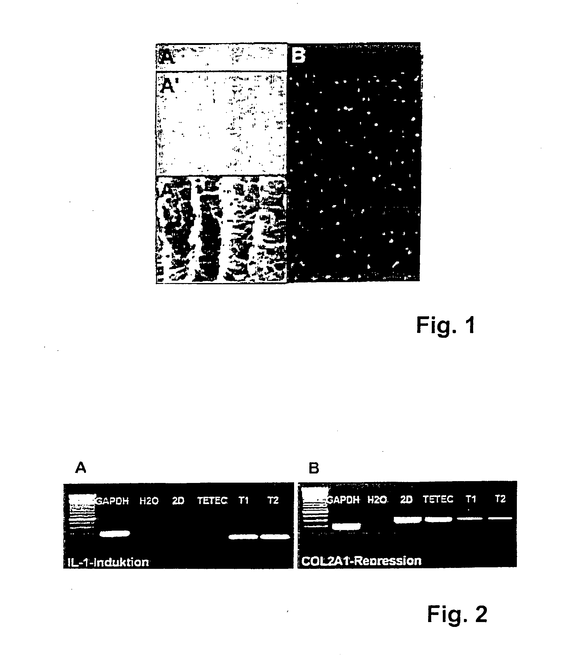
[0084] With respect to the biocompatibility of the implants, induction of Interleukin IL-1 expression and reduction of collagen type II expression of chondrocytes seeded into different implants was assessed in vitro.

[0085] The results of these tests are shown in FIG. 2. In FIG. 2A (“IL-1 Induktion”) it is shown that IL-1 induction in chondrocytes seeded on an implant according to the invention (“TETEC”) is lower than IL-1 expression in chondrocytes seeded on commercially available implants (“T1” and “T2”). When IL-1 expression was increased hypertrophy and degeneration of chondrocytes seeded into the implants could be observed in vitro. A marker and controls “GAPDH” and “H2O” are displayed in lanes 1 to 3 respectively.

[0086] Further, expression of collagen type II, which is an essential structural protein in cartilage, was remarkably reduced in chondrocytes seeded into commercially available implants (“T1” and “T2”) in comparison to chondrocytes seeded into implants according to t...

example 3

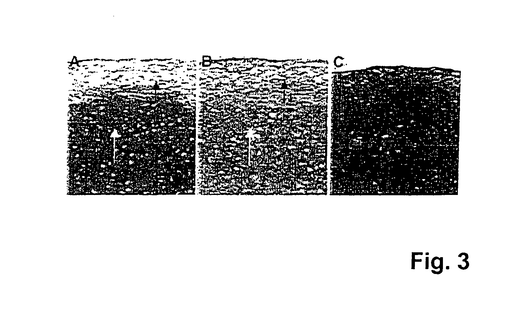
[0087] The implant prepared as mentioned above (see Example 1) was tested in animal studies. Experiments were conducted in SCID (“severe combined immunodeficiency”) mice, into which human cells can be transplanted without rejection of these cells, since in the mentioned mice the enzyme adenosine deaminase is deficient and—as a result—T or B cells are not being developed.

[0088] It was previously shown in SCID mice that human articular chondrocytes do only produce solid hyaline cartilage when the transplanted cells express certain marker genes, the fact of which has to be proofed in molecular / biological quality assays. Chondrocytes not expressing collagen type II, BMP-2 (bone morphogenetic protein 2) and FGFR-3 (fibroblast growth factor receptor 3) any more are not able to regenerate high quality cartilage.

[0089] In test group A, human articular chondrocytes expressing relevant marker genes were seeded on an implant according to the invention (5×10⁵ cells / cm2 carrier layer, i.e. sec...


PUM

PropertyMeasurementUnit
Lengthaaaaaaaaaa
Lengthaaaaaaaaaa
Lengthaaaaaaaaaa

Abstract

The present invention relates to an implant for repairing a cartilage defect comprising a first layer and a second layer. The first layer comprises a membrane-like structure and the second layer comprises a sponge-like structure with directional and/or interconnected pores. The first layer is facing the synovial space and the second layer is located towards bone.

Description

CROSS-REFERENCE TO RELATED APPLICATIONS [0001] This application is a continuation of pending international application PCT / EP2004 / 012109, filed Oct. 27, 2004, designating the United States. The content of the above-referenced application is incorporated herein by this reference in its entirety.FIELD OF THE INVENTION [0002] The invention relates to an implant for repairing a cartilage defect, and more particularly for repairing load-bearing cartilage, such as articular cartilage, comprising a first layer and a second layer, the first layer facing the synovial space and the second layer being located towards bone. The invention further relates to a method for treating a cartilage defect. BACKGROUND OF THE INVENTION [0003] Cartilage suffers from a very limited ability for repair of joint surface damage. It typically fails to heal on its own and cartilage defects can be associated with pain, loss of function and disability. [0004] Articular cartilage forms a layer at the surface joints....

Claims


Application Information

Patent Timeline
IPC IPC(8): A61F2/28
CPCA61F2/30756A61F2310/00383A61F2/44A61F2002/0086A61F2002/2817A61F2002/30004A61F2002/30011A61F2002/30032A61F2002/30062A61F2002/30324A61F2002/30677A61F2002/30761A61F2002/30762A61F2002/30764A61F2002/30766A61F2002/3093A61F2002/30971A61F2210/0004A61F2240/001A61F2250/0014A61F2250/0023A61F2250/003A61F2250/0036A61F2310/00365A61F2/38
Inventor GAISSMAIER, CHRISTOPHFRITZ, JUERGENAICHER, WILHELM
Owner TETEC TISSUE ENG TECH
Why Eureka
Try Eureka