Eureka AIR delivers breakthrough ideas for toughest innovation challenges, trusted by R&D personnel around the world.

Endoluminal medical device for local delivery of cathepsin inhibitors, method of making and treating

Inactive Publication Date: 2007-12-20
COOK INC +1
View PDF48 Cites 34 Cited by
  • Summary
  • Abstract
  • Description
  • Claims
  • Application Information

AI Technical Summary

Benefits of technology

[0012] The preferred embodiments described herein can provide devices and methods able to stop and / or reverse the progression of endovascular disease preventing further weakening and dilation of the vessel wall.

Problems solved by technology

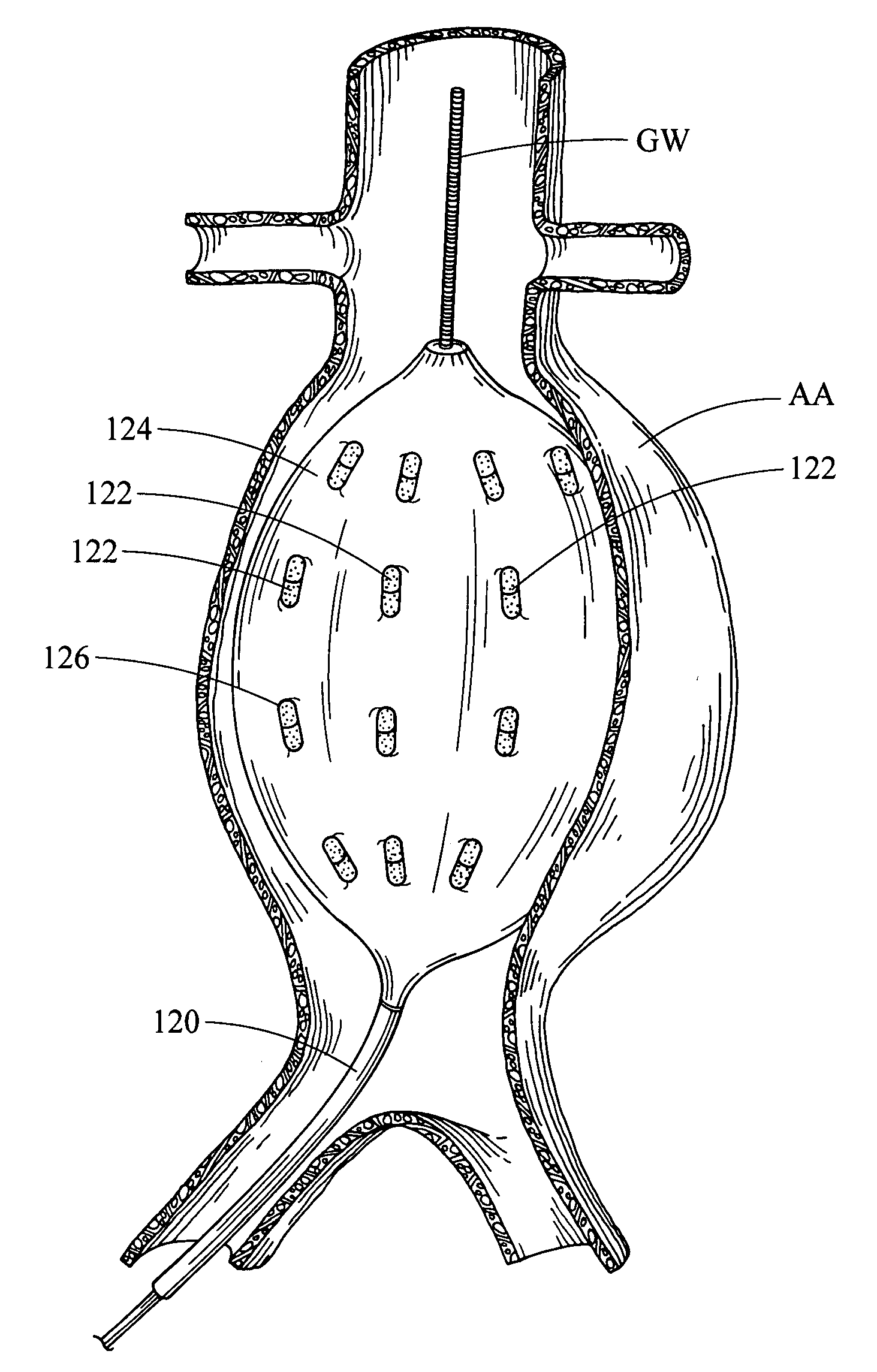
Endovascular disease may be characterized by weakened vessels due to elastin breakdown, which results in dilation of vessels and aneurysm.
When a local dilatation of a vessel occurs, irregular blood flow patterns result in the lumen of the vessel, typically leading to clot formation.
Typically, the wall of the vessel also progressively dilates and weakens, often resulting in vessel rupture.
Vessel rupture, in turn, often causes dramatic negative consequences such as a stroke, when a cerebral vessel ruptures, or even death, when an abdominal aortic aneurysm (“AAA”) ruptures.
In addition, balloons such as angioplasty or dilation balloons are expanded to open a body passage or vessel lumen, thereby causing potential trauma or injury to the expanded passage or vessel.
Endoleaks arising after such grafting may be caused by incomplete sealing between the endovascular prosthesis and the aortic wall or by defects within the endovascular prosthesis.

Method used


Image

  • Endoluminal medical device for local delivery of cathepsin inhibitors, method of making and treating
  • Endoluminal medical device for local delivery of cathepsin inhibitors, method of making and treating
  • Endoluminal medical device for local delivery of cathepsin inhibitors, method of making and treating

Examples


example 1

Testing Compounds as Cathepsin Inhibitors

[0577] The cathepsin inhibitory effects of the compound of the invention can be determined in vitro by measuring the inhibition of, e.g., recombinant human cathepsins B, K, L and S. The buffer for use in the cathepsin B, L and S assays is a 0.1 M pH 5.8 phosphate buffer containing EDTA (1.33 mM), DTT (2.7 mM) and Brij (0.03%). The in vitro assays are carried out as follows:

[0578] (a) For cathepsin B:

[0579] To a microtiter well is added 100 uL of a 20 uM solution of inhibitor in assay buffer followed by 50 uL of a 6.4 mM solution of Z-Arg-Arg-AMC substrate (Peptides International) in assay buffer. After mixing, 50 uL of a 0.544 nM solution of recombinant human cathepsin B in assay buffer is added to the well, yielding a final inhibitor concentration of 10 uM. Enzyme activity is determined by measuring fluorescence of the liberated aminomethylcoumarin at 440 nM using 380 nM excitation, at 20 minutes. % Enzyme inhibition is determined by comp...


PUM


Abstract

An endoluminal medical device comprises a drug release system that releases a cathepsin inhibitor at a predetermined location within a lumen of a patient. The endoluminal devices and methods of treatment of disease can treat illnesses such as aneurysms and aortic dissections.

Description

RELATED APPLICATIONS [0001] The present patent document is a continuation-in-part of PCT Application Serial No. PCT / US2006 / 048865, filed Dec. 22, 2006, designating the United States and which will be published in English, which claims the benefit of the filing date under 35 U.S.C. § 119(e) of Provisional U.S. Patent Application Ser. No. 60 / 755,961 filed Jan. 3, 2006. All of the foregoing applications are hereby incorporated by reference.BACKGROUND [0002] 1. Technical Field [0003] This invention relates generally to methods and medical devices and, more particularly, to medical devices incorporating drugs, bioactive agents, therapeutic agents or diagnostic agents for treating an aneurysm and an aortic dissection. The invention also relates to kits and to treating an aorta wall adjacent to an aortic aneurysm as a preventive measure. [0004] 2. Background of the Invention [0005] Endovascular disease may be characterized by weakened vessels due to elastin breakdown, which results in dila...

Claims


Application Information

Patent Timeline
IPC IPC(8): A61F2/06A61K38/00A61P9/00
CPCA61L27/34A61L27/54A61L29/085A61L2300/608A61L31/10A61L31/16A61L2300/434A61L29/16A61P9/00
Inventor BIGGS, DAVID P.GREWE, DAVID D.FEARNOT, NEAL E.RUANE, PATRICK H.RAGHEB, ANTHONY O.
Owner COOK INC
Why Eureka
Eureka Blog
Learn More