Hemangio-colony forming cells
a technology of hemangiocolony and forming cells, which is applied in the direction of immunological disorders, extracellular fluid disorders, antibody medical ingredients, etc., can solve the problems of not preventing rejection, limited hscs sources, and limited hscs
- Summary
- Abstract
- Description
- Claims
- Application Information
AI Technical Summary
Benefits of technology
Problems solved by technology
Method used
Image
Examples
example 1
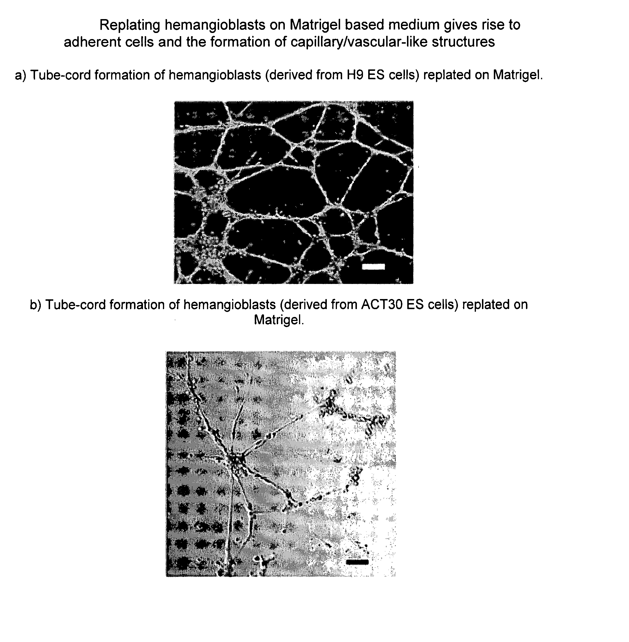
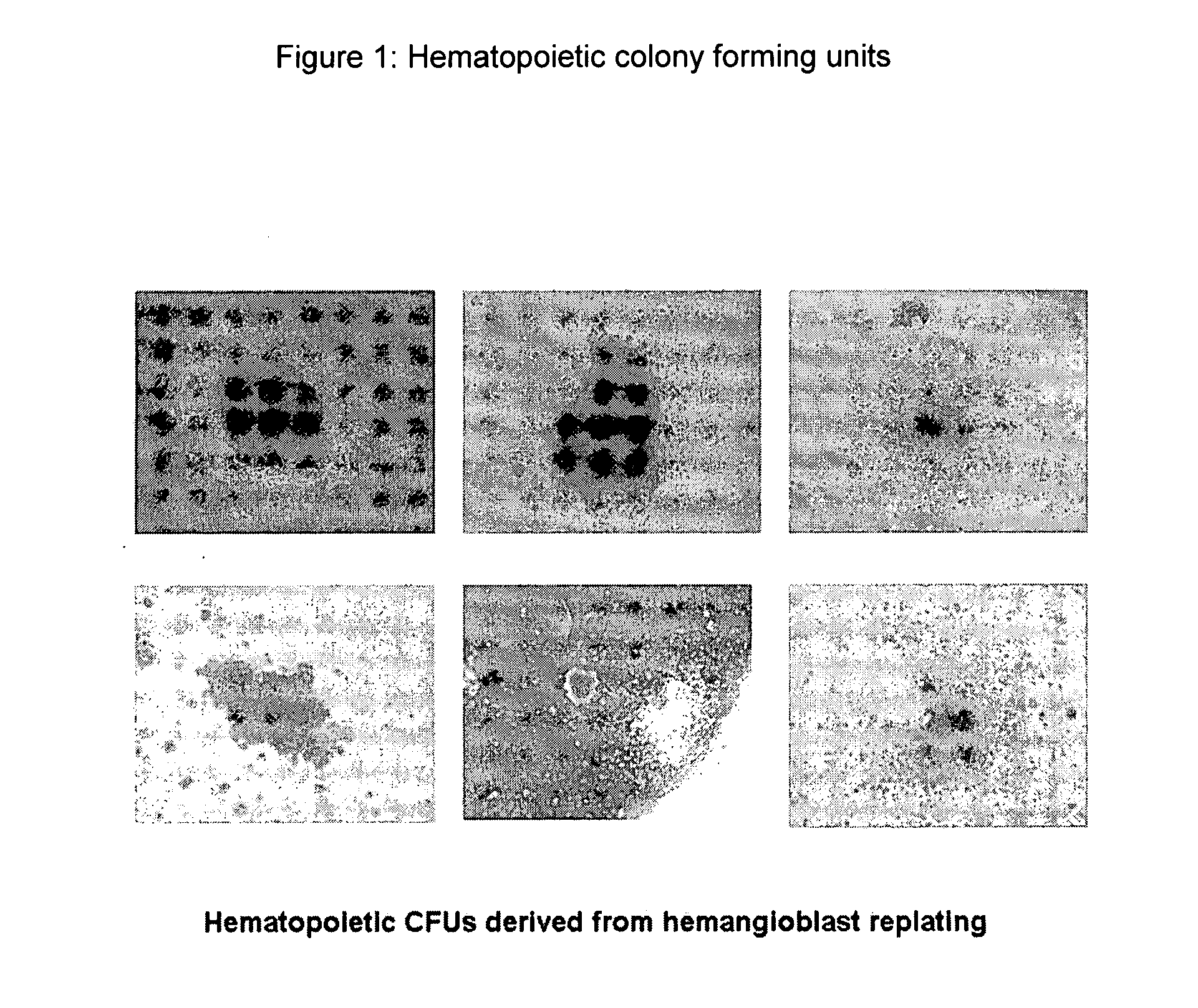
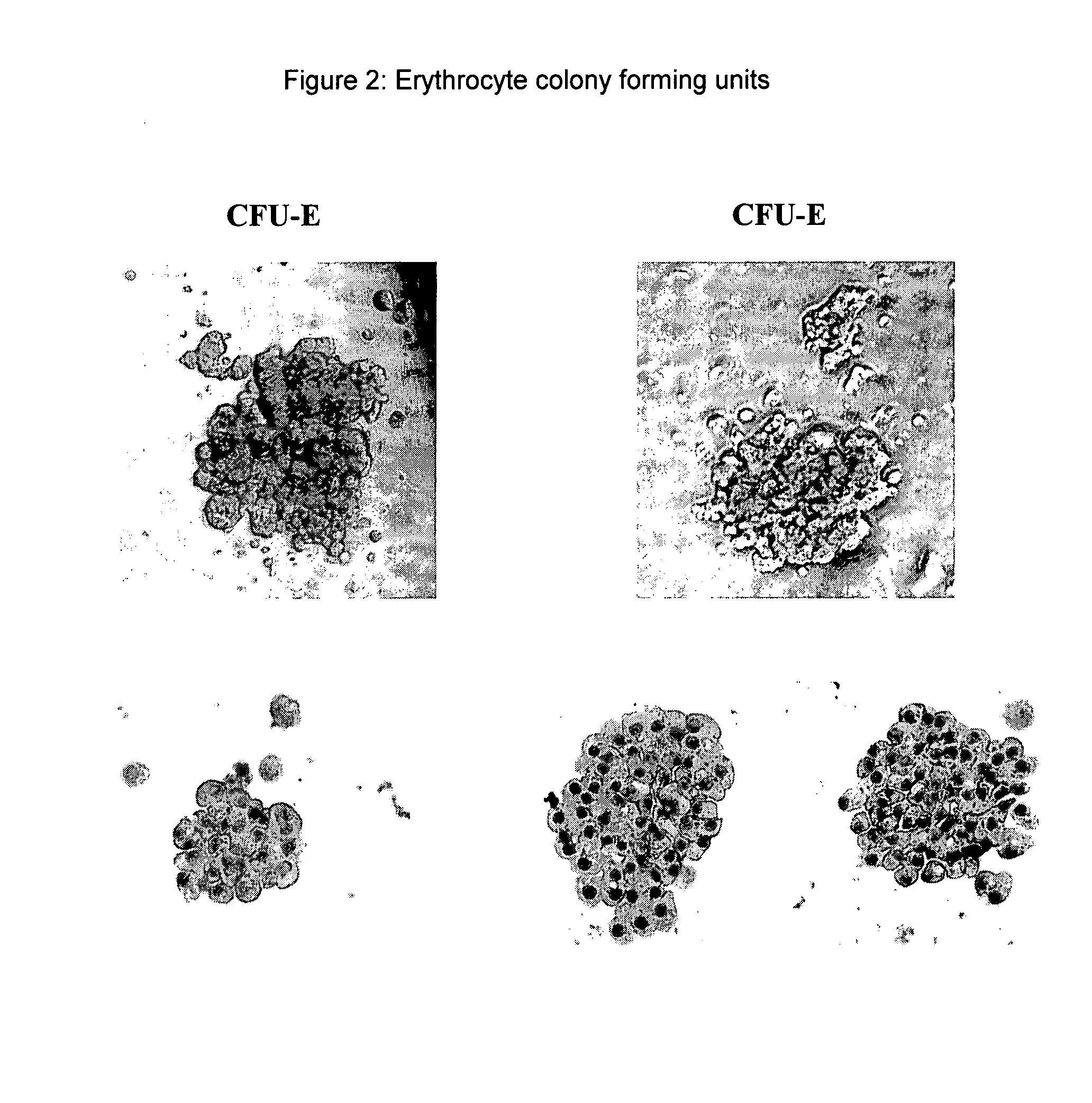
Hemangioblasts Derived from Human ES Cells Exhibit Both Hematopoietic and Endothelial Potential
[0392] The human embryonic stem cell lines H1 and H9 are cultured in ES cell media with mouse embryonic fibroblasts (MEFs). Cultured human ES cells are detached, pelleted by centrifugation at 200 g, and resuspended in embryoid body (EB) formation medium. EB formation medium comprises serum-free Stemline media (Sigma®) supplemented with BMP-4 and VEGF. Cells are then plated on ultra-low attachment culture dishes and cultured in a CO2 incubator. After 24-48 hours, early hematopoietic cytokines (TPO, Flt-3 ligand, and SCF) are added and the cells incubated for another 24-48 hours to form EBs.
[0393] Embryoid bodies (EB) are cultured for 2-6 days or optionally 2-5 days and then collected, washed with PBS and disaggregated into single cell suspensions using Trypsin / EDTA. The numbers of EBs are determined and approximately 5-10×106 cells / mL are cultured in serum-free methylcellulose medium that...
example 2
Additional Characterization of hESC-Derived BC Cells or Hemangioblasts
[0400] Human ES cell culture. The hES cell lines used in this study were previously described HI, H7, and H9 (NIH-registered as WA01, WA07, and WA09) cell lines and four lines (MA01, MA03, MA40, and MA09) derived at Advanced Cell Technology. Undifferentiated human ES cells were cultured on inactivated (mitomycin C-treated) mouse embryonic fibroblast (MEF) cells in complete hES media until they reached 80% confluence (Klimanskaya & McMahon; Approaches of derivation and maintenance of human ES cells: Detailed procedures and alternatives, in Handbook of Stem Cells. Volume 1: Embryonic Stem Cells, ed. Lanza, R. et. al. (Elsevier / Academic Press, San Diego, 2004). Then the undifferentiated hES cells were dissociated by 0.05% trypsin-0.53 mM EDTA (Invitrogen™) for 2-5 min and collected by centrifugation at 1,000 rpm for 5 minutes.
[0401] EB formation. To induce hemangioblast precursor (mesoderm) formation, hES cells (2 ...
example 3
Method of Producing and Purifying an Exemplary HOXB4 Protein
[0432] An exemplary HOXB4 protein is made and purified. This is a TAT-HA-HOXB4 fusion protein.
[0433] A pTAT-HA-HoxB4 expression vector was generated by cloning a PCR fragment (sense primer 5′-TAC CTA CCC ATG GAC CAC TCG CCC-3′ (SEQ ID NO: 16) and antisense primer 5′-TCG TGG CTC CCG AAT TCG GGG GCA-3′ (SEQ ID NO: 17) encompassing the HoxB4 open reading frame with and without N-terminal 32 amino acids into the NcoI and EcoRI sites of pTAT-HA expression vector (a gift from Dr. S F. Dowdy, University of California San Diego, La Jolla, Calif.), which contains an N-terminal TAT PTD. The generated pTAT-HA-HoxB4 plasmid was confirmed by sequence analysis and transformed into BL21(DE3)pLysS bacteria (Invitrogene™).
[0434] Bacterial cells growing at log phase were induced with 1 mM IPTG at 30 C for 4 hours and collected by centrifugation. Since the 6×His-fused recombinant TAT-HoxB4 protein was found to be sequestered into inclusion...
PUM
| Property | Measurement | Unit |
|---|---|---|
| Fraction | aaaaa | aaaaa |
| Fraction | aaaaa | aaaaa |
| Fraction | aaaaa | aaaaa |
Abstract
Description
Claims
Application Information
 - R&D
- Intellectual Property
- Life Sciences
- Materials
- Tech Scout
- Unparalleled Data Quality
- Higher Quality Content
- 60% Fewer Hallucinations
Browse by: Latest US Patents, China's latest patents, Technical Efficacy Thesaurus, Application Domain, Technology Topic, Popular Technical Reports.
© 2025 PatSnap. All rights reserved.Legal|Privacy policy|Modern Slavery Act Transparency Statement|Sitemap|About US| Contact US: help@patsnap.com


