Bioactive wound dressings and implantable devices and methods of use

Inactive Publication Date: 2008-01-24
MEDIVAS LLC
View PDF3 Cites 82 Cited by
  • Summary
  • Abstract
  • Description
  • Claims
  • Application Information

AI Technical Summary

Benefits of technology

[0017]FIG. 1 is a graph showing the ATP generated in 48 hours by smooth muscle cells plated in Cambrex-complete-media (growth factors plus 5% fetal bovine serum (FBS)), Cambrex-minus-growth-factors (gf)-plus-5% FBS, or Cambrex-minus-gf-

Problems solved by technology

Based on this concept, it follows that optimal repair of wounds and lesions cannot be achieved by the administration of single proteins, or single genes whose encoded products

Method used


Image

  • Bioactive wound dressings and implantable devices and methods of use
  • Bioactive wound dressings and implantable devices and methods of use
  • Bioactive wound dressings and implantable devices and methods of use

Examples


example 1

Delivery of Growth / Survival Components by Alginic Acid

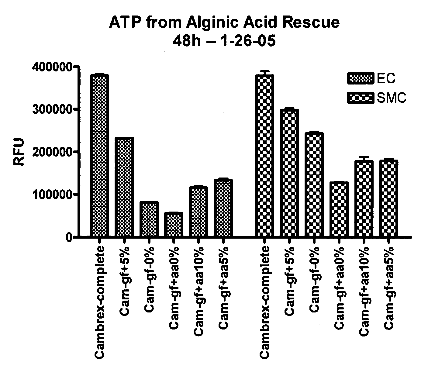
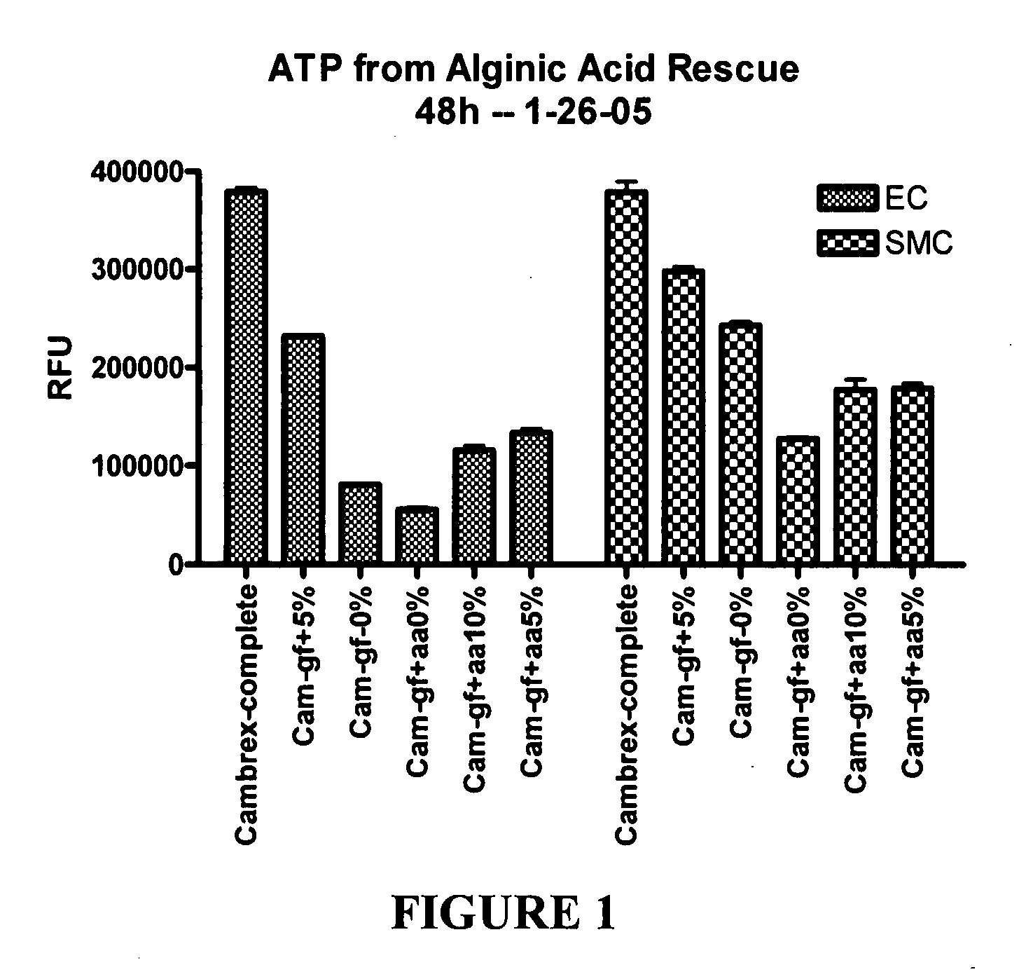
[0260] This example illustrates experiments that were conducted to test the ability of hydrogel particles to deliver components required for cell growth and survival. The hydrogel particles were formed using low viscosity alginic acid (AA) (Sigma Chemicals, A2158) prepared using an adaptation of a published protocol (J Raymond et al., Am J Neuroradiol (2003) 24:1214-1221).

[0261] Preliminary experiments demonstrated that 4% low viscosity alginate made up in 0.5% sodium chloride can absorb about one third of its volume of the desired media, such as conditioned medium from progenitor cell cultures that had been isolated from human blood, and then be crosslinked by adding 5% calcium chloride. Polymerization occurs very rapidly and AA particles form, encapsulating the media within the alginate. The rate of release of media from the particles during the first 24 hours is very rapid, with about half of the media being released. The r...

example 2

Encapsulation of Cells in Hydrogel

[0275] A highly purified and well-characterized sodium alginate with very low levels of endotoxins and proteins, optimal for in vitro and in vivo applications, was used to encapsulate cells (NovaMatrix, Pronova UltraPure (UP) LVM alginate). This alginate is made up in 2% HEPES with some sonication, to help the AA go into complete suspension, and then filter sterilized. Cell-containing-particles are made by mixing cells plus fetal bovine serum (FBS) with the alginate, drawing the mixture up into a 1 cc syringe and forming particles drop wise into a 5% calcium chloride solution. This process produces about 100 particles / ml with about 2000 cells / particle. The cells used for this experiment were SMCs, which are a robust, anchorage-dependent cell line normally not grown in suspension.

[0276] To determine cell viability within the AA particles, two viability dyes were used: (1) Calcein AM, which is retained in cells that have intact membranes. Calcein A...

example 3

Polymer Coating of Hydrogel Encapsulated Cells

[0279] PEA conjugated to fluorescent dansyl-lysine was used to coat particles to visualize the polymer coating for consistency and completeness of coating. The results of this experiment indicated that a reliable method to coat AA particles to achieve consistency and completeness of coating was to suspend them in the polymer and allow the polymer to flow through a mesh that captures the particles. The particles were then rapidly removed from the mesh by resuspending in the appropriate buffer.

[0280] Then, to determine if the SMCs contained in AA particles can survive coating with PEA.H polymer, AA particles were formed containing 2000 smooth muscle cells / particle. Test AA particles were coated with a 10% (w / v ethanol) polymer solution and incubated in the presence of the two viability dyes shown in Example 2 above as able to enter the encapsulated cells. Control particles encapsulated the SMCs, but were also exposed to the dyes, but we...


PUM

PropertyMeasurementUnit
Timeaaaaaaaaaa
Timeaaaaaaaaaa
Biological propertiesaaaaaaaaaa

Abstract

The present invention provides wound dressings, optionally surgically implantable, containing biodegradable, polymers and hydrogels having allogenic or autologous precursor cells, such as stem cells and progenitor cells dispersed within. Alternatively, the wound dressings can have conditioned medium obtained from the precursor cells dispersed within. The wound dressings promote tissue restoration processes at a site of application or implantation. Additional bioactive agents can also be dispersed within the polymer/hydrogel matrix, which can be formulated to biodegrade at a controlled rate by adjusting the composition. Methods are also provided for using such biodegradable wound dressings as a delivery device or carrier for the precursor cells, conditioned medium and bioactive agents, or as coatings on implantable medical devices, to promote tissue restoration at a lesion site.

Description

CROSS REFERENCE TO RELATED APPLICATIONS [0001] This is a continuation-in-part application of PCT Application No. PCT / US2005 / 038925 filed Oct. 27, 2005 which claims the benefit under 35 USC §119(e) to U.S. application Ser. No. 60 / 623,446 filed Oct. 28, 2004 and this application is a continuation-in-part of U.S. application Ser. No. 10 / 362,848, filed Oct. 14, 2003 which claims the benefit under 35 USC §120. The disclosure of the prior applications is considered part of and is incorporated by reference in the disclosure of this applicationFIELD OF THE INVENTION [0002] The invention relates generally to compositions used in healing wounds and lesions, and in particular to biodegradable polymer dressings that promote natural healing processes at wound sites. BACKGROUND INFORMATION [0003] In general, wounds and lesions can be divided into two types: acute and chronic. In cases where a wound is not initially surgically closed (delayed primary closure), the wound is left open for a time suf...

Claims


Application Information

Patent Timeline
IPC IPC(8): A61K35/12A61F13/00A61K9/00A61P17/02A61K35/22A61K35/28A61K35/30
CPCA61F13/00012A61F13/069A61F2013/00221A61F2013/0054A61K35/30A61F2013/00927A61K35/22A61K35/28A61F2013/0091A61P17/02
Inventor CARPENTER, KENNETH W.TURNELL, WILLIAM G.DEFIFE, KRISTIN M.GRAKO, KATHRYN A.KATSARAVA, RAMAZ
Owner MEDIVAS LLC
Why Eureka
Try Eureka