Method and apparatus for precise selection and extraction of a focused component in isoelectric focusing performed in micro-channels
a microchannel and focused component technology, applied in the field of methods and equipment, can solve the problems of unavoidable component zone broadening, cross-contamination of closely migrating peaks, and inability to achieve more conventional separatory methods
- Summary
- Abstract
- Description
- Claims
- Application Information
AI Technical Summary
Benefits of technology
Problems solved by technology
Method used
Image
Examples
experimental examples
Example 1
[0058] An IMED having the structure shown in FIG. 3 was used to selectively extract different hemoglobin variants of a mixture. An imaged cIEF system (Model iCE280, Convergent Bioscience Ltd., Toronto, Canada) was used as the real-time, whole separation channel UV detection apparatus, and protein detection was achieved through UV absorption at 280 nm. An autosampler from Prince CE system (Prince. Technologies, Emmen, The Netherlands) was used for sample injection and fluid manipulation along the separation channel. Methyl cellulose (MC), hourse heart myoblobin, human transferrin, and pH 3-10 Pharmalyte were obtained from Sigma (St Louse, Mo.). Hemoglobin control AFSC was obtained from Helena Laboratories (Beaumont, Tex.). HPLC grade water was from J. T. Baker (Phillipsburg, N.J.), and was used for all solutions. Sample mixture solution was prepared by mixing protein with 8% pH 3-10 Pharmalytes and 0.35% MC.
[0059] The extraction channel of the IMED was first filled with HP...
example 2
[0063]FIG. 5 shows the separation and extraction of Hemoglobin S in the IMED with the device according to FIG. 4. The pI difference between Hemoglobin F and S and between Hemoglobin S and C was about 0.1. It can be seen that the separation pattern was not affected significantly upon hemoglobin s extraction, while the peak area of Hemoglobin S was reduced following extraction.
example 3
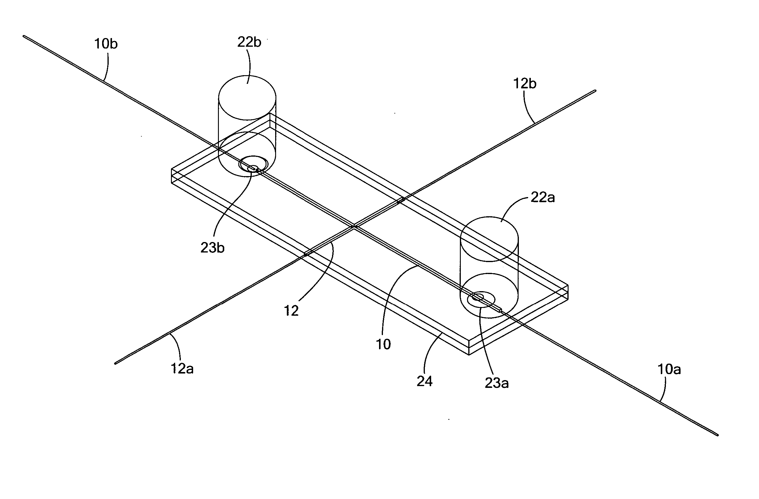
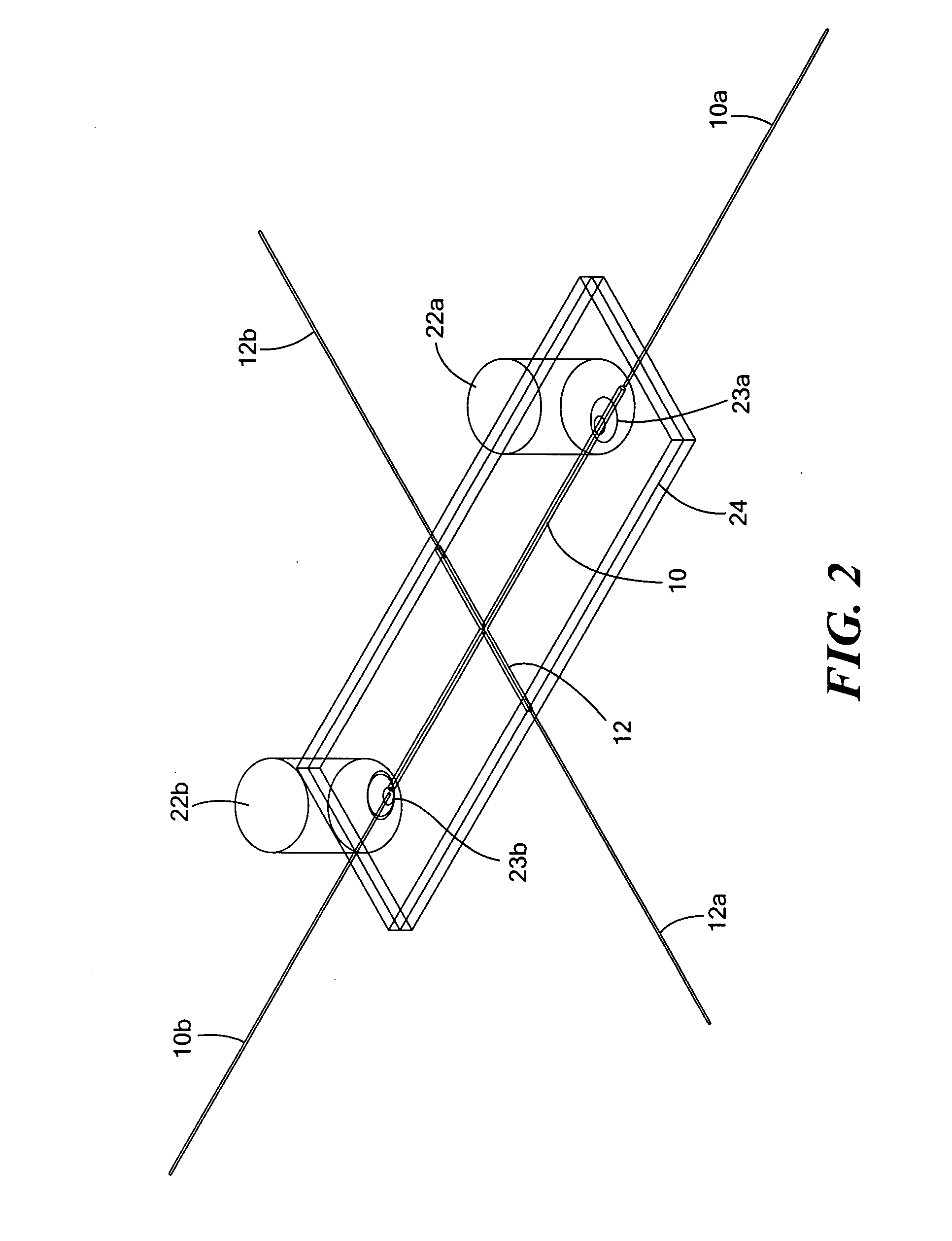
[0064] An IMED having the structure substantially represented in FIG. 2 was used to separate and selectively extract target protein component for off line MALDI-TOF characterization. FIG. 6 shows the cIEF electrophoregram of myoglobin and transferrine in 8% 5-8 Pharmalytes, 0.35% MC solution. It can be seen that the major component of transferrine and the major component of myoglobin were very well separated. Upon cIEF separation, the extracted individual protein was diluted to 100 μL in DI water, and 1 μL of this diluted sample was further diluted at either 1:2 or 1:10 ratio with 0.2% (v / v) trifluoroacetic acid in water. The further diluted protein sample of 1 μL was mixed with 1 μL of sinapinic acid (10 mg / mL), and 1 μL of this sample mixture was spotted for MALDI TOF MS (Voyager STR, Applied Biosystems, Foster City, Calif.).
[0065]FIGS. 7 and 8 shows the mass spectrograms of extracted myoglobin and transferrine. From the mass spectrogram, it can be seen that the presence of carri...
PUM
| Property | Measurement | Unit |
|---|---|---|
| pore size | aaaaa | aaaaa |
| thickness | aaaaa | aaaaa |
| optical whole column imaging detection | aaaaa | aaaaa |
Abstract
Description
Claims
Application Information
 - R&D
- Intellectual Property
- Life Sciences
- Materials
- Tech Scout
- Unparalleled Data Quality
- Higher Quality Content
- 60% Fewer Hallucinations
Browse by: Latest US Patents, China's latest patents, Technical Efficacy Thesaurus, Application Domain, Technology Topic, Popular Technical Reports.
© 2025 PatSnap. All rights reserved.Legal|Privacy policy|Modern Slavery Act Transparency Statement|Sitemap|About US| Contact US: help@patsnap.com


