Methods for Selecting Proteins that are Readily Presented as Antigens on Dendritic Cells

a dendritic cell and protein technology, applied in the field of protein selection that is readily presented as antigen on dendritic cells, can solve the problems of insufficient efficacy of dendritic cell vaccine therapy, inability to achieve an activation that is efficient, and inability to achieve an effective activation

Inactive Publication Date: 2008-03-13
MEDICAL & BIOLOGICAL LAB CO LTD +1
View PDF3 Cites 3 Cited by
  • Summary
  • Abstract
  • Description
  • Claims
  • Application Information

AI Technical Summary

Problems solved by technology

However, activation of cellular immunity cannot be separated from induction of immunological tolerance.
Therefore, how to achieve an activation that is efficient and specific to the target is a major issue in tumor immunity.
However, for the reason that effective cancer antigens differ depending on the patient, and so on, dendritic cell vaccine therapies are not necessarily effective therapeutic methods for all patients.
However, at present there is no practical evaluation system for assessing the effectiveness of cancer antigen proteins.
Therefore, for antigenic proteins in general, constructing an evaluation system similar to the above evaluation system has to be considered completely impossible.
However, the kind of intracellular compartments the antigenic proteins are taken into from the outside, the way the antigenic proteins taken up or their degradation products pass through the membrane to translocate to the cytoplasm, and such have not been solved yet.

Method used


Image

  • Methods for Selecting Proteins that are Readily Presented as Antigens on Dendritic Cells
  • Methods for Selecting Proteins that are Readily Presented as Antigens on Dendritic Cells
  • Methods for Selecting Proteins that are Readily Presented as Antigens on Dendritic Cells

Examples


example 1

Proteasome-Dependent Degradation of Accumulated OVA

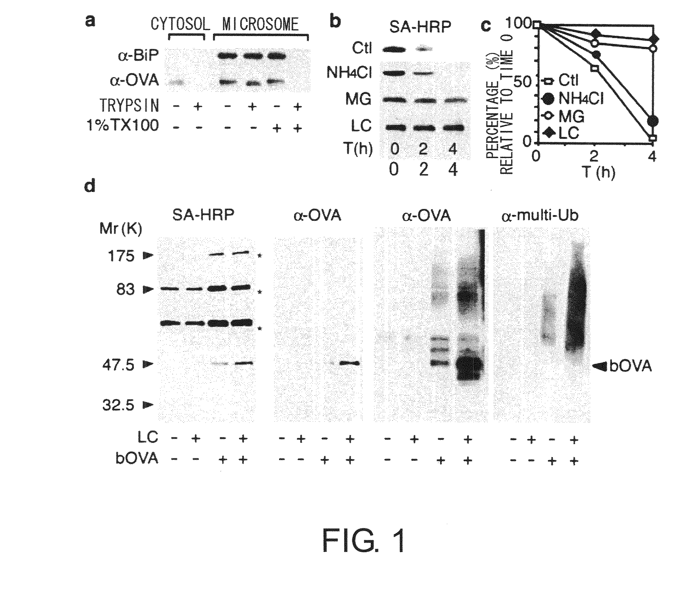
[0060] Intracellular localization of foreign antigens was examined by cell fractionation methods. Chicken ovalbumin (OVA) was used as an antigen model, and DC2.4 (haplotype H-2 Kb), a cultured cell line of mouse dendritic cells, was used as a model for dendritic cells. The DC2.4 cell line is derived from dendritic cells, and was established while retaining many of the properties of dendritic cells, such as cross-presentation. BMDCs with a haplotype of H-2 Kb, the same as that of DC2.4, were used as the control. BMDCs were induced from the bone marrow cells of C57BL / 6 mice (SLC) in RPMI-1640 supplemented with 5% FCS using 5 ng / mL GM-CSF (MBL), and those cells purified by magnetic beads after five days of culturing were used.

[0061] DC2.4 cells made to take up bOVA antigens were suspended in 10 mM Tris-HCl (pH7.4) containing 250 mM sucrose, this was homogenized using glass beads, centrifuged for 30 minutes at 250,000×g, and then frac...

example 2

Relationship Between Accumulated OVA and ERAD-Related Proteins

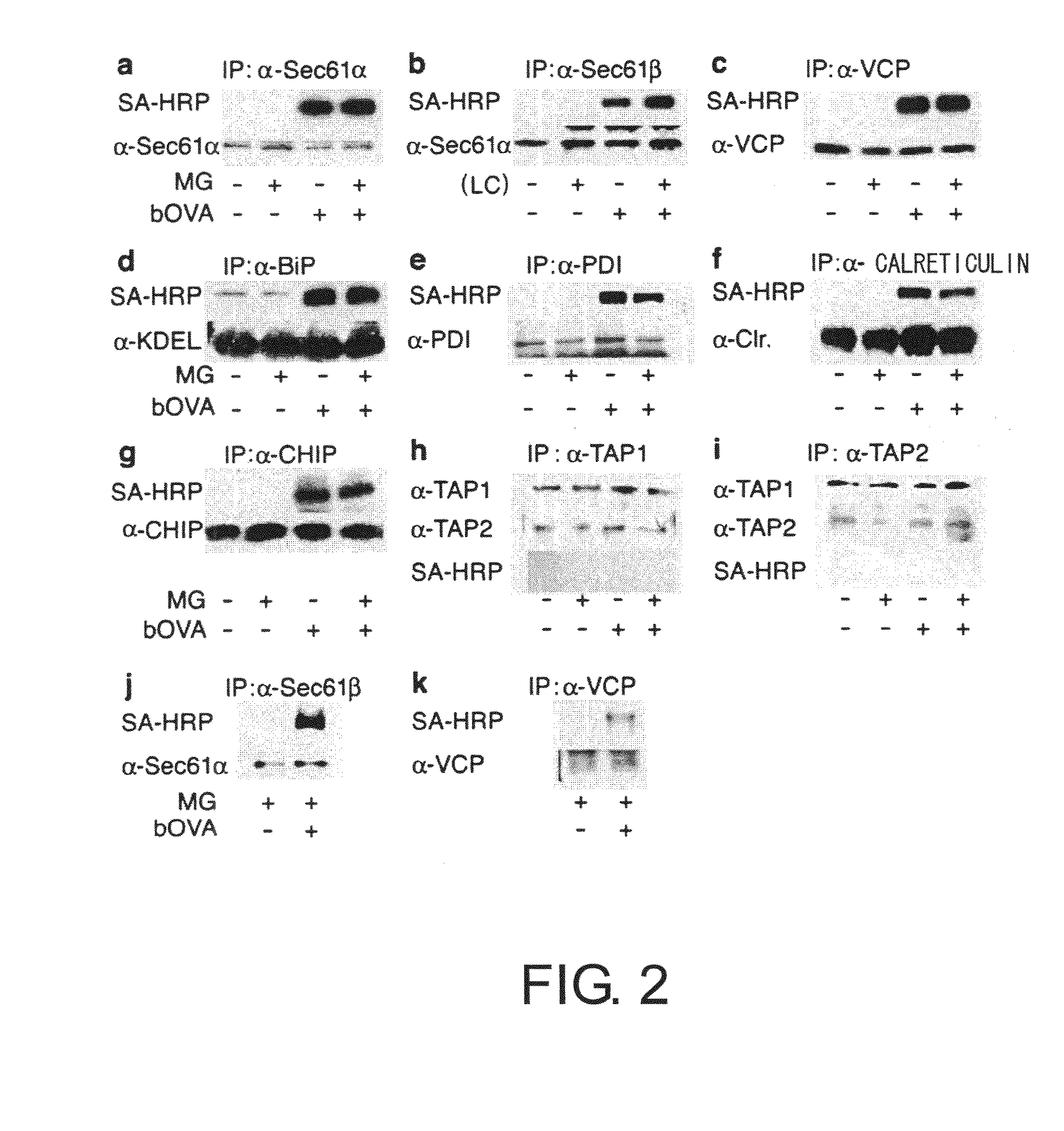
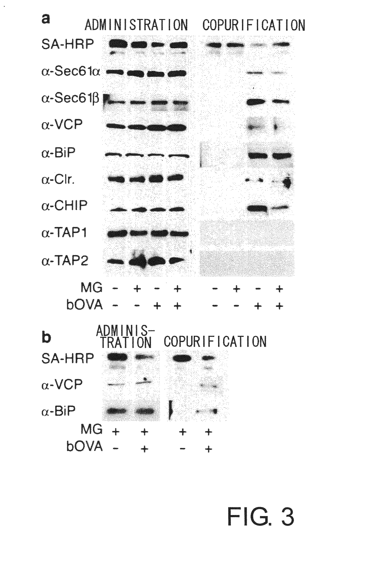
[0066] To prove the above-mentioned hypothesis, first, the relationship between accumulated OVA and substances involved in retrograde transport from the ER to the cytoplasm was examined by co-immunoprecipitation. 2×10⁷ DC2.4 or BMDC cells were cultured for four hours in a medium containing 10 μM MG-132 in the presence of bOVA at 250 μg mL−1. Cells were lysed in 20 mM HEPES pH7.6 containing 1% digitonin and protease inhibitors to collect biotin-labeled samples. The samples were cleared of impurities in advance using protein G sepharose (Amersham Pharmacia Biotech), and after incubation with antibodies for precipitation (anti-Sec61β, anti Sec61α, anti-VCP, anti-BiP, anti-PDI, anti-calreticulin, anti-TAP1, and anti-TAP2), the immunoprecipitates were collected using protein G. When chicken anti-CHIP antibody was used, immunoprecipitates were collected using a rabbit anti-IgY column (Mr. S. Seki, MBL). The precipitated sample...

example 3

Decrease Caused by ERAD Inhibition of the Degradation of Accumulated OVA

[0071] Next, to further confirm the involvement of ERAD, the effect on bOVA accumulation of ERAD inhibition, for example by Ca2+ depletion or thapsigargin treatment, was examined. Depletion of Ca2+ in the ER is known to inhibit ERAD. Thapsigargin (Tg) is known to decrease the Ca2+ concentration in the ER lumen by binding to the ER membrane and functioning as an ionophore and to inhibit ERA D. Thus, in the presence of Tg, bOVA degradation by ERAD is predicted to be suppressed. DC2.4 cells were pulsed with bOVA in the presence of MG-132, then washed with PBS or with PBS containing 1 mM EGTA, and then chased for four hours. As shown in FIG. 4a, accumulated bOVA did not decrease with Ca2+ depletion. Re-addition of Ca2+ to cells pre-exposed to EGTA restored bOVA degradation (FIG. 4a). This eliminates the possibility that Ca2+ depletion caused irreversible damage to DC2.4 cells. Thapsigargin also inhibited the degrad...


PUM

PropertyMeasurementUnit
concentrationsaaaaaaaaaa
temperatureaaaaaaaaaa

Abstract

The present inventors worked on elucidating the mechanism of antigen cross-presentation by dendritic cells, and revealed that foreign antigens taken up in dendritic cells are degraded by proteasomes after undergoing polyubiquitination. They discovered a positive correlation between the uptake amount of an antigenic protein by dendritic cells or the level of polyubiquitination and the level of antigen presentation of the antigenic protein. Based on these findings, methods were developed for predicting the intensity of cross-presentation of an anti genic protein by measuring the amount of antigenic protein taken up or polyubiquitinated protein. The use of these methods allows selecting antigenic proteins that are readily presented as antigens from among a number of proteins. The methods of the present invention enable suitable cancer immune vaccines to be provided through selection of more suitable cancer-specific proteins.

Description

TECHNICAL FIELD [0001] The present invention relates to methods for selecting proteins that are readily presented as antigens on dendritic cells, wherein the methods are based on cross-presentation mechanisms by dendritic cells. BACKGROUND ART [0002] When cells in a living body undergo cancerization, normally, immune defense mechanisms function to remove the cancerized cells. Cancer is thought to develop when the cancerized cells escape the surveillance of the immune defense mechanisms. The cancerized cells are mainly removed through cellular immunity. [0003] If a cellular immune mechanism is activated by a suitable procedure, it can act to strongly attack and remove cancers that have formed. Therefore, activation of cellular immunity may become a method for efficient removal of cancer cells. Cancer immunotherapy has already drawn attention as a means to treat cancer. Various methods have been researched developed so far, such as cell introduction therapies in which LAK cells, CTL c...

Claims


Application Information

Patent Timeline
Patent Type & Authority Applications(United States)
IPC IPC(8): G01N33/567C12N5/02C12N5/0784
CPCG01N33/5047C12N5/0639
Inventor IMAI, JUNMARUYA, MIKAKOKOYASU, SHIGEOHASEGAWA, HIRONORIYAHARA, ICHIRO
Owner MEDICAL & BIOLOGICAL LAB CO LTD
Why Eureka
Try Eureka