Thermal strain imaging of tissue

a tissue and thermal strain technology, applied in the field of thermal strain imaging of tissue, can solve the problems of vascular disease, significant health problems, invasive techniques, etc., and achieve the effect of improving imaging resolution and contrast, and improving detection and diagnosis

Inactive Publication Date: 2008-04-03
RGT UNIV OF MICHIGAN
View PDF7 Cites 11 Cited by
  • Summary
  • Abstract
  • Description
  • Claims
  • Application Information

AI Technical Summary

Benefits of technology

[0008] Vascular disease is a significant health issue. Consequently, there is a need for a non-invasive method for detecting plaques on arterial and vascular wall tissue. Furthermor

Problems solved by technology

However, these techniques are either invasive or lack sufficient spatial and contrast resolution

Method used


Image

  • Thermal strain imaging of tissue
  • Thermal strain imaging of tissue
  • Thermal strain imaging of tissue

Examples


Embodiment Construction

Rabbit Kidney Model

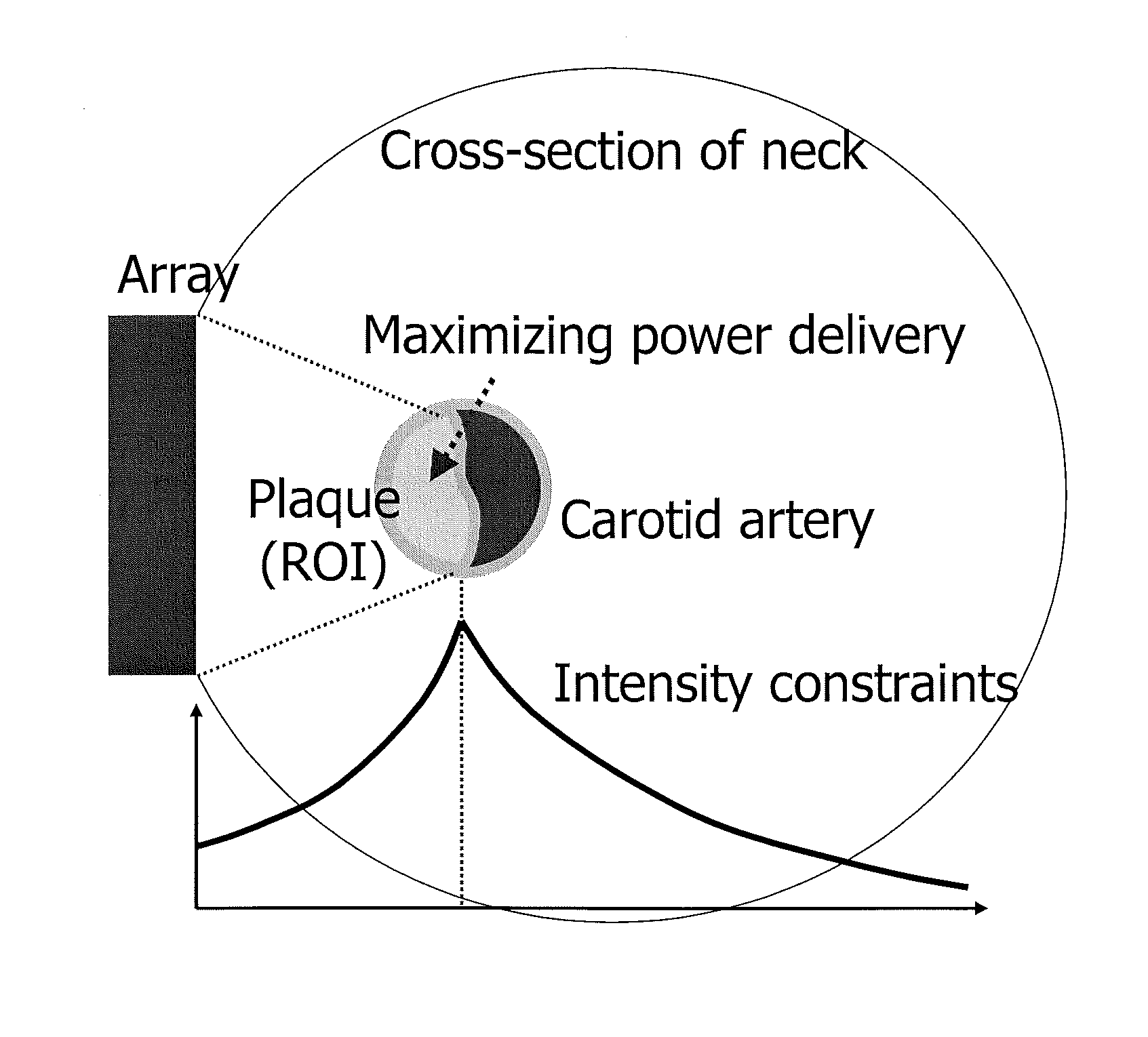
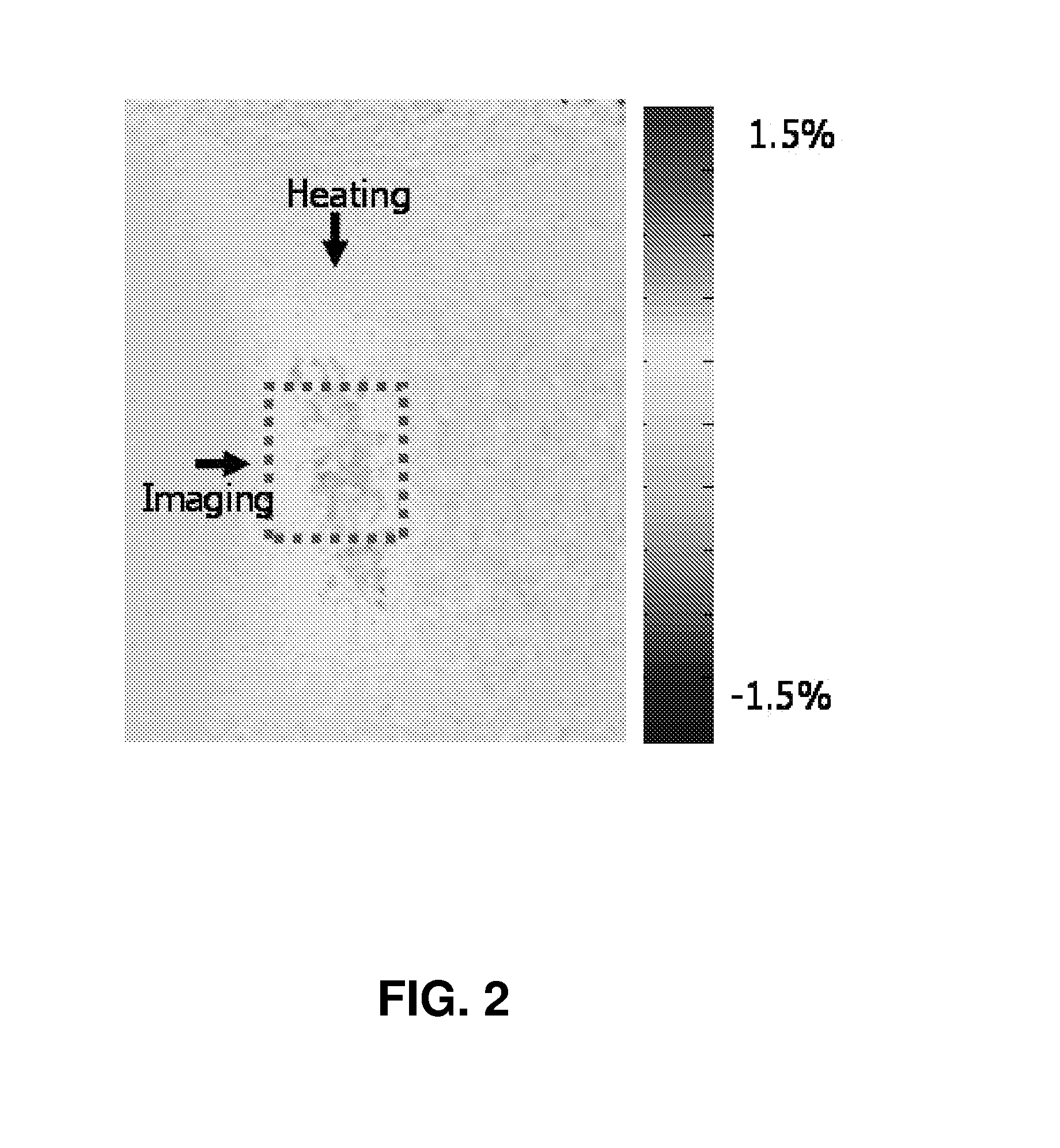
[0034] It is well known that lipids have a negative temperature dependence of the sound speed, whereas water-based tissues have positive temperature dependence [F. A. Duck, Physical Properties of Tissue. London: Academic, 1990]. Controlled local temperature modulation can be used to image the spatial distribution of temporal strain produced by changes in the sound speed [T. Bowen, “Radiation-induced Thermoacoustic Soft-Tissue Imaging,” IEEE Transactions on Sonics and Ultrasonics, vol. 29, pp. 187-187, 1982]. The opposite sign of the two different tissue types creates the contrast required for resolving the fatty tissue from surrounding water-based tissue.

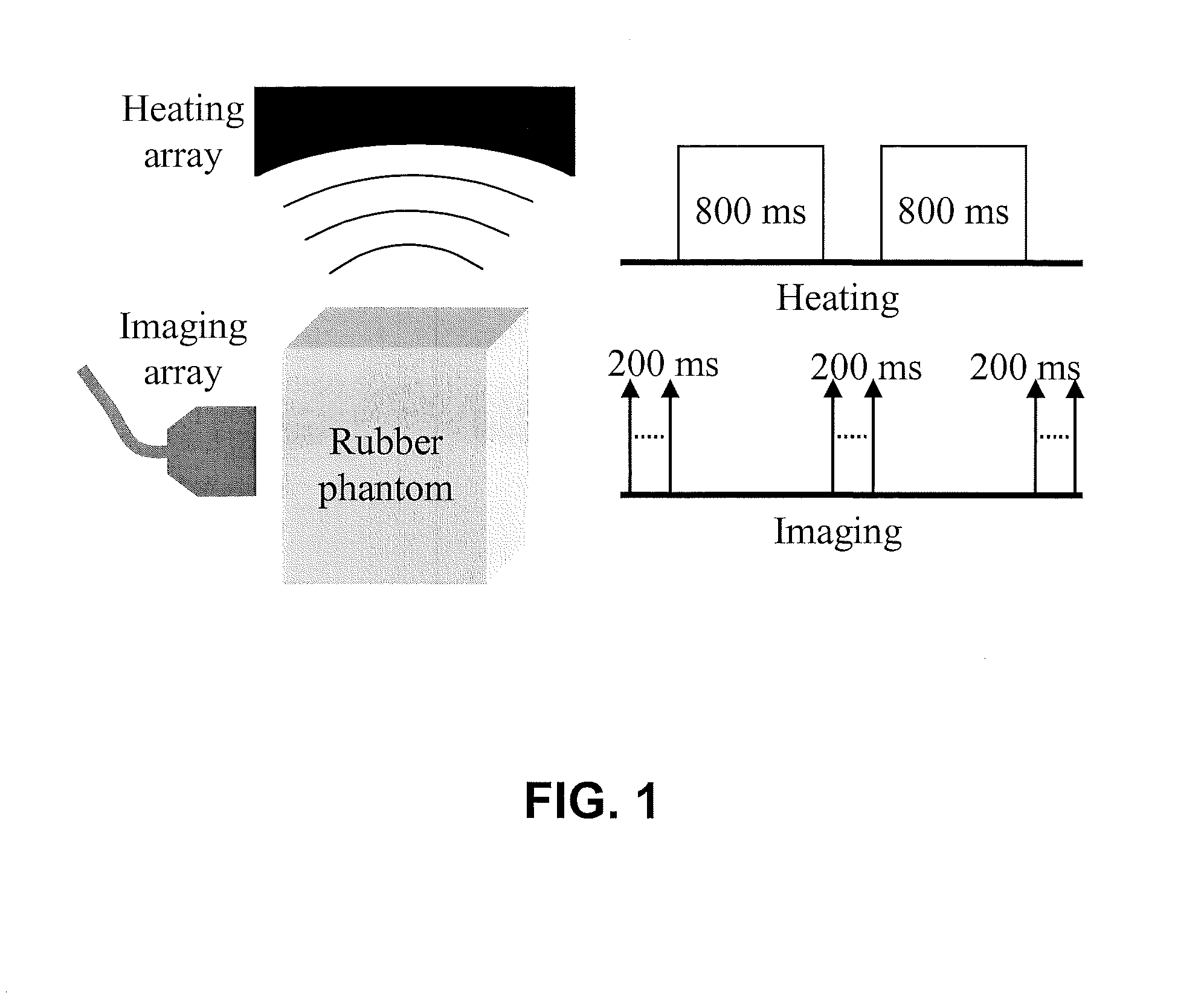
[0035] Using a 2-D phased array in combination with a conventional ultrasound scanner, the feasibility of ultrasound inducing and imaging of thermal strain is demonstrated. Among other heating sources, including microwave [Y. Shi, R. S. Witte, and M. O'Donnell , Identification of Vulnerable Plaque Atheroscleroti...


PUM


Abstract

Thermal strain imaging can be used to identify vascular plaques. Methods include heating and imaging with ultrasound to identify vulnerable plaques, which typically consist of a large lipid-rich core, in peripheral arteries of a patient. Lipid-bearing tissue has a negative temperature dependence of sound speed, whereas water-based tissue has a positive one, allowing thermal strain imaging to differentiate the two different types of tissues with high contrast to characterize plaque composition. Apparatus and methods of the present teachings allow noninvasive and reliable plaque identification.

Description

CROSS-REFERENCE TO RELATED APPLICATIONS [0001] This application claims priority to U.S. Provisional Application No. 60 / 849,083 filed on Oct. 3, 2006, which is incorporated by reference. [0002] All literature and similar materials cited in this disclosure, including but not limited to, patents, patent applications, articles, books, treatises, and internet web pages, regardless of the format of such literature and similar materials, are expressly incorporated by reference in their entirety for any purpose. In the event that one or more of the incorporated literature and similar materials differs from or contradicts this disclosure, including but not limited to defined terms, term usage, described techniques, or the like, this disclosure controls.GOVERNMENT RIGHTS [0003] Portions of the present disclosure were made with U.S. Government support under National Institutes of Health grant EB003451. The U.S. Government has certain rights in this disclosure.FIELD [0004] The present disclosur...

Claims


Application Information

Patent Timeline
IPC IPC(8): A61B8/12
CPCA61B8/587A61B8/0858
Inventor KIM, KANGHUANG, SHENG-WENWITTE, RUSSELL S.O'DONNELL, MATTHEW
Owner RGT UNIV OF MICHIGAN
Why Eureka
Try Eureka