Methods of Regenerating Cartilage

a cartilage surface and cartilage technology, applied in the direction of biocide, unknown materials, drug compositions, etc., can solve the problems of deterioration, deformation, and difficulty in repair of cartilage lesions, and achieve the effect of not delay or prevent further deterioration of cartilage surfa

Inactive Publication Date: 2008-06-12
SMITH & NEPHEW INC
View PDF16 Cites 18 Cited by
  • Summary
  • Abstract
  • Description
  • Claims
  • Application Information

AI Technical Summary

Problems solved by technology

These articular cartilage lesions are difficult to repair and as such represent a major challenge to surgeons.
Moreover, the current treatments available today yield a fibrous repair wherein a fibrocartilage is formed that provides only temporary relief The fibrocartilage tissue does not have the same mechanical properties as the natural hyaline cartilage found in the joint surfaces and degrades faster over time as a consequence of wear.
Patients typically have to undergo repeated surgical procedures to relieve reoccurring symptoms, though this type of surgery does not delay or prevent further deterioration of the cartilage surface.
In addition to creating a fibrocartilage type tissue and therefore only providing temporary relief, this may require the creation of a large osteochondral defect within which the implant is inserted.
The creation of this large defect causes damage to the underlying subchondral bone.
This manual labor-intense process is extremely costly and time consuming.
In addition to the prohibitive cost, there is a traumatic impact to the patient of having two surgical procedures to the knee.
Furthermore, the quality of the autologous cells and their minimum expansion capability limits the benefits of performing this procedure.
Unfortunately, fibrocartilage tissue is also formed by the use of this approach.
However, these cells do not differentiate into chondrocytes and only promote poor quality fibrous cartilage repair.

Method used


Image

  • Methods of Regenerating Cartilage
  • Methods of Regenerating Cartilage
  • Methods of Regenerating Cartilage

Examples


example

[0052]Bone marrow aspirate was harvested from an allogeneic donor goat. A bone marrow stromal cell (BMSC) fraction was obtained following plastic adherence of the cells and subsequent culture expansion was performed using standard culture conditions (37° C. / 5% CO2) and medium (alpha-MEM / 10% FCS). Cells were passaged on reaching 80% confluence (up to P3) and cryopreserved prior to use.

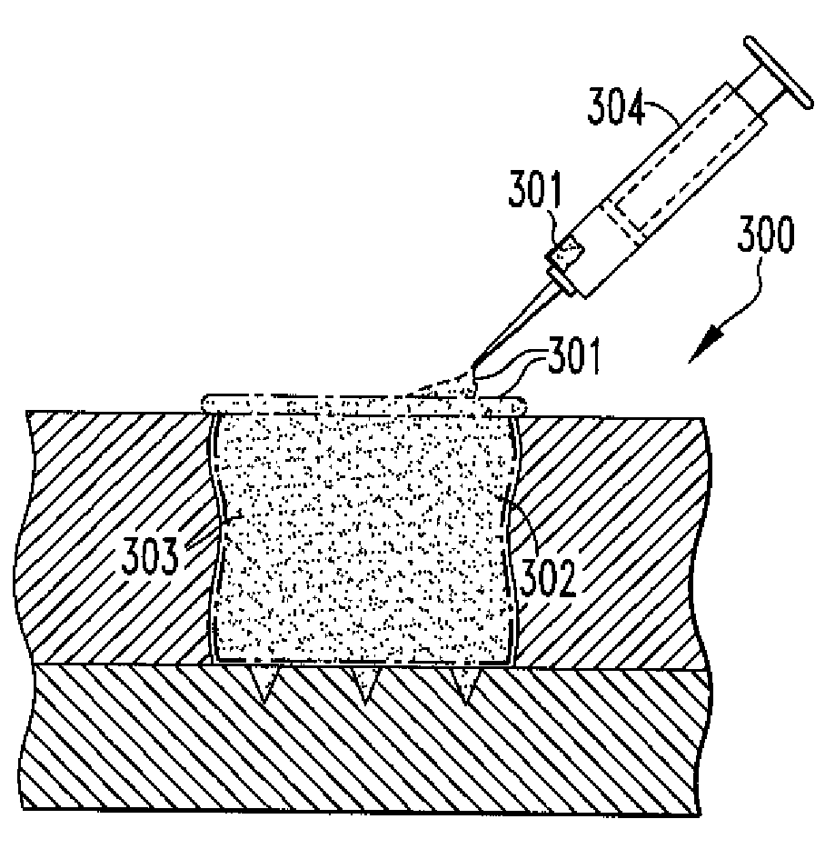
[0053]Treatment recipient goats, approximately 2.5 years old and 50-90 kg were used in the study. X-ray analysis confirmed normal bone mineral density. Micro fracture was performed on each goat (Group 1: N=3 micro fracture only, Group 2: N=3 micro fracture plus cell injection) as follows: A single annular defect (about 8 mm diameter) was generated in the medial femoral condyle of the stifle joint at a depth equivalent to the subchondral bone layer. A chondral pick (about 1 mm diameter) and mallet was used to perform the micro fracture procedure (about 3 mm depth, average of 7 holes per defect). After wo...


PUM


Abstract

The present disclosure relates to methods of regenerating cartilage. In an embodiment, a method includes initiating a release of precursor cells, including bone marrow cells and progenitor cells, into a cartilage defect; and applying a population of exogenous cells to the cartilage defect. The exogenous cells, selected from a group including chondrocytes, synoviocytes, fat pad cells, chondroprogenitor cells, mesenchymal stem cells, and any combination thereof, induce the precursor cells to form cartilage tissue through a release of factors by the exogenous cells. The factors stimulate the precursor cells to form cartilage cells. The cartilage cells then form cartilage tissue. The factors are selected from a group including transforming growth factors, fibroblast growth factors, platelet-derived growth factors, insulin-like growth factors, epidermal growth factors, interleukins, and any combination thereof. Other methods of regenerating cartilage are also disclosed.

Description

CROSS REFERENCE TO RELATED APPLICATIONS[0001]This application claims the benefit of U.S. Provisional Application No. 60 / 869,123, filed Dec. 8, 2006, the disclosure of which is incorporated herein by reference in its entirety.BACKGROUND OF THE INVENTION[0002]1. Field of the Invention[0003]The present disclosure relates to the treatment and repair of defects or lesions in cartilage. More specifically, the disclosure relates to methods of regenerating cartilage to form a more hyaline-like repair of a cartilage defect or lesion.[0004]2. Related Art[0005]Articular cartilage defects within the knee are frequently observed in a broad spectrum of patients. These articular cartilage lesions are difficult to repair and as such represent a major challenge to surgeons. Moreover, the current treatments available today yield a fibrous repair wherein a fibrocartilage is formed that provides only temporary relief The fibrocartilage tissue does not have the same mechanical properties as the natural ...

Claims


Application Information

Patent Timeline
Patent Type & Authority Applications(United States)
IPC IPC(8): A61K35/00A61P19/00A61K35/28A61K35/32
CPCA61K35/28A61K35/32A61K2300/00A61P19/00
Inventor HUCKLE, JAMESFREESTONE, SARAHLANGFORD, KELLYBURDON, DREW
Owner SMITH & NEPHEW INC
Why Eureka
Try Eureka