Integrated Beam Former And Isolation For An Ultrasound Probe

a beam former and ultrasound probe technology, applied in the field of medical diagnostic systems and methods, can solve the problems of heavy cabling and equipment, difficult positioning next to patients, and stiff cabling from the ultrasound system to the catheter's proximal connector, etc., to enhance safety and improve imaging performan

Inactive Publication Date: 2008-06-19
ST JUDE MEDICAL ATRIAL FIBRILLATION DIV
View PDF99 Cites 30 Cited by
  • Summary
  • Abstract
  • Description
  • Claims
  • Application Information

AI Technical Summary

Benefits of technology

[0004]The present invention is directed toward providing compact, portable ultrasound systems which can improve imaging performance and enhance safety—particularly in connection with intrabody, percutaneous ultrasound probes, such as catheters and endoscopes containing ultrasound transducer arrays.

Problems solved by technology

While ultrasound imaging catheter systems have proven to be invaluable diagnostic tools, the associated cabling and equipment present difficulties for clinicians.
For example, the cabling from the ultrasound system to the catheter's proximal connector is heavy, stiff, and limited in length.
The large cart which normally contains the ultrasound system takes up space in the operating room or catheterization lab, and may be difficult to position next to the patient.

Method used


Image

  • Integrated Beam Former And Isolation For An Ultrasound Probe
  • Integrated Beam Former And Isolation For An Ultrasound Probe
  • Integrated Beam Former And Isolation For An Ultrasound Probe

Examples


Embodiment Construction

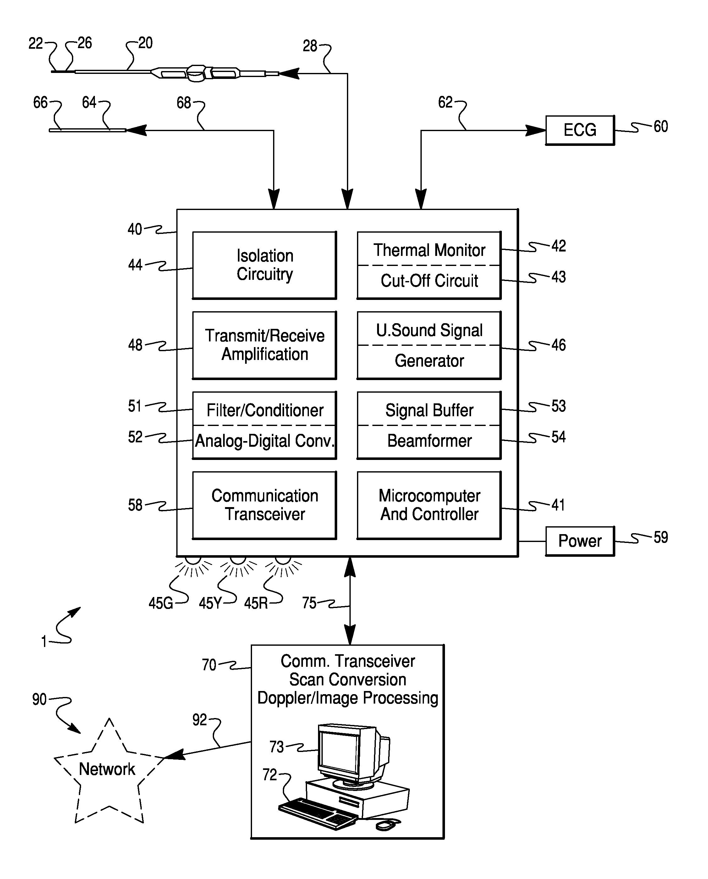
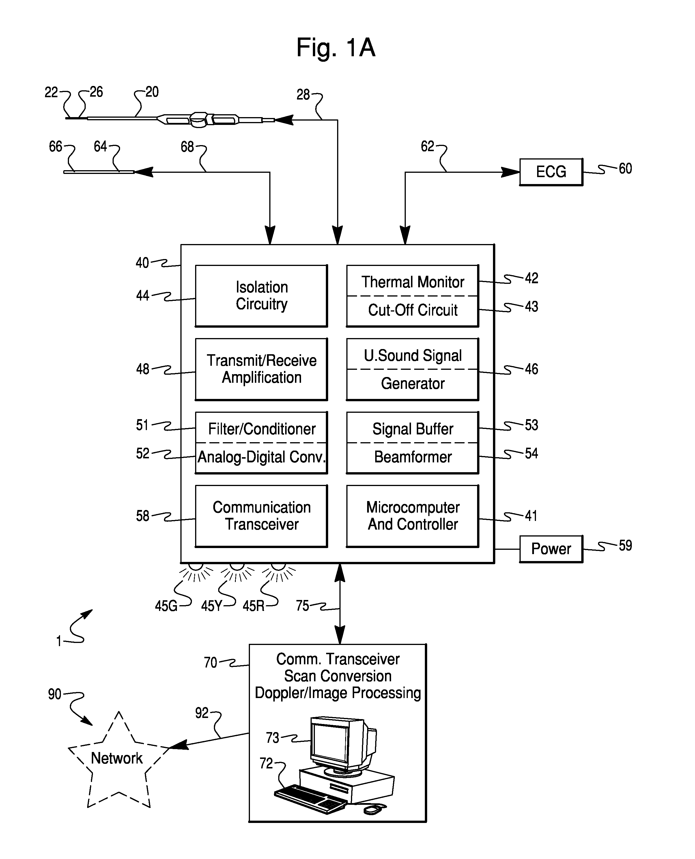
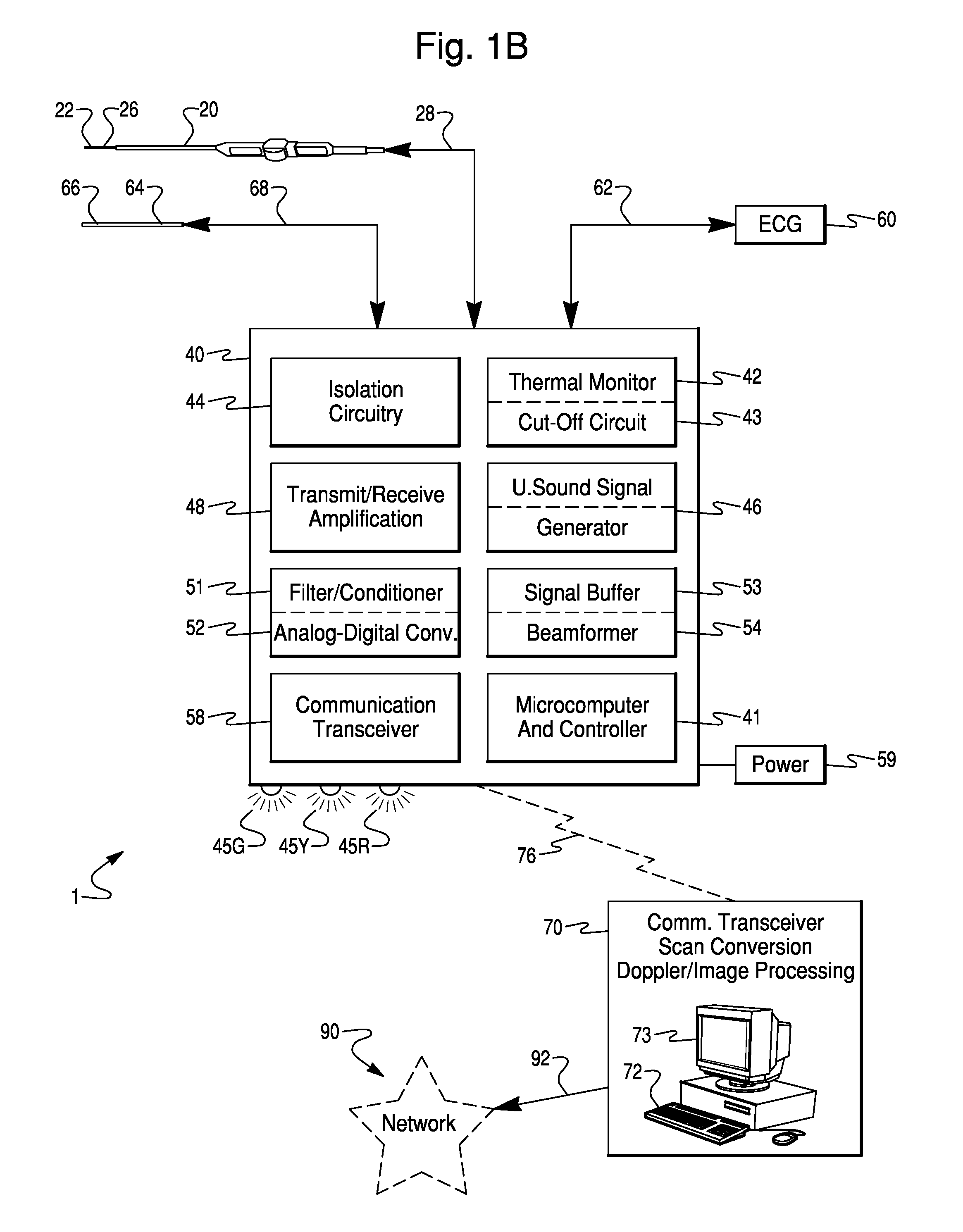
[0018]Various embodiments of the present invention will be described in detail with reference to the accompanying drawings. Wherever possible, the same reference numbers will be used throughout the drawings to refer to the same or like parts.

[0019]As used herein, the terms “about” or “approximately” for any numerical values or ranges indicate suitable dimensional tolerances that allow the part or collection of components to function for their intended purposes as described herein. Also, as used herein, the terms “patient”, “host”, and “subject” refer to any human or animal subject and are not intended to limit the systems or methods to human use. Further, embodiments of the invention will be described for use with an intracardiac ultrasound transducer array catheter. However, the embodiments may be applicable to any medical ultrasound transducer.

[0020]The equipment and cabling historically associated with ultrasound imaging present ergonomic challenges for clinicians. For example, t...


PUM


Abstract

A compact, portable ultrasound imaging system includes a combination of an isolation circuit, an ultrasound signal generator, and a beam former within a single unit. The isolation circuit may limit unintended, leakage current from the ultrasound system through the transducer array of the ultrasound system. The signal generator and beam-former may be implemented using large scale integration semiconductor chips. Connections are provided for connecting an ultrasound transducer array, for outputting ultrasound data and for receiving user input and commands.

Description

FIELD OF THE INVENTION[0001]The present invention is a medical diagnostic system and method, and more particularly is directed to a compact ultrasound imaging catheter system.BACKGROUND OF THE INVENTION[0002]Recent advancements in miniaturization of ultrasound technology has enabled the commercialization of catheters including phased array ultrasound imaging transducers small enough to be positioned within a patient's body via intravenous cannulation. By imaging vessels and organs, including the heart, from the inside, such miniature ultrasound transducers have enabled physicians to obtain diagnostic images available by no other means.[0003]While ultrasound imaging catheter systems have proven to be invaluable diagnostic tools, the associated cabling and equipment present difficulties for clinicians. For example, the cabling from the ultrasound system to the catheter's proximal connector is heavy, stiff, and limited in length. The length of the cabling required to reach from the ult...

Claims


Application Information

Patent Timeline
Patent Type & Authority Applications(United States)
IPC IPC(8): A61B8/00
CPCA61B5/042A61B8/12A61B8/4405A61B8/4427A61B8/565A61B8/56A61B2560/0437A61B5/7232A61B8/4472A61B5/283
Inventor JENKINS, DAVID A.BYRD, CHARLES BRYANDALA-KRISHNA, PRAVEEN
Owner ST JUDE MEDICAL ATRIAL FIBRILLATION DIV
Why Eureka
Try Eureka