Isolation And Use Of Solid Tumor Stem Cells
a technology of stem cells and solid tumors, applied in the field of cancer diagnosis and treatment, can solve the problems of significantly improving the outcome of treatment, and achieve the effect of increasing proliferation and reducing proliferation
- Summary
- Abstract
- Description
- Claims
- Application Information
AI Technical Summary
Benefits of technology
Problems solved by technology
Method used
Image
Examples
example 1
Isolation of Breast Cancer Stem Cells
[0264]The purpose of this EXAMPLE is to show structural and cell functional characterization of breast cancer stem cells.
[0265]We have developed both a tissue culture and a mouse model to identify the breast tumor clonogenic cell. In the mouse model, NOD / SCID mice Lapidot et al., Nature 367(6464): 645-8 (1994)) are treated with VP-16 (Etoposide) (available from commercial sources, such as Moravek Biochemicals, Brea, Calif., USA), and implanted with primary human breast cancer tissue (obtained from mastectomy or lumpectomy specimens). Three of five primary tumors formed tumors in this system.
[0266]The tumor cells isolated from malignant pleural effusions obtained from two patients (see, Zhang et al., Invasion & Metastasis 11(4): 204-15 (1991)) were suspended in Matrigel™ (available from Becton Dickinson, Franklin Lakes, N.J., USA), then were injected into mice. Tumors formed in the injected mice.
[0267]By this method, we can generate enough tumor c...
example 2
Role for Notch in Breast Cell Proliferation
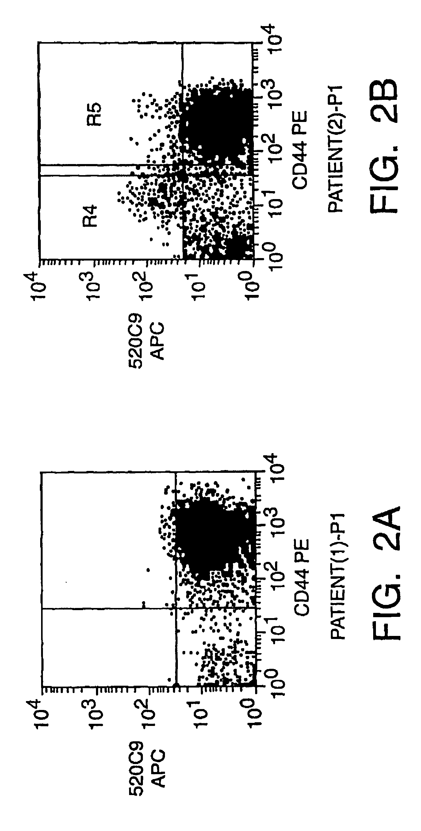
[0278]The purpose of this EXAMPLE is to provide preliminary evidence that in at least two different tumors, Notch 4 is expressed by a minority of the tumorigenic cells. Cells from tumor T1 and tumor T2 from EXAMPLE 1 were analyzed for expression of Notch 4. Cells were stained with an anti-Notch 4 polyclonal antibody and analyzed by FACS. 5-15% of cells expressed detectable levels of Notch 4. Furthermore, different populations of non-tumorigenic cells express different Notch ligands and members of the Fringe family. To determine which Notch RNAs are expressed by normal breast tissue and breast tumor tissue, we performed RT-PCR using primers specific for each Notch mRNA. Interestingly, Notch 1, Notch 3 and Notch 4, but not Notch 2, were expressed by both normal breast cells and breast tumor cells. We prepared RNA from 100,000 cells. RT-PCR of RNA from normal breast cells or breast tumor cells was performed using primers specific for Notch 1, ...
example 3
Mouse Xenograft Model
[0287]We have developed a xenograft model in which we have been able to establish tumors from primary breast tumors via injection of tumors in the mammary gland of severely immunodeficient mice. Xenograft tumors have been established from mastectomy specimens of all five patients that have been tested to date. We have also been able to establish tumors from three malignant pleural effusions. NOD / SCID mice were treated with VP-16, and implanted with primary human breast cancer tissue. Tumor cells isolated from three malignant pleural effusions suspended in Matrigel™ were injected into mice and also formed a tumors. This enabled us to generate enough malignant tumor cells to facilitate analysis by flow-cytometry and assay for the ability of different subsets of cells to form tumors. We have extensively studied the tumors formed by one of the primary tumors and one of the malignant pleural effusion cells. Furthermore, in the three tumors that we have attempted to d...
PUM
| Property | Measurement | Unit |
|---|---|---|
| volume | aaaaa | aaaaa |
| volume | aaaaa | aaaaa |
| diameter | aaaaa | aaaaa |
Abstract
Description
Claims
Application Information
 - R&D
- Intellectual Property
- Life Sciences
- Materials
- Tech Scout
- Unparalleled Data Quality
- Higher Quality Content
- 60% Fewer Hallucinations
Browse by: Latest US Patents, China's latest patents, Technical Efficacy Thesaurus, Application Domain, Technology Topic, Popular Technical Reports.
© 2025 PatSnap. All rights reserved.Legal|Privacy policy|Modern Slavery Act Transparency Statement|Sitemap|About US| Contact US: help@patsnap.com


