Method for the Detection of Disease-Related Prion

a prion and disease technology, applied in the field of prion diseases, can solve the problems of a pre-treatment with proteases, a strong limitation of diagnostic capabilities, and a lack of protease sensitive prpsup>sc/sup>,

Inactive Publication Date: 2008-09-11
PRIONICS
View PDF3 Cites 5 Cited by
  • Summary
  • Abstract
  • Description
  • Claims
  • Application Information

AI Technical Summary

Benefits of technology

[0013]Such a method would offer the advantage to detect carriers of a transmissible spongiform encephalopathy by screening blood for the presence of disease related PrP and could e.g. prevent vCJD from being accidentally transmitted by blood transfusion.DEFINITIONS

Problems solved by technology

Due to the slow kinetics of accumulation of PrPSc in the preclinical stage of the disease, the current diagnostic capabilities are strongly limited with respect to the detection of the disease early in the incubation period.
However, such assays will not detect the protease sensitive form of PrPSc.
A major drawback of this method is the use of a pre-treatment with proteases to hydrolyse protease sensitive PrP.

Method used


Image

  • Method for the Detection of Disease-Related Prion
  • Method for the Detection of Disease-Related Prion
  • Method for the Detection of Disease-Related Prion

Examples


example 1

[0044]The graph in FIG. 1 shows the results of a first experiment. The data shown illustrate that BSE positive plasma samples can be distinguished from negative samples using the sandwich immunoassay. The mean of double measurements are shown, whereas the error bars indicate the difference to the higher value.

[0045]96-well plates were coated with 2 μg / ml anti-IgM antibody diluted in phosphate buffered saline (PBS) pH 7.4, 0.1% bovine serum albumin (BSA) for 1 hour at room temperature. Unbound antibody was removed by washing the plates three times with PBS pH 7.4 containing 0.05% Tween-20. Thereafter, the plates were blocked for 2 hours and washed three times with PBS pH 7.4 containing 0.05% Tween-20. Conformation specific antibody 15B3 in PBS pH 7.4 at a concentration of 2 μg / ml was bound to anti-IgM for 1 hour at room temperature. After washing the plate three times with PBS pH 7.4 containing 0.05% Tween 20, 110 μl Plasma from BSE negative and positive cattle diluted 1:1 with Tris ...

example 2

[0046]The graph in FIG. 2 shows the results of a second experiment. The data shown illustrate that Sheep scrapie positive plasma samples can be distinguished from negative samples using the sandwich immunoassay. The mean of double measurements are shown whereas the error bars indicate the difference to the higher value.

[0047]Coating of plate with 15B3 antibody was performed as described in example 1. 110 μl of Plasma from scrapie negative and positive sheep diluted 1:1 with TBS containing 0.2% Sarcosyl was added to the plate and incubated for 1.5 hours at room temperature. Unbound proteins were washed away with TBS containing 0.1% Sarcosyl. POD labelled monoclonal antibody 805 recognizing amino acids 25-40 of PrP was diluted to 10 ng / ml in a buffer containing PBS pH 7.4, 0.02% Casein, 0.1% Tween-20 and added for 1 hour to wells. Unbound antibody was removed by washing for three times with PBS pH 7.4 containing 0.05% Tween-20. 100 μl of chemiluminescent substrate solution was added t...

example 3

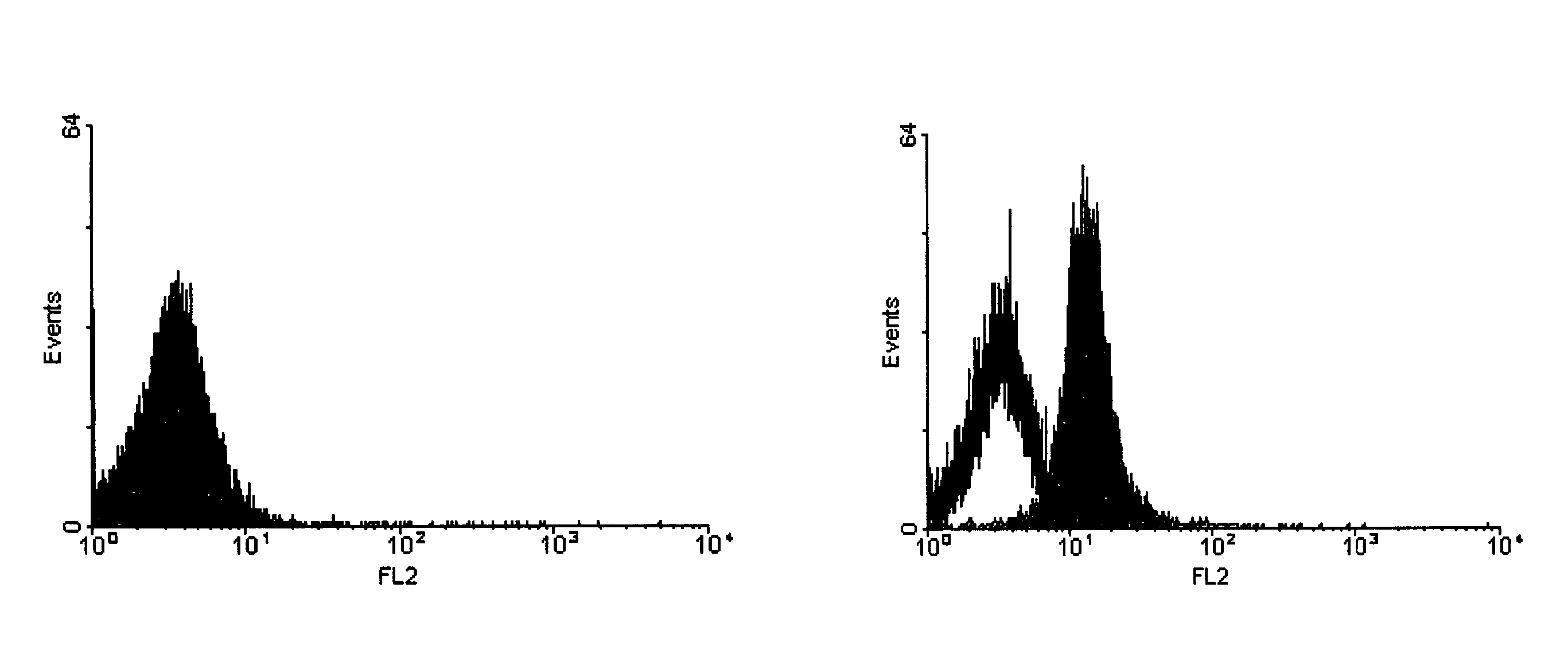
[0048]The graph in FIG. 3 shows the results of a third experiment. The data shown illustrate that scrapie positive brain homogenate can be distinguished from negative samples using FACS analysis. A histogram is shown whereas y-axis indicates the events and x-axis the relative intensity of fluorescence measured by the FL-2 detector.

[0049]60 ng monoclonal 15B3 antibody or Isotype control IgM antibody (Becton Dickinson 550963) were coated on 3 μl magnetic anti-IgM Dynabeads (Dynal 110.15) in phosphate buffer saline (PBS) pH 7.4 containing 0.1% bovine serum albumin (BSA) for 1 hour at room temperature on a rotating wheel. Unbound antibody was removed by washing the beads three times with PBS pH 7.4 containing 0.1% BSA. The beads were added to 1 ml Tris buffered saline (TBS) containing 0.25% Sarkosyl and 10 μl of a 10% scrapie positive or negative brain homogenate and incubated over night at 4° C. on a rotating wheel. Thereafter, unbound proteins were removed by washing the beads three t...


PUM

PropertyMeasurementUnit
molecular massaaaaaaaaaa
pHaaaaaaaaaa
concentrationaaaaaaaaaa

Abstract

Methods for the detection of the disease associated conformation of the prion protein as an indication of transmissible spongiform encephalopathies (TSEs), including preclinical detection of infected live animals and humans, and post-mortem detection methods are disclosed. In one aspect, the tissue or body fluid sample of a test subject is contacted with an antibody that binds only the disease related conformation of the prion protein under non-denaturing conditions.

Description

FIELD OF THE INVENTION[0001]The present invention is directed to methods for the detection of prion diseases in animals and humans.BACKGROUND OF THE INVENTION[0002]Transmissible spongiform encephalopathies (TSEs) or prion diseases are fatal neurodegenerative diseases in animals and man. The onset of clinical illness is preceded by a long incubation period of months to decades. Clinical symptoms of TSEs include dementia and loss of movement coordination. In the 1980s it was found that a common hallmark of TSEs was the accumulation of an abnormal protease resistant isoform (PrPres or PrPSc) of the host-encoded prion protein (PrPC) in affected animals and humans. The discovery of PrPSc provided a molecular marker that was shown to be specific for all prion diseases as well as the major and very likely the only, constituent of the infectious particle, denominated prion.[0003]The protease resistant core of PrPSc, designated PrP27-30, was discovered by analyzing proteinase K-treated fract...

Claims


Application Information

Patent Timeline
Patent Type & Authority Applications(United States)
IPC IPC(8): G01N33/53G01N33/566G01N33/68
CPCG01N2800/2828G01N33/6896
Inventor PURRO, MARIOZWALD, DANIELSCHMID, JAQUELINEBIFFIGER, KARINKUHN, FRANZISKAOESCH, BRUNORABER, ALEX
Owner PRIONICS
Why Eureka
Try Eureka