

Methods, Compositions and Systems for Analyzing Imaging Data
- Summary
- Abstract
- Description
- Claims
- Application Information
AI Technical Summary
Problems solved by technology
Method used
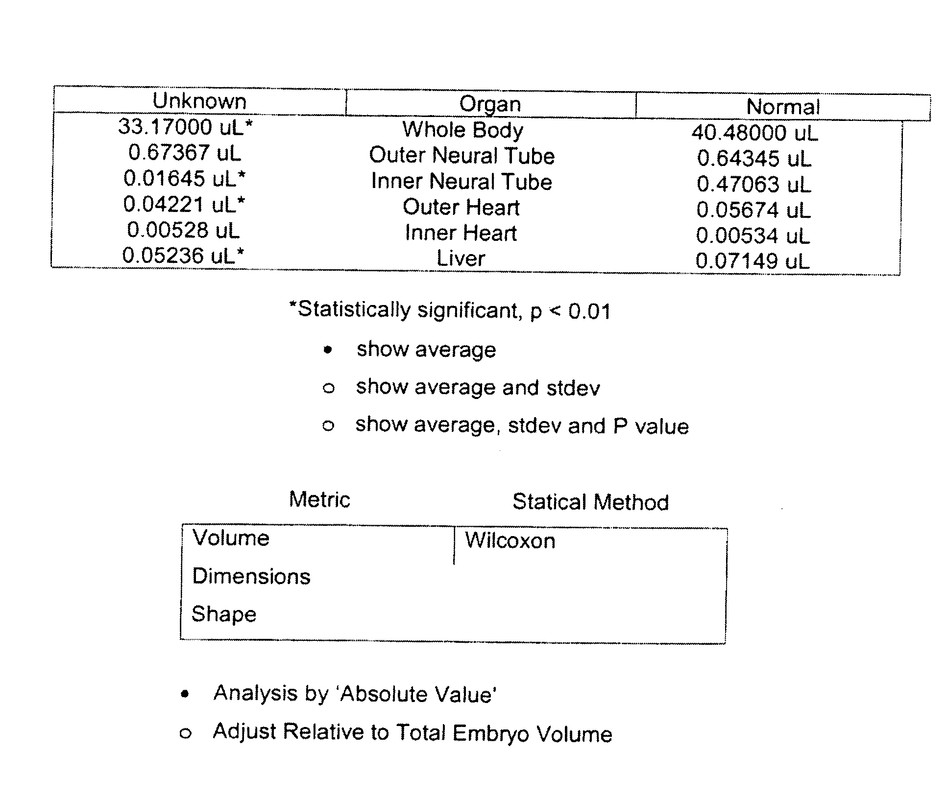
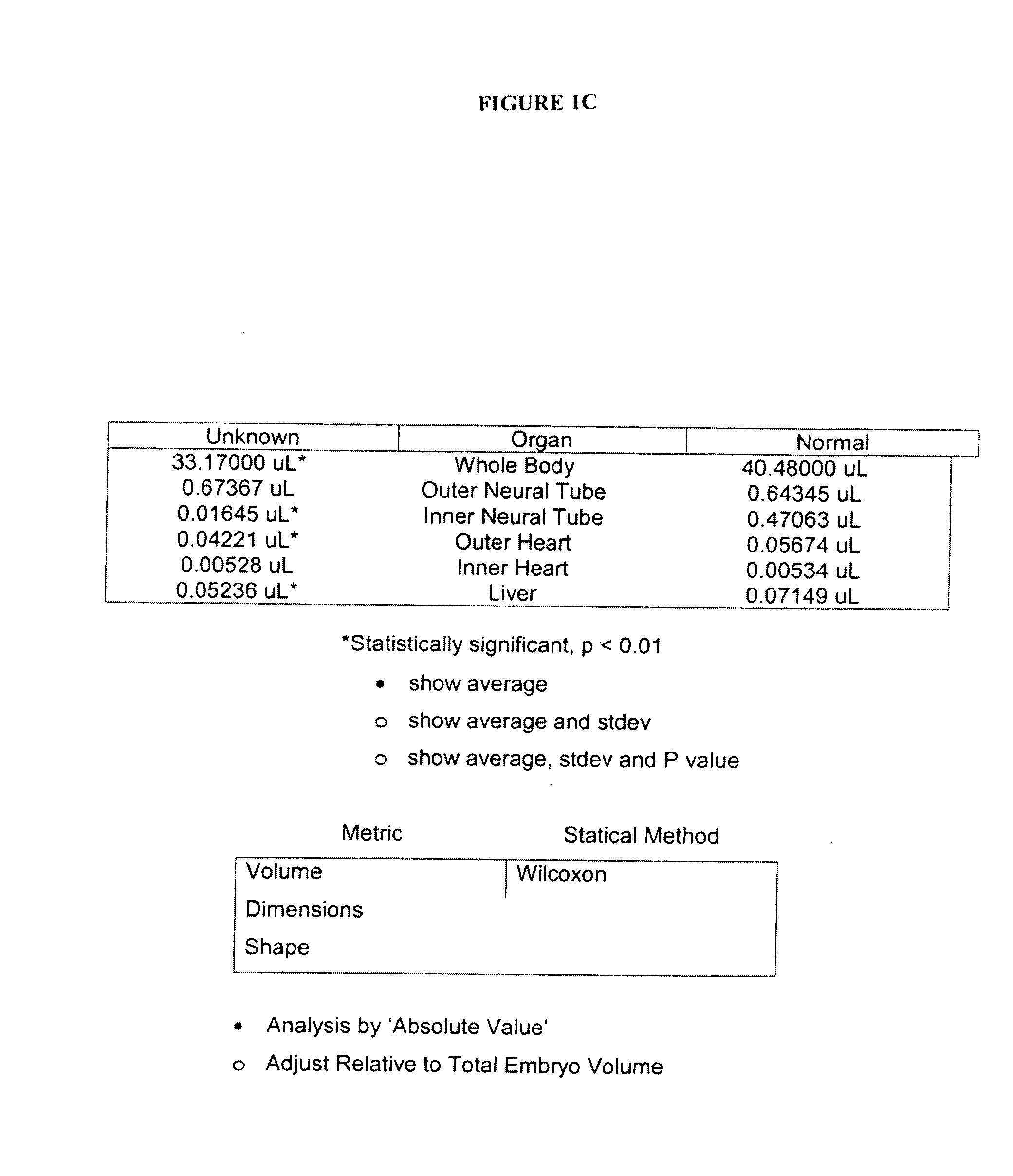
Image

Examples
example 1
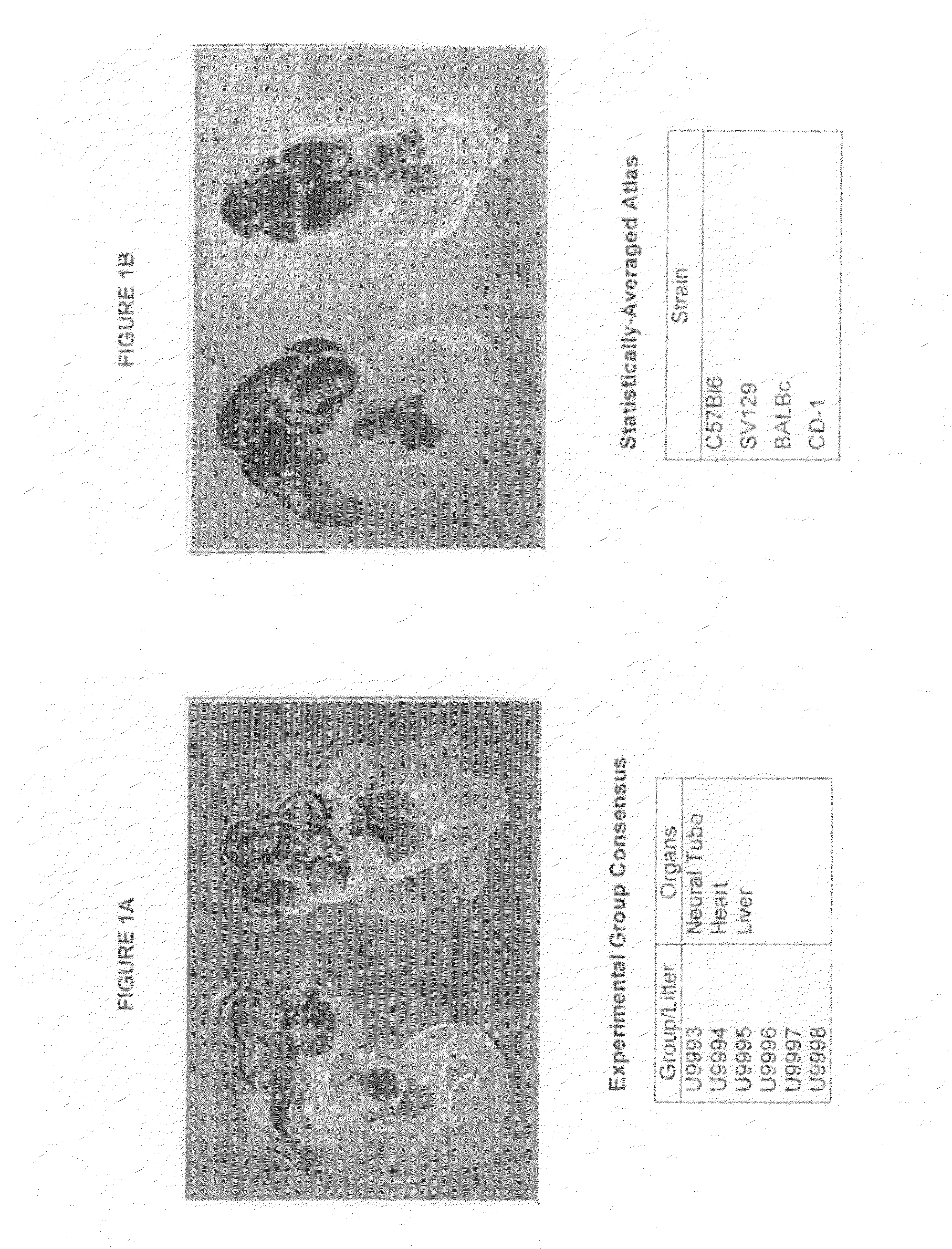
Collecting Mouse Embryo MicroCT Data
Virtual Histology
[0135]Litters of pure-strain C57BL / 6 mice are generated using standard husbandry techniques. Mating cages of one sire and two dams are established, and dams are monitored each morning for the presence of cervical mucous plugs. The presence of a cervical plug is taken as evidence of successful impregnation, and the morning that a cervical plug is found is designated embryonic day E0.5. Mouse litters are harvested on embryonic day E12.5 following euthanasia of the pregnant dam. Amnion and placenta are dissected away from the embryos under a dissecting microscope. Embryos are then fixed in 10% buffered formalin overnight at 4° C. Generally, sixty embryos are harvested for microCT imaging.
[0136]For microCT-based virtual histology, formalin-fixed embryos are stained to saturation overnight in a solution of 0.1M sodium cacodylate (pH 7.2), 1% glutaraldehyde, and 1% osmium tetroxide rocking at room temperature. Embryos are then washed fo...
example 2
Analysis of MicroCT Data Sets
[0138]A modified version of an existing watershed-based segmentation software system (Cates et al., (2005) Med Image Anal. 9(6):566-78) is used for segmenting features of interest (forebrain, midbrain, hindbrain, heart, and liver) from mouse-embryo MicroCT data. A set of robust landmark locations are used to grossly locate each feature of interest. These landmark locations are a subset of the full point distribution model (PDM). The landmark points generally meet certain requirements, including: they are easily identifiable in the data scans (e.g. a well-defined junction or cusp), they span the feature (e.g. several points on each side), and the number of landmarks are limited (i.e. only as many as an expert can label in under five minutes).
[0139]Based on the locations of the landmarks, the rest of the PDM points are distributed across the rest of the features' surface. An interactive software system, which modifies a commercially available application s...
PUM
 Abstract
Description
Claims
Application Information