Combined X-ray detector and ultrasound imager

a detector and x-ray technology, applied in the field of dual-mode imaging systems, can solve the problems of affecting the accuracy of the overall scan, and unable to produce instantaneous or real-time images of the region of interest of the object, so as to facilitate drug delivery and remove speckles and shadows.

Inactive Publication Date: 2008-10-02
GENERAL ELECTRIC CO
View PDF38 Cites 87 Cited by
  • Summary
  • Abstract
  • Description
  • Claims
  • Application Information

AI Technical Summary

Benefits of technology

[0008]In certain embodiments, the imaging panel may include a sensor array coupled to an electronics array. The electronics array may include a plurality of integrated circuit chips stacked on top of the sensor array. The plurality of integrated chip may include an aperture control block, image processing block, power management block, transmit / receive block, beam formation block, audio block, display block, or a combination thereof. Furthermore, the first and second 2D matrices of sensors may electronically scan the volume via changing the size and shape of a group of sensors and / or by using different transmit and receive delays in coordination with each group of sensors.
[0009]In certain embodiments, the imaging panels may be configured to perform through transmission imaging which includes transmitting a signal from one panel through an imagining volume to one or more additional panels that then receive the signal. The process may be repeated in a specified or calculated manner (i.e., an image cycle) with additional panels transmitting and receiving through the imaging volume. The image cycle may follow a predetermined sequence and time interval or may be adjusted by the operator. Once the image cycle is complete, a multiple volumetric image, from current or previous studies, may be co-registered and the signals processed to remove speckle and shadowing artifacts. Embodiments of through volume transmission may include using the first and second 2D matrices of sensors to measure the time to transmit and receive between the imaging panels, as well as, to measure the attenuation of each transmit beam as it passes from one imaging panel to another.
[0010]In certain embodiments, the imaging panels may be configured to compare differences in volumetric shear and bulk strain images and B-mode images prior to and after compressing the imaging volume. Furthermore, one imaging panel may transmit a high-energy acoustic pulse to compress the tissue and one or more imaging panels may measure volumetric strain and B-mode images prior to and after the high-energy pulse. Similarly, on subsequent volumetric imaging cycles a different panel could provide the high-energy pulse. Additionally, the first and second 2D matrices of sensors may be configured to produce acoustic energy that is capable of ablating tissue, eliminating tumors, or destroying microbubbles in a tumor region to facilitate drug delivery.

Problems solved by technology

Unfortunately, existing sensors must be physically moved across the object to generate an image, and thus, measurement errors may result due to movement of the object among other things.
Moreover, as a result of the movement delay time, the overall scan does not produce an instantaneous or real-time image of the region of interest of the object.
These problems can complicate registration with images from other modalities.
Again, the foregoing problems with existing ultrasound systems can complicate registration between these two modalities.

Method used


Image

  • Combined X-ray detector and ultrasound imager
  • Combined X-ray detector and ultrasound imager
  • Combined X-ray detector and ultrasound imager

Examples


Embodiment Construction

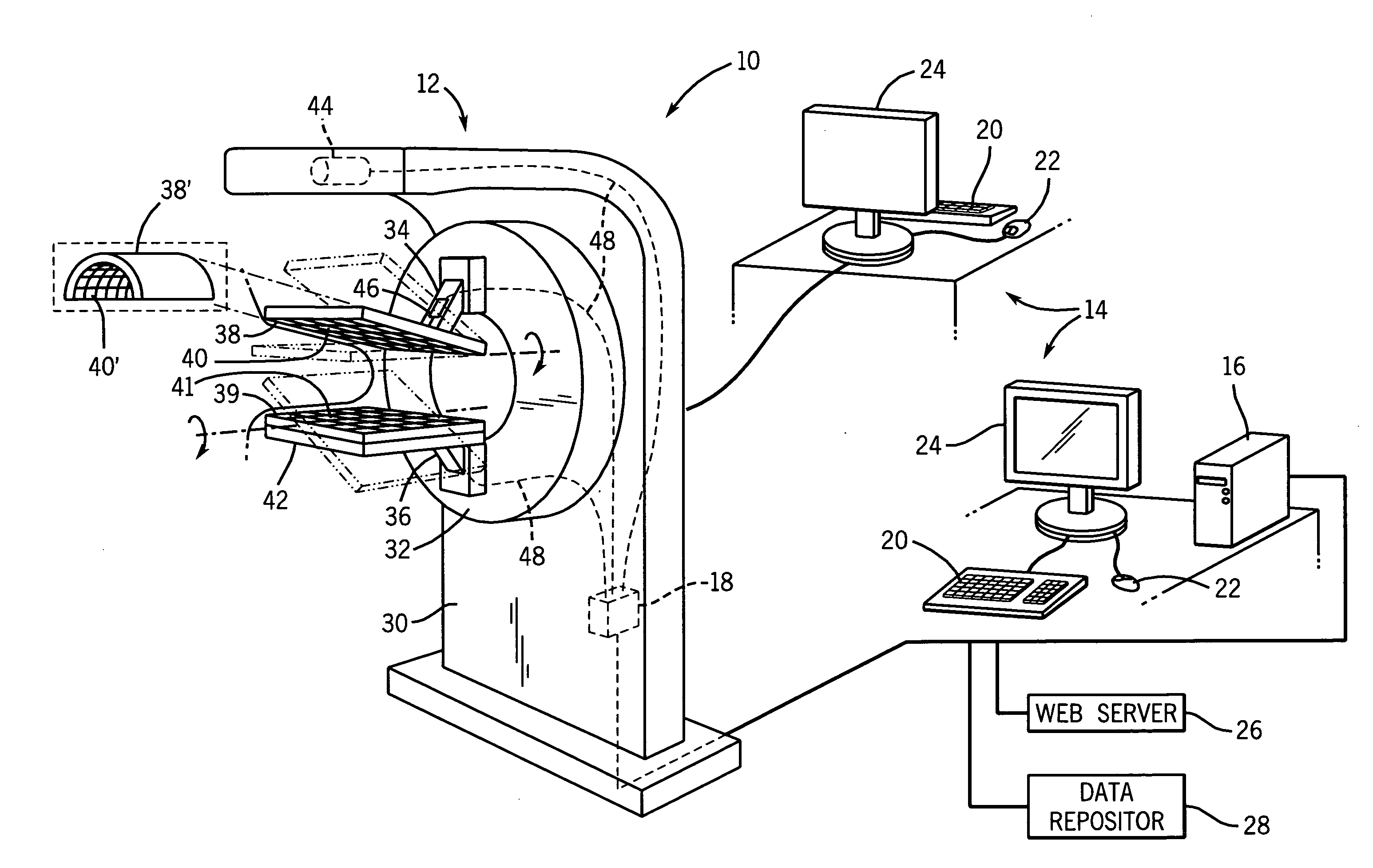
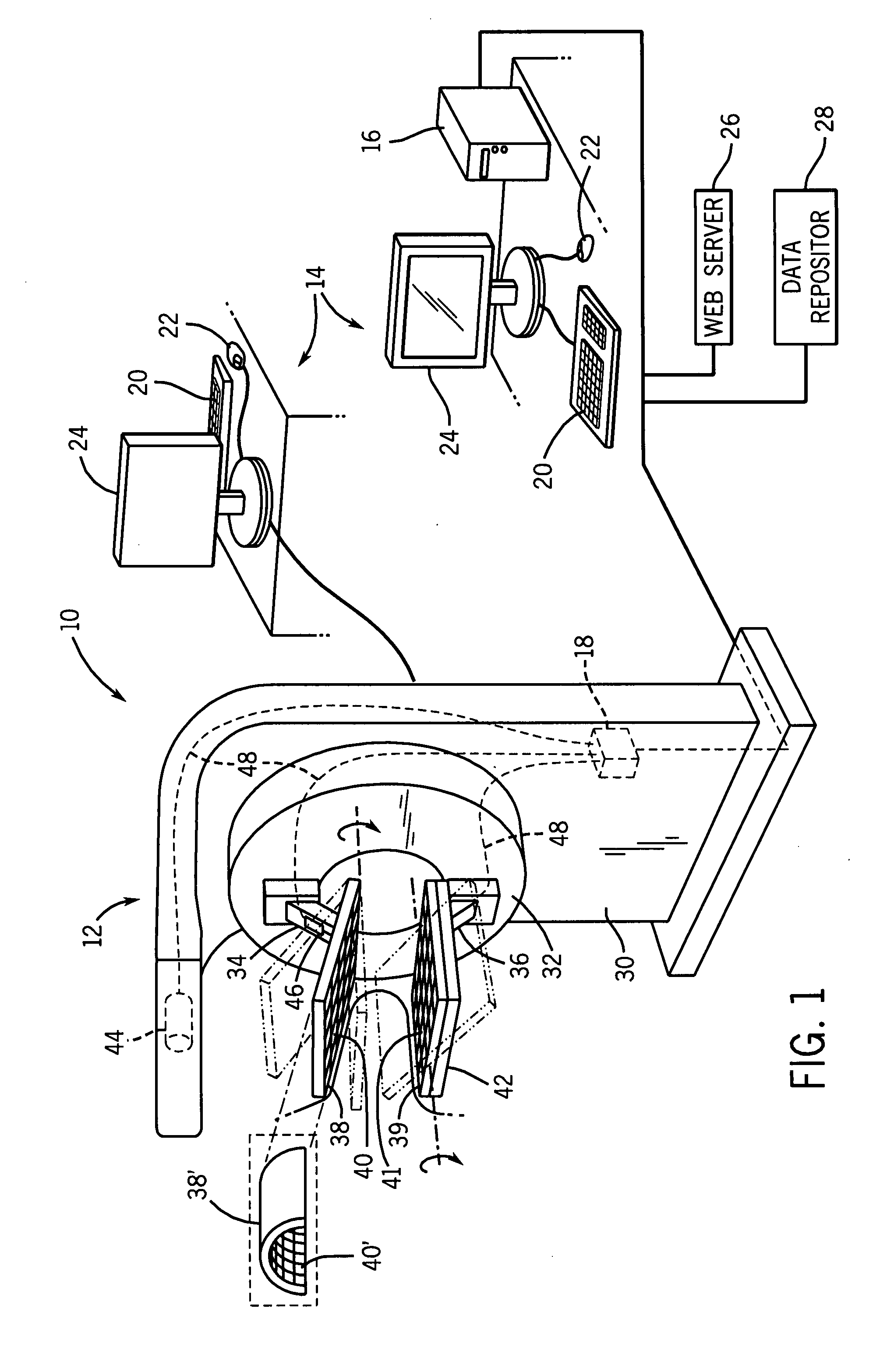
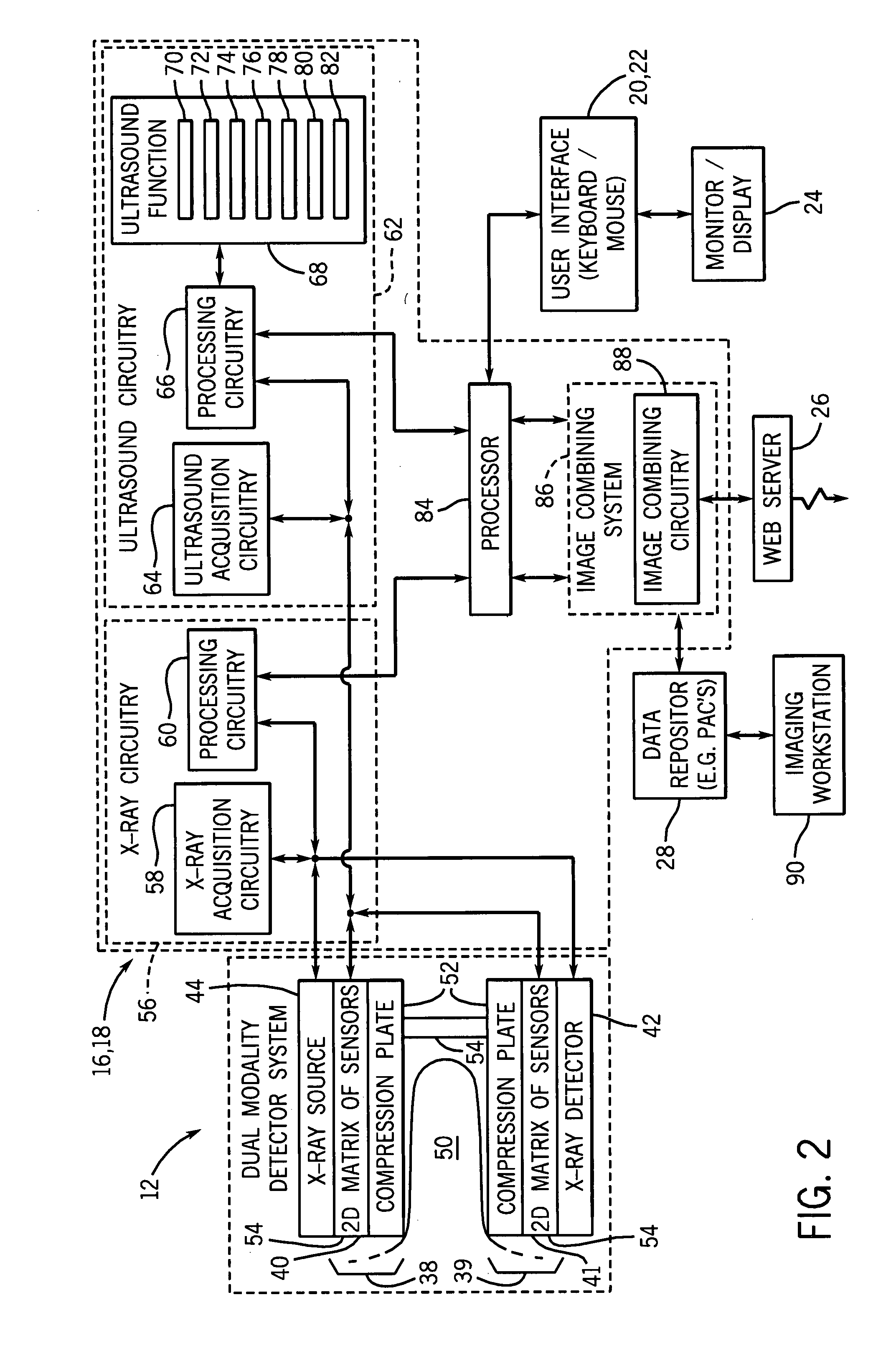
[0029]As discussed in further detail below, various embodiments of an imaging system are provided. The imaging system may include multiple modalities and may be configured to produce a co-registered dual modality image without requiring the relocation of the imaging volume. The modalities may include an ultrasound systems, X-ray imaging systems (including mammography system), molecular imaging systems, computed tomography (CT) systems, positron emission tomography (PET) systems, magnetic resonance imaging (MRI) systems, and electric impedance imaging systems. The imaging system may be configured to electronically scan the image volume without moving the sensors via a first imaging panel and a second imaging panel disposed about the imaging volume. The imaging panels may include a 2D matrix of sensors that may be configured to transmit, receive, or both, (e.g., ultrasound) alone or in various combinations with one another. The 2D matrix may be further subdivided into groups of sensor...


PUM


Abstract

An imaging system is disclosed that includes a first imaging panel and a second imaging panel disposed about an imaging volume. The imaging panels may be configured to image the entire imaging volume and may include panels using any form of acoustic or electromagnetic energy such as ultrasound panels, optical panels, electrical impedance panels, field emitter/x-ray detector panels, or a combination thereof. In one embodiment, a first group of sensors are included in a 2D matrix of sensors configured to transmit ultrasound through the imaging volume to a second group of sensors included in a second 2D matrix of sensors and vice versa. In a second embodiment the system may further include a second imaging system having a transmitter, a receiver, or both, disposed adjacent the first imaging panel, the second imaging panel, or both. A third embodiment may include at least one additional imaging panel.

Description

BACKGROUND[0001]The present invention relates to a dual modality imaging system. More specifically, embodiments of the present invention relate to a combined ultrasound and X-ray imaging system.[0002]In modern healthcare facilities, medical diagnostic and imaging systems are used for identifying, diagnosing, and treating diseases. Diagnostic imaging refers to any visual display of structural or functional patterns of organs or tissues for a diagnostic evaluation. One diagnostic imaging technique is ultrasound. An ultrasound imaging system uses an ultrasound sensor or transducer for transmitting ultrasound signals into an object, such as the breast of the patient being imaged, and for receiving reflected ultrasound signals back into the same transducer. The reflected ultrasound signals received by the ultrasound sensor are processed to reconstruct an image of the object. Unfortunately, existing sensors must be physically moved across the object to generate an image, and thus, measure...

Claims


Application Information

Patent Timeline
Patent Type & Authority Applications(United States)
IPC IPC(8): A61B6/00
CPCA61B5/0091A61B5/0536A61B6/4233A61B6/4488A61B6/502A61B8/0825A61B8/4416A61B6/5247A61B6/4417
Inventor FISHER, RAYETTE ANNTHOMENIUS, KAI ERIKKORMAN, CHARLES STEVEN
Owner GENERAL ELECTRIC CO
Why Eureka
Try Eureka