Isolation and growth of stem cells from hemangiomas

a technology of stem cells and hemangiomas, which is applied in the field of stem cells, can solve the problems of creating cosmetic problems, not always cosmetically acceptable, and each conventional treatment option carries potential side effects

Inactive Publication Date: 2008-12-04
NEVADA CANCER INST
View PDF4 Cites 10 Cited by
  • Summary
  • Abstract
  • Description
  • Claims
  • Application Information

AI Technical Summary

Benefits of technology

[0016]In another aspect, the step of determining an effect of a test agent on a stem or progenitor cell includes generating global maps for SALL4 promoter binding in the presence and absence of the agent.
[0017]In one aspect, the method includes determining the effect of the test agent on a stem cell or progenitor cell in the presence and absence of an angiogenesis inhibitor, where the inhibitor includes VEGFR-1, NRP-1, angiopoietin 2, TSP-1, TSP-2, angiostatin, endostatin, vasostatin, calreticul

Problems solved by technology

Most disappear spontaneously (immature hemangiomas), while others persist and create cosmetic problems.
While many hemangiomas eventually “involute,” the result is not always cosmetically acceptable.
Each conventional treatment option carries potential side effects.
Clearly, surgery always presents risks, whether for infection, unexpected patient reaction to anesthesia, and/or unexpected aesthetic results.
While systemic corticosteroid treatment is suspected of certain side eff

Method used


Image

  • Isolation and growth of stem cells from hemangiomas
  • Isolation and growth of stem cells from hemangiomas
  • Isolation and growth of stem cells from hemangiomas

Examples


example 1

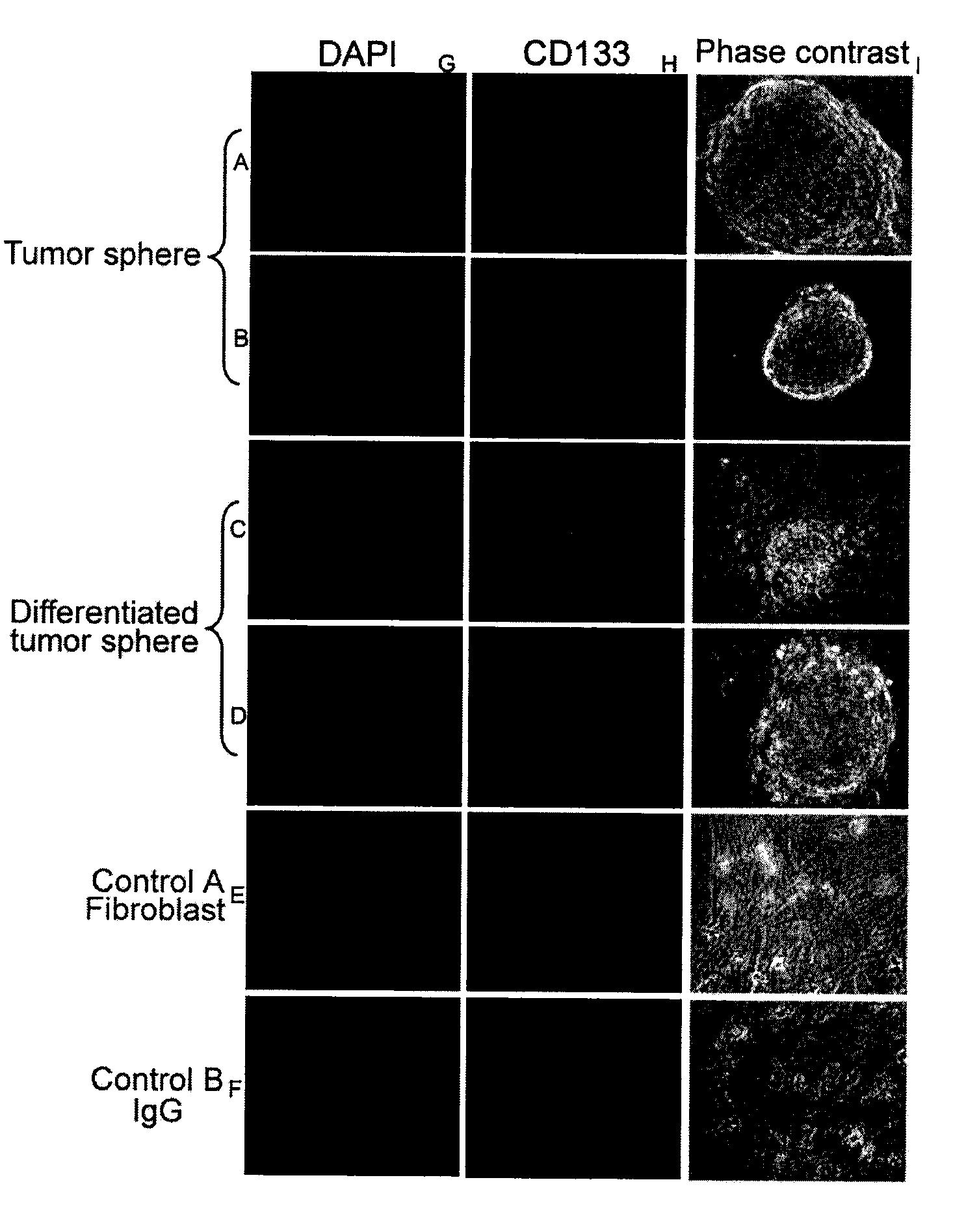
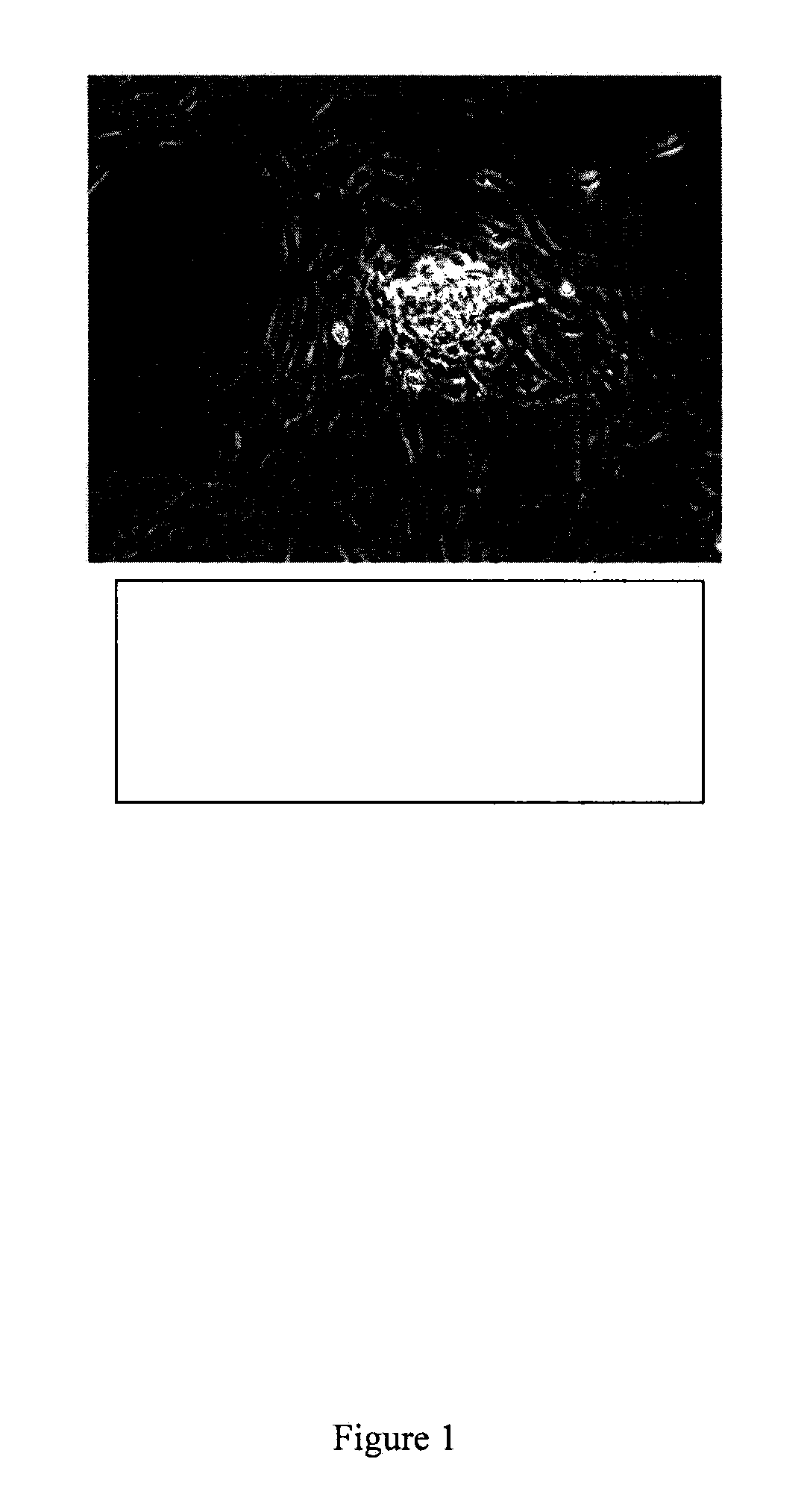
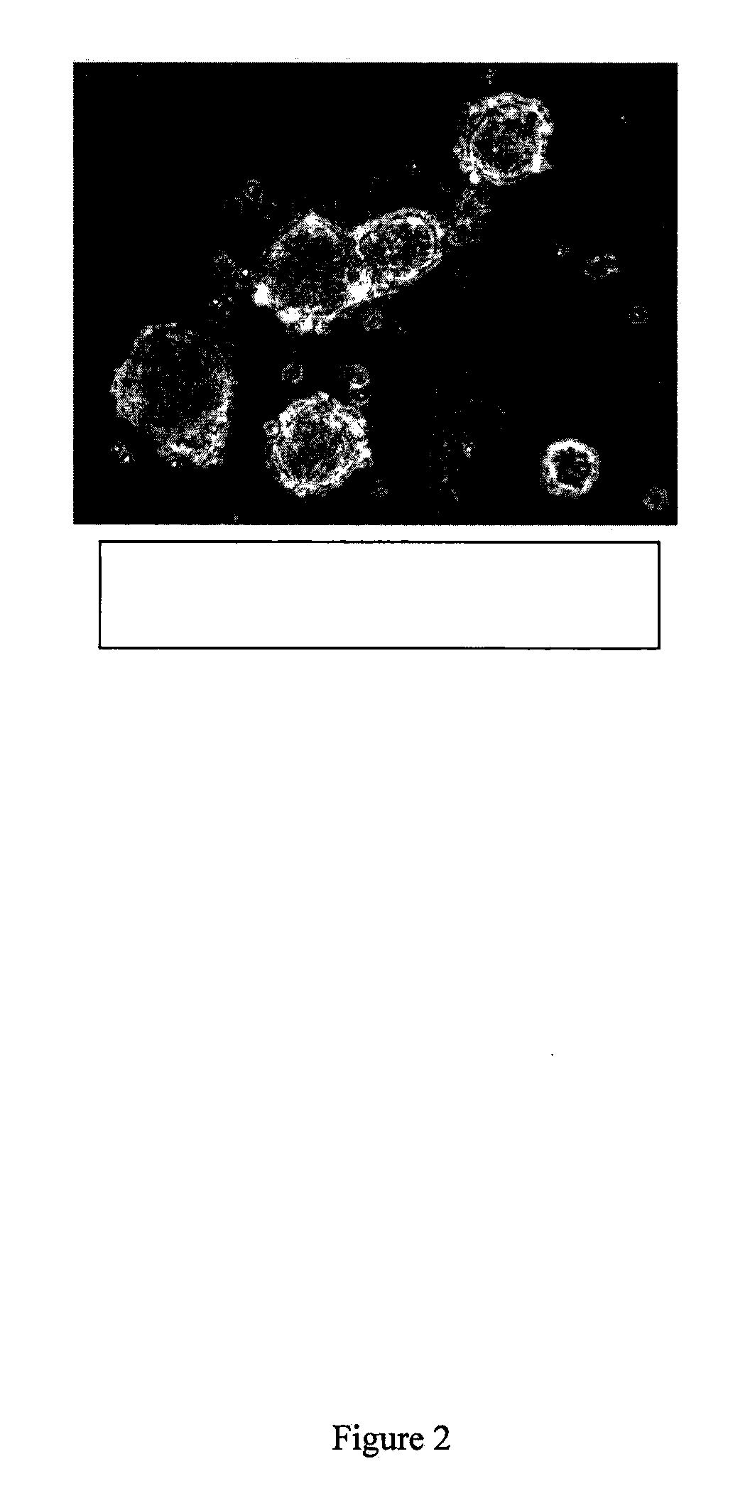
Isolation of Tumor Spheres

[0161]Tissue samples were isolated from superficial raised patches on the skin of male and female infants (less than 1 year), prior to involution. Upon receipt, the tissue was minced and digested with collagenase for 2 hours at 37° C. Following digestion and several wash steps, the tissue mixture was then filtered through a 100 μm cell strainer and grown in culture using KNOCKOUT™ Dulbecco's Modified Eagle's Medium (DMEM), 10% KNOCKOUT™ Serum, 1× non-essential amino acids (NEAA), and 20 μg / ml basic-fibroblast growth factor (bFGF). Following growth of this primary culture for approximately three days, tumor spheres will spontaneously form from the homogenous mixture of primary culture. These spheres are collected using collagenase digestion, washed, and plated at a density of 200 cells per milliliter with DMEM-F12 nutrient mix, 20 μg / ml bFGF and NEAA on low-attachment plates. The tumor spheres form and propagate using collagenase digestion, which digest the ...

example 2

ChIP Assay

[0167]Isolated cells from tumor spheres (1×10⁶ cells / well in 6-well plates), are processed using a ChIP Assay Kit (Upstate, Charlottesville, Va.) following the manufacture's protocol. Briefly, cells are cross-linked by adding formaldehyde (27 μl of 37% formaldehyde / ml) and incubated for 10 min. Then, chromatin is sonicated to an average size of approximately 500 bp and immunoprecipitated with SALL4 antibodies, preimmune serum, or anti-HA (hemagglutination) antibody. Antibodies for histone modifications, histone H3 trimethy K4 and histone H3 dimethy K79, may be purchased from Abeam (Cambridge, Mass.). Histone-DNA crosslinks are reversed by heating at 65° C. followed by digestion with proteinase K (Invitrogen, Carlsbad, Calif.). DNA is recovered by using a PCR purification kit (Qiagen, Valencia, Calif.) and then used for PCR or QRT-PCR (quantitative real time polymerase chain reaction).


PUM

PropertyMeasurementUnit
Compositionaaaaaaaaaa
Cell proliferation rateaaaaaaaaaa

Abstract

The present invention describes stem cells and progenitor cells derived from hemangiomas, including testing of angiogenic inhibitors using these cells. The invention as described is useful in providing a process to culture and propagate hemangioma stem cells and generate xenograft models to develop treatments for infantile hemangiomas and other types of vascular lesions.

Description

CROSS REFERENCE TO RELATED APPLICATION[0001]This application claims priority under 35 U.S.C. §120 as a continuation-in-part of U.S. application Ser. No. 11 / 809,871, filed Jun. 1, 2007, and claims benefit of priority under 35 U.S.C. §119(e) to U.S. Provisional Application No. 61 / 050,131, filed May 2, 2008, the disclosures, each of which, are incorporated by reference in their entireties.FIELD OF THE INVENTION[0002]The present invention relates generally to stem cells, and more specifically to progenitor cells and stem cells isolated from human infantile hemangiomas, methods for culturing such cells, and methods of treating disorders associated with hemangiomas.BACKGROUND INFORMATION[0003]Infantile hemangiomas, the most common tumors of infancy, are vascular tumors characterized by rapid proliferation of endothelial cells during the first few months of postnatal life followed by slow spontaneous involution. Most disappear spontaneously (immature hemangiomas), while others persist and ...

Claims


Application Information

Patent Timeline
IPC IPC(8): C12N5/08C12N5/06C12Q1/68C12Q1/02G01N33/53A01K67/02A61K38/20
CPCA01K67/0275A01K2217/05A01K2227/105A01K2267/0331A01K2267/0381C07K14/4705C12N15/8509C12N2800/30C12N2830/008C12Q1/6886C12Q2600/136G01N33/5011G01N33/5073A61P35/00
Inventor MA, YUPOFINK, LOUIS M.WARD, DAVID C.WANER, MILTON
Owner NEVADA CANCER INST
Why Eureka
Try Eureka