
Medical imaging facility, in particular for producing image recordings in the context of a treatment of cardiac arrhythmias, and associated method

a medical imaging and recording technology, applied in the field of medical imaging facilities, can solve the problems of unsatisfactory cardiac arrhythmia treatment, unfavorable patient safety, and relatively high risk of patients, and achieve the effects of improving patient safety, improving patient safety, and improving patient comfor

Inactive Publication Date: 2009-03-19
SIEMENS AG
View PDF15 Cites 22 Cited by
  • Summary
  • Abstract
  • Description
  • Claims
  • Application Information

AI Technical Summary

Benefits of technology

[0013]This has the advantage that it is possible to produce both computed tomography recordings and intravascular magnetic resonance recordings using a single system, in other words without having to move the patient and carry out a new registration. Also an intravascular facility is used for magnetic resonance imaging, so there is no need for an expensive separate magnetic resonance tube or facility.
[0047]It is thus possible with the inventive hybrid system and / or integrated medical facility with the option of producing computed tomography recordings and IVMRI recordings to improve the medical workflow with more reliable and faster ablation of tissue parts with a lower patient risk.

Problems solved by technology

In the case of ventricular tachycardias interference means that instead of a complete contraction of the cardiac muscle there is incomplete contraction, with the result that the heart pumps inadequately.
This has the disadvantage of involving a relatively high level of risk for the patient.
This method is deployed for example for supraventricular tachycardias and also increasingly in the field of ventricular tachycardias, particularly given that drug treatment with antiarrhythmic agents has only a low success rate and on the other hand implantable defibrillators have unpleasant side effects for the patient.
However it remains difficult to identify and locate the correct points for the ablation during the intervention.
However the ultrasound catheter only supplies two-dimensional recordings, so that only limited image information with low resolution is available during the procedure, since the high-resolution recordings with three-dimensional data do not show the current situation during therapy.
The electrophysiological potentials do not allow actual tissue display, in particular any display of the scar tissue resulting in the heart for example in the context of a cardiac infarction.
However ventricular tachycardias for example originate from what are known as reentrant circuits, which generally occur at and in the outer edges of the electrically inactive myocardial scar tissue, so the lack or inadequacy of the tissue display is problematic for the therapy.

Method used


Image

  • Medical imaging facility, in particular for producing image recordings in the context of a treatment of cardiac arrhythmias, and associated method
  • Medical imaging facility, in particular for producing image recordings in the context of a treatment of cardiac arrhythmias, and associated method
  • Medical imaging facility, in particular for producing image recordings in the context of a treatment of cardiac arrhythmias, and associated method

Examples


Embodiment Construction

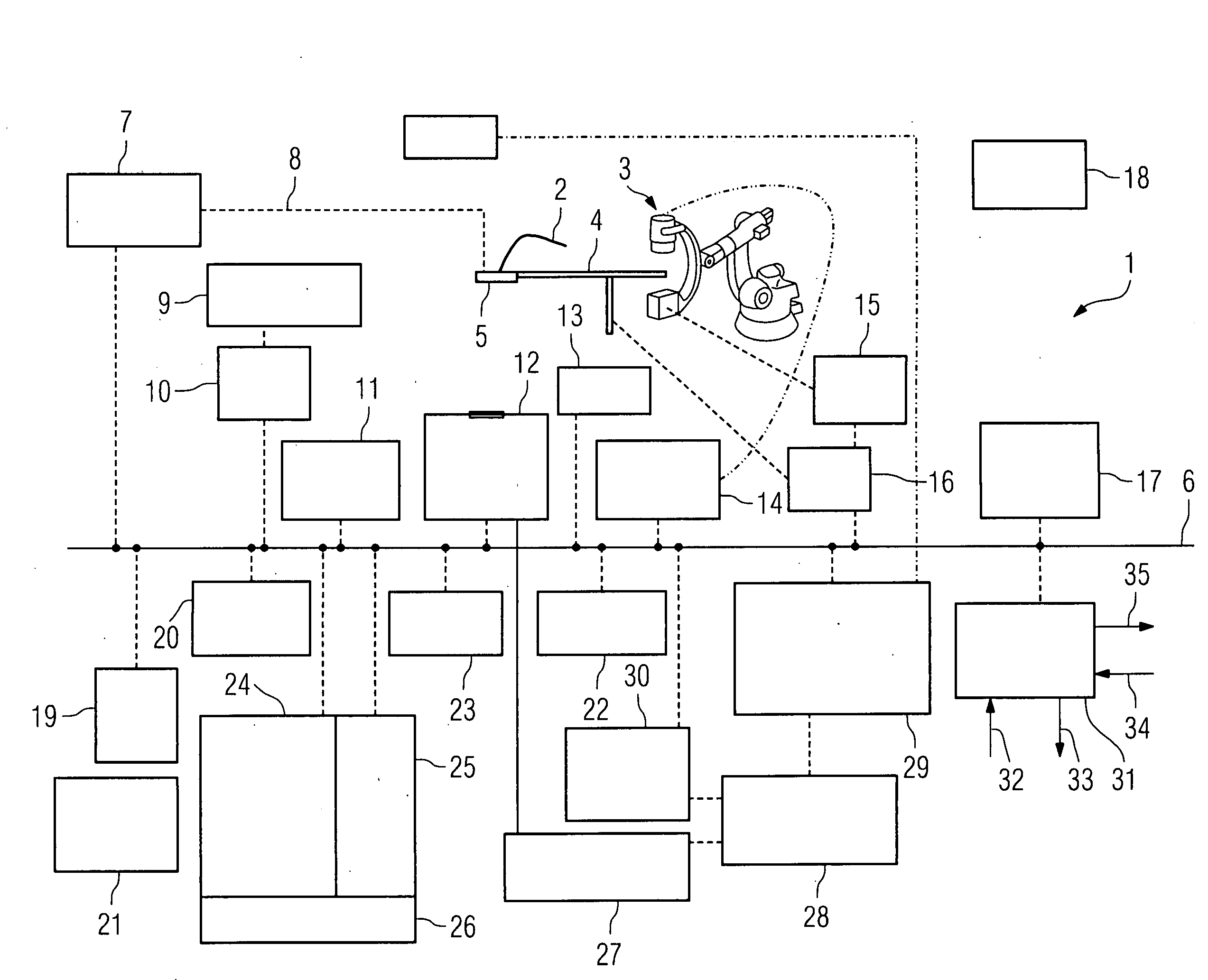
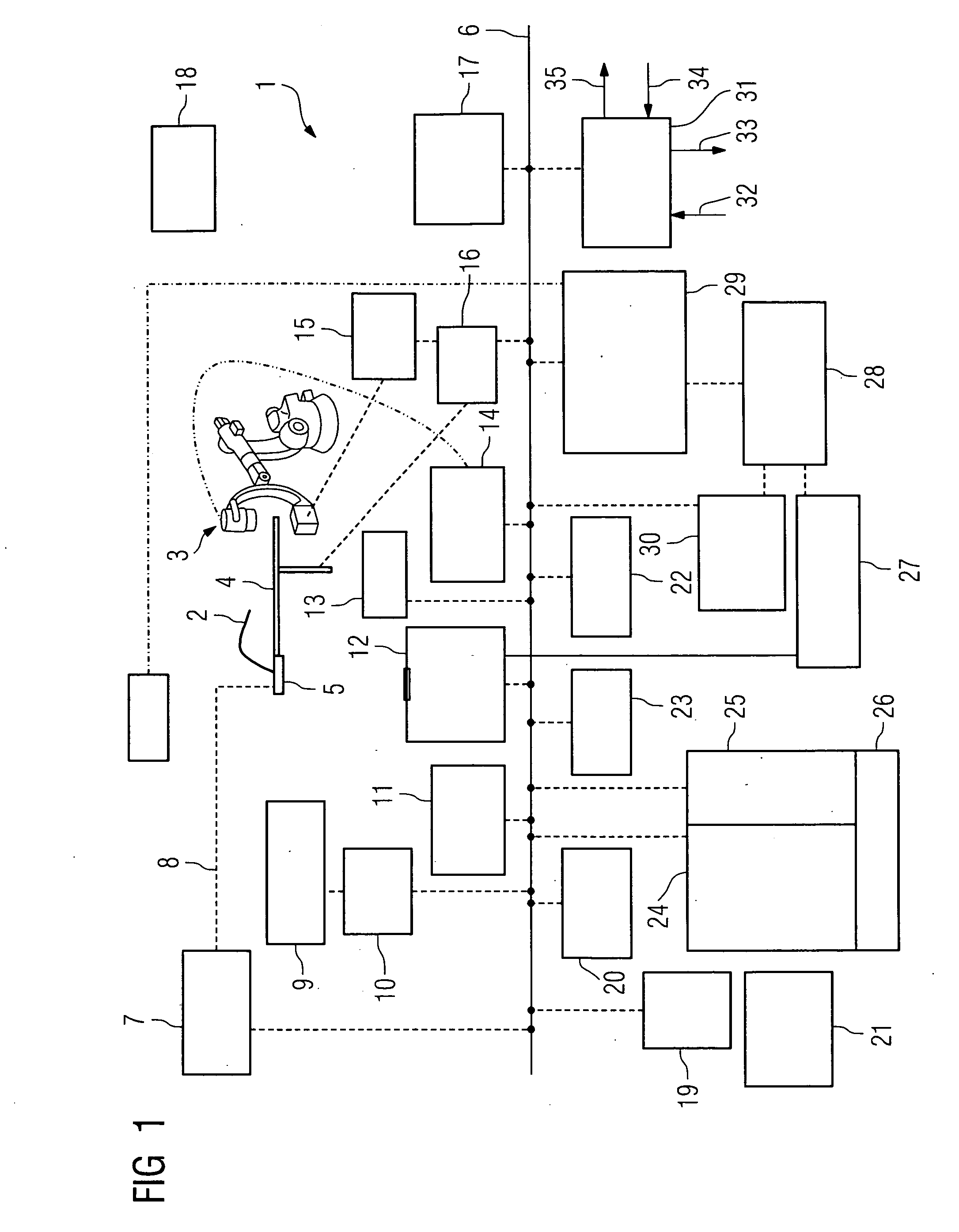
[0053]FIG. 1 shows an inventive medical imaging facility 1, configured as a hybrid system with an IVMRI catheter 2 with corresponding IVMRI image recording elements and a computed tomography device 3, here in the form of a buckling arm robot with at least 4 degrees of movement or degrees of freedom of movement.

[0054]A patient support facility 4 is also provided. An operating and interface facility 5 is used to record and forward the IVMRI date and allows operation of the IVMRI image recording elements in proximity to the table.

[0055]An IVMRI preprocessing and control unit 7 is connected to a data bus 6.

[0056]A connection 8 for an ultrasound catheter is also provided on the patient support facility 4 to produce ultrasound images. A signal interface 9 for the ultrasound data is connected to a preprocessing unit 10 for ultrasound data, which in turn is connected to the data bus 6.

[0057]The catheters of the system are provided with position sensors, to which a preprocessing unit 11 is a...


PUM


Abstract

The invention relates to a medical imaging facility, in particular for producing image recordings in the context of a treatment of cardiac arrhythmias, the medical imaging facility being configured as an integrated facility with at least one computed tomography device configured as a C-arm computed tomography device and configured to produce soft-tissue recordings and at least one image recording element based on intravascular magnetic resonance imaging, the medical imaging facility having at least one common control facility for both image recording methods, being configured for the automatic evaluation and display of at least one image recording of at least one computed tomography facility and / or at least one image recording element based on intravascular magnetic resonance imaging and / or for the display of identified scar tissue and / or regions of impaired and / or increased electrophysiological activity by means of at least one program means in real time.

Description

CROSS REFERENCE TO RELATED APPLICATIONS[0001]This application claims priority of German application No. 10 2007 043 731.7 filed Sep. 13, 2007, which is incorporated by reference herein in its entirety.FIELD OF THE INVENTION[0002]The invention relates to a medical imaging facility, in particular for producing image recordings in the context of a treatment of cardiac arrhythmias, and associated method.BACKGROUND OF THE INVENTION[0003]Cardiac arrhythmias are disruptions of the normal heartbeat sequence, whose causes can be found in disturbances of the formation or conduction of stimuli in the cardiac muscle.[0004]One example of a cardiac arrhythmia is what is known as bradycardia, with which there is a drop in heart rate. For many years cardiac pacemakers have been used to treat bradycardia, these being intended to restore a normal sinus rhythm.[0005]Other cardiac arrhythmias are tachycardial problems such as for example atrial fibrillation. With this type of cardiac arrhythmia interfe...

Claims


Application Information

Patent Timeline
IPC IPC(8): A61B5/055
CPCA61B6/032A61B6/4441A61B18/1492A61B19/5244A61B6/4458G01R33/285G01R33/287G01R33/4812A61B6/5247A61B2019/5289A61B34/20A61B2090/364
Inventor MASCHKE, MICHAEL
Owner SIEMENS AG
Learn More