Methods for treating macular edema and ocular angiogenesis using an Anti-inflammatory agent and a receptor tyrosine kinase inhibitor

a technology of ocular angiogenesis and anti-inflammatory agents, which is applied in the field of prevention and treatment of macular edema and/or ocular angiogenesis, can solve the problems of pathologic ocular angiogenesis, vision loss, pathologic changes,

Inactive Publication Date: 2009-04-23
NOVARTIS AG
View PDF14 Cites 11 Cited by
  • Summary
  • Abstract
  • Description
  • Claims
  • Application Information

AI Technical Summary

Problems solved by technology

Although PDR is the major cause of legal blindness in patients with diabetes mellitus, macular edema is the most common cause of vision loss.
These molecules can alter the retinal microvasculature and cause pathologic changes such as extracellular matrix remodeling, retinal vascular leakage leading to edema, and ultimately pathologic ocular angiogenesis.
In addition, removal of fluid from the diabetic retina may be impaired.
Regardless of disease etiology, when the edema involves the fovea, visual acuity is threatened.
Although posterior segment NV (PSNV) and macular / retinal edema are major causes of vision loss in adults, viable, approved treatment options are currently limited.
Laser photocoagulation alone or photodynamic therapy (PDT) with VISUDYNE® are therapies that involve laser-induced occlusion of the affected vasculature, which can result in localized damage to the retina.
Unfortunately, laser photocoagulation is a cytodestructive procedure and the visual field of the treated eye is irreversibly compromised.
Although glucocorticoids are effective in treating many ocular conditions, there are significant side effects associated with the available products.
Although some side effects are due to the glucocorticoid itself, some may result from, or be exacerbated by, excipients in the formulations and the method of delivery.
Furthermore, there are no NSAIs developed for treating persons suffering from ocular edema and / or NPDR.

Method used


Image

  • Methods for treating macular edema and ocular angiogenesis using an Anti-inflammatory agent and a receptor tyrosine kinase inhibitor
  • Methods for treating macular edema and ocular angiogenesis using an Anti-inflammatory agent and a receptor tyrosine kinase inhibitor
  • Methods for treating macular edema and ocular angiogenesis using an Anti-inflammatory agent and a receptor tyrosine kinase inhibitor

Examples


example 1

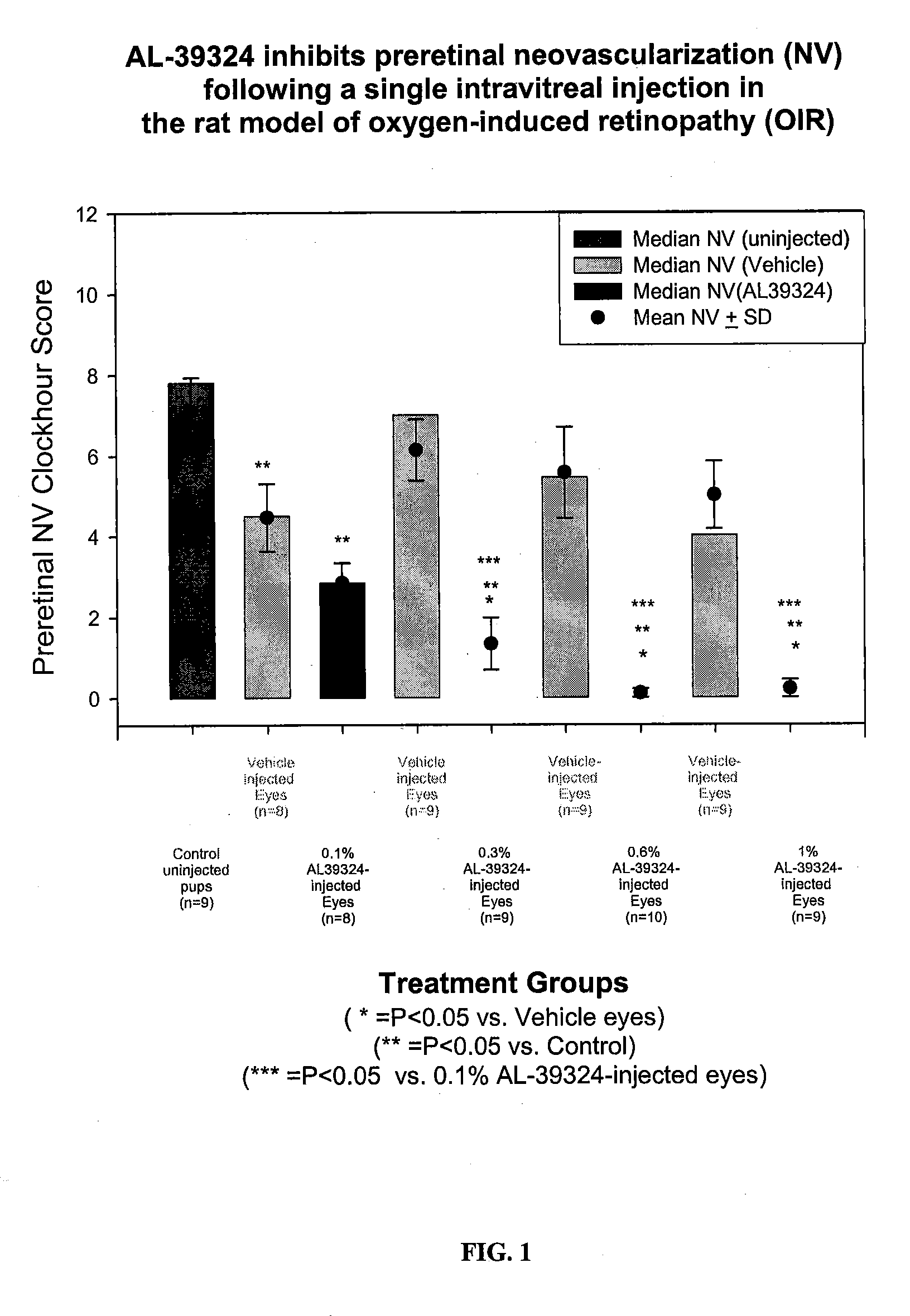
Prevention of Preretinal Neovascularization Following Intravitreal Delivery of the Receptor Kinase Tyrosine Inhibitor (RTKi), Compound 86, in the Rat Model of Oxygen-Induced Retinopathy

[0090]METHODS: Pregnant Sprague-Dawley rats were received at 14 days gestation and subsequently gave birth on Day 22±1 of gestation. Immediately following parturition, pups were pooled and randomized into separate litters (n=17 pups / litter), placed into separate shoebox cages inside oxygen delivery chamber, and subjected to an oxygen-exposure profile from Day 0-14 postpartum. Litters were then placed into room air from Day 14 / 0 through Day 14 / 6 (days 14-20 postpartum). Additionally on Day 14 / 0, each pup was randomly assigned as an oxygen-exposed control or into various treatment groups. For those randomized into an injection treatment group: one eye received a 5 μl intravitreal injection of 0.1%, 0.3%, 0.6%, or 1% RTKi and the contralateral eye received a 5 μl intravitreal injection of vehicle. At Day...

example 2

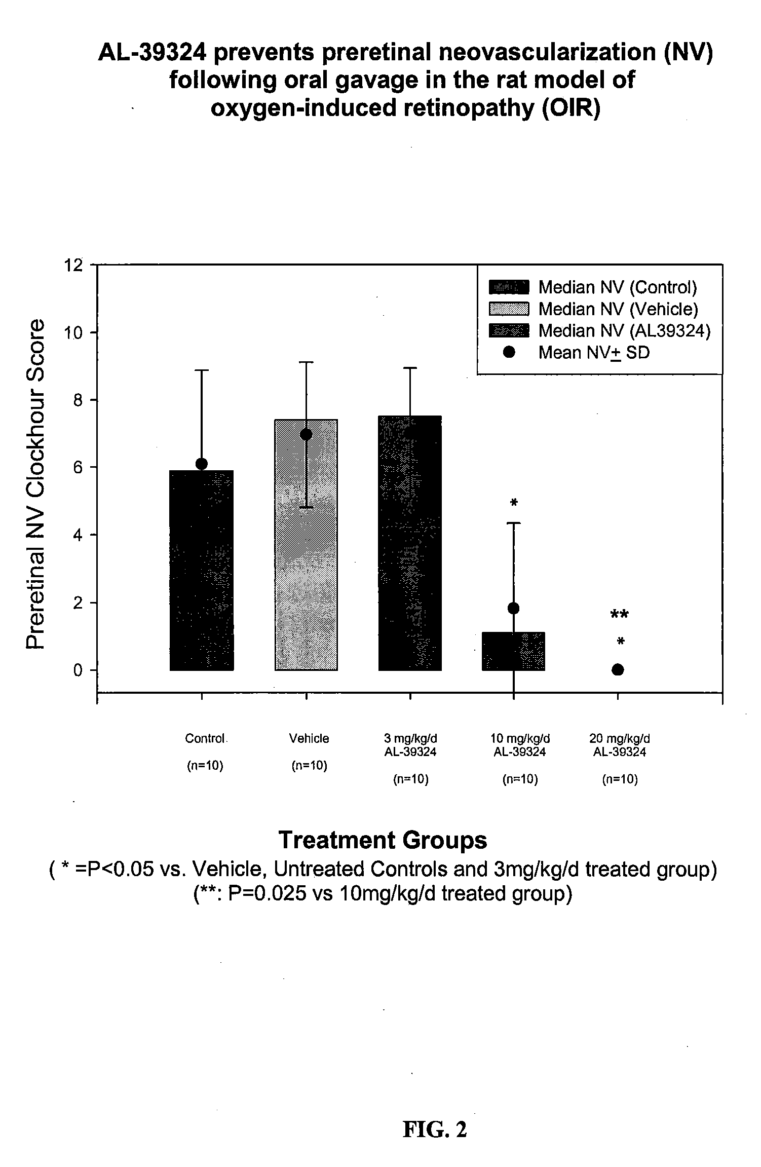
Systemic Administration of RTKi (Compound 86) Potently Prevents Preretinal Neovascularization in the Rat OIR Model

[0093]METHODS: Pregnant Sprague-Dawley rats were received at 14 days gestation and subsequently gave birth on Day 22±1 of gestation. Immediately following parturition, pups were pooled and randomized into separate litters (n=17 pups / litter), placed into separate shoebox cages inside oxygen delivery chamber, and subjected to an oxygen-exposure profile from Day 0 to Day 14 postpartum. Litters were then placed into room air from Day 14 / 0 through Day 14 / 6 (days 14-20 postpartum). Additionally on Day 14 / 0, each pup was randomly assigned as oxygen-exposed controls, vehicle treated, or drug-treated at 1.5, 5, 10 mg / kg, p.o., BID. At Day 14 / 6 (20 days postpartum), all animals in both studies were euthanized and retina whole mounts were prepared as described in Example 1 above.

[0094]RESULTS: Systemic administration of RTKi provided potent efficacy in the rat OIR model, where 20 m...

example 3

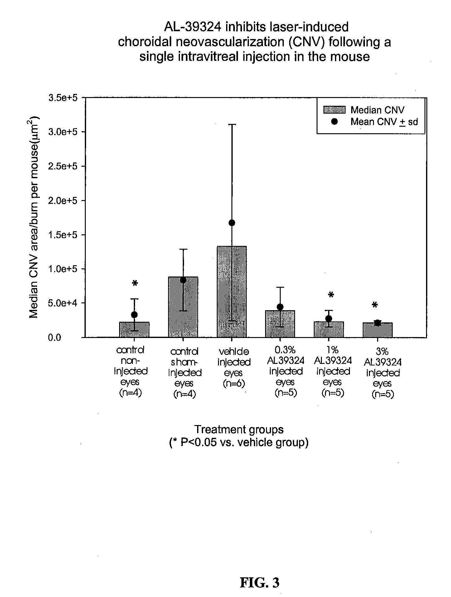
Prevention of Laser-Induced Choroidal Neovascularization (CNV) Following a Intravitreal Delivery of the Receptor Kinase Tyrosine Inhibitor (RTKi), Compound 86, in the Mouse

[0095]Methods. CNV was generated by laser-induced rupture of Bruch's membrane. Briefly, 4 to 5 week old male C57BL / 6J mice were anesthetized using intraperitoneal administration of ketamine hydrochloride (100 mg / kg) and xylazine (5 mg / kg) and the pupils of both eyes dilated with topical ocular instillation of 1% tropicamide and 2.5% Mydfin®. One drop of topical cellulose (Gonioscopic®) was used to lubricate the cornea. A hand-held cover slip was applied to the cornea and used as a contact lens to aid visualization of the fundus. Three to four retinal burns were placed in randomly assigned eye (right or left eye for each mouse) using the Alcon 532 nm EyeLite laser with a slit lamp delivery system. The laser burns were used to generate a rupture in Bruch's membrane, which was indicated opthalmoscopically by the form...


PUM

PropertyMeasurementUnit
spot sizeaaaaaaaaaa
spot sizeaaaaaaaaaa
pHaaaaaaaaaa

Abstract

The present invention provides methods for inhibiting increased vascular permeability and / or pathologic ocular angiogenesis via administration of a combination of one or more molecules that potently inhibit select receptor tyrosine kinases (RTKs) or vascular endothelial growth factor (VEGF) and one or more anti-inflammatory agents.

Description

RELATED APPLICATION[0001]The present application claims priority to U.S. Provisional Patent Application No. 60 / 871,409 filed Dec. 21, 2006.BACKGROUND OF THE INVENTION[0002]1. Field of the Invention[0003]The present invention is directed to the prevention and treatment of macular edema and / or ocular angiogenesis, specifically diabetic macular edema, exudative age-related macular degeneration and / or proliferative diabetic retinopathy. In particular, the present invention is directed to the use of certain formulations including a COX-II inhibitor, such as a derivative of 3-benzoylphenylacetic acid, and a receptor tyrosine kinase (RTK) inhibitor to treat such disorders.[0004]2. Description of the Related Art[0005]Diabetes mellitus is characterized by persistent hyperglycemia that produces reversible and irreversible pathologic changes within the microvasculature of various organs. Diabetic retinopathy (DR), therefore, is a retinal microvascular disease that is manifested as a cascade of...

Claims


Application Information

Patent Timeline
Patent Type & Authority Applications(United States)
IPC IPC(8): A61K31/5377A61K31/423A61K31/416
CPCA61K31/416A61K31/423A61K31/5377A61K45/06A61K2300/00
Inventor BINGAMAN, DAVID P.
Owner NOVARTIS AG
Why Eureka
Try Eureka