
Apparatus and Method for Vascular Access

a technology of vascular access and apparatus, which is applied in the field of guided cannulation of veins and arteries, can solve the problems of increasing workflow complexity, wasting time, and wasting patient time, and avoiding the use of current guided cannulation devices

Inactive Publication Date: 2009-05-07
TELEFLEX LIFE SCI LTD
View PDF105 Cites 160 Cited by
  • Summary
  • Abstract
  • Description
  • Claims
  • Application Information

AI Technical Summary

Benefits of technology

[0010]Some embodiments of the invention provide devices and methods to substantially reduce the amount of time required to place an intravascular device using conventional devices and methods. Some embodiments of the invention provide devices and methods to substantially reduce the need for X-ray imaging related to placing such device. Some embodiments of the invention provide devices and methods to increase placement reliability and accuracy and to verify device location post-placement.
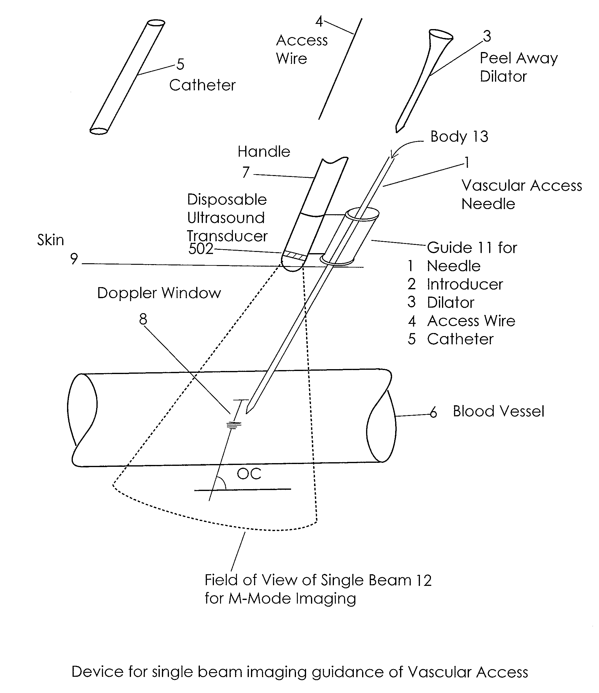
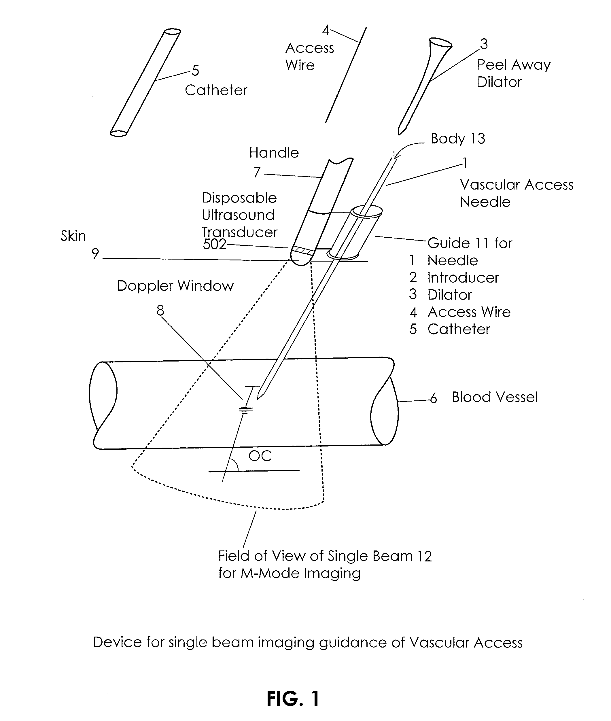
[0013]Another aspect of the invention includes an endovascular device guide attached to the imaging device capable of guiding several types of endovascular devices comprising a needle, a stylet, a catheter, and an introducer. The device may include adaptors to match the outer diameter of the endovascular guided device to the inner diameter of the guide. The device having the ability to slide or otherwise move with respect to the imaging device as to provide single hand deployment capability of the endovascular guided device.
[0022]Another aspect of the invention includes a system for tracking clinical procedures and improve workflow efficiency comprising: a workflow processor; an input interface; an output interface; a code reader; a communication component; and a database interface. The workflow processor stores information about procedure times, device information, patient and operator information, calculates parameters of the procedure like time duration and elapsed time between activities, and provides statistical data analysis of such parameters. The information about the endovascular procedure can be input into the system through a dedicated user interface guiding data acquisition. The output interface presents results of procedure workflow analysis. The code reader can be an RFID reader, a bar code reader or a reader of any computer readable label. The communication component can communicate over the network (wired or wireless) with the hospital information system. The communication component can communicate with other systems for tracking clinical procedures and establish a network of such systems. The database interface allows the procedure and workflow information to be archived.
[0036]Numerous alternatives are possible such as being constructed as a single-use device or where the information output is a scrolling chart. Additionally, the Doppler information can be bidirectional and / or the Doppler acquisition can be pulsed wave or continuous wave. Additionally, the guide attached to the imaging device is configured to guide one of the endovascular device selected from the group consisting of: a needle; a stylet; a catheter; and an introducer. There may also be an adaptor to match the outer diameter of the endovascular guided device to the inner diameter of the guide. The endovascular device may also be configured to slide or move with respect to the imaging device as to provide single hand deployment capability of the endovascular guided devices described herein.
[0045]In another aspect, there is a system for tracking clinical procedures and workflow having one or more of: a workflow processor; an input interface; an output interface; a code reader; a communication component; and a database interface. The workflow processor may store information about procedure times, device information, patient and operator information, calculate parameters of the procedure like time duration and elapsed time between activities, and provide statistical data analysis of such parameters. The information about the endovascular procedure may be input into the system through a dedicated user interface guiding data acquisition. The output interface may present results of procedure workflow analysis. The code reader can be an RFID reader, a bar code reader or a reader of any computer readable label. The communication component can communicate over a wired network or a wireless network with a hospital information system. The communication component can communicate with other systems for tracking clinical procedures and establish a network of such systems. The database interface allows the procedure and workflow information to be archived.

Problems solved by technology

Currently, preparing the patient for and performing vein and artery cannulation is time consuming, challenging in terms of locating the blood vessels and, under circumstances, ensuring the desired vessel is accessed (e.g., vein vs. artery).
Current guided cannulation devices are either too expensive or difficult to use.
General purpose imaging systems are gaining acceptance but they are expensive and represent an increase in workflow complexity because they are not sterile.
In addition, general imaging systems are limited in terms of their ability to image in near field, i.e., closed to the surface of the skin.
Additional challenges remain unaddressed in many areas related to endovascular devices.
One challenge that remains is for devices and methods endovascular positioning within or towards the center of a vessel.
Another challenge that remains are devices and methods that rely on acoustic triangulation or positioning to localize and place endovascular devices.
Another challenge related to work flow efficiency and monitoring of the placement and confirmation of endovascular device locations.

Method used


Image

  • Apparatus and Method for Vascular Access
  • Apparatus and Method for Vascular Access
  • Apparatus and Method for Vascular Access

Examples


Embodiment Construction

[0097]An aspect of the invention includes a transcutaneous ultrasound vascular access guiding system comprising: a single element ultrasound device providing A-Mode imaging, Doppler and correlation-based blood velocity estimation; a processor to process and correlate ultrasound information; and a system for information output. The transcutaneous ultrasound vascular access guiding system may also comprise a lens which controls the single element ultrasound beam shape. The transcutaneous ultrasound vascular access guiding system may also comprise a lens which provides a matching layer between the ultrasound transducer and the skin. transcutaneous ultrasound vascular access guiding system comprising can be constructed as a single-use device. Also, the information can be output as a scrolling chart. The Doppler information can be bidirectional. The Doppler acquisition can be pulsed or continuous wave (PW or CW).

[0098]Another aspect of the invention includes an endovascular device guide ...


PUM


Abstract

In an aspect, embodiments of the invention relate to the effective and accurate placement of intravascular devices such as central venous catheters, in particular such as peripherally inserted central catheters or PICC. One aspect of the present invention relates to vascular access. It describes devices and methods for imaging guided vascular access and more effective sterile packaging and handling of such devices. A second aspect of the present invention relates to the guidance, positioning and placement confirmation of intravascular devices without the help of X-ray imaging. A third aspect of the present invention relates to devices and methods for the skin securement of intravascular devices and post-placement verification of location of such devices. A forth aspect of the present invention relates to improvement of the workflow required for the placement of intravascular devices.

Description

CROSS-REFERENCE TO RELATED APPLICATIONS[0001]This application claims the benefit of U.S. Provisional Patent Application No. 60 / 937,280 filed on Jun. 26, 2007 by Sorin Grunwald et al., entitled “Apparatus and Method for Vascular Access” which is incorporated herein by reference in its entirety.[0002]This application is also a continuation-in-part of U.S. Non-Provisional patent application Ser. No. 11 / 431,140 filed on May 8, 2006 by Sorin Grunwald et al., entitled “Endovenous Access and Guidance System Utilizing Non-Image Based Ultrasound”, now publication no. 2007-0016072-A1; U.S. Non-Provisional patent application Ser. No. 11 / 431,118 filed on May 8, 2006 by Sorin Grunwald et al., entitled “Endovascular Access and Guidance System Utilizing Divergent Beam Ultrasound”, now publication no. 2007-0016070-A1; U.S. Non-Provisional patent application Ser. No. 11 / 431,093 filed on May 8, 2006 by Sorin Grunwald et al., entitled “Ultrasound Sensor”, now publication no. 2007-0016069-A1; and U.S. ...

Claims


Application Information

Patent Timeline
Patent Type & Authority Applications(United States)
IPC IPC(8): A61B8/00A61B7/00G06Q50/00G16H20/40
CPCA61B5/0402A61B8/445A61B5/1459A61B5/411A61B7/00A61B8/06A61B8/0841A61B8/12A61B8/42A61B8/461A61B19/201A61B19/5244A61B2017/00106A61B2017/003A61B2019/5276A61B2019/5278A61B2019/5429G06Q50/22A61B5/06A61B2090/3782A61B34/20A61B90/11A61B2090/378A61B2090/3929G16H20/40A61B5/318
Inventor GRUNWALD, SORINSANDER, FIONA MARIASAMSON, WILFRED J.HILL, BRADLEY
Owner TELEFLEX LIFE SCI LTD
Learn More