

X-ray ct apparatus and image processing apparatus
- Summary
- Abstract
- Description
- Claims
- Application Information
AI Technical Summary
Benefits of technology
Problems solved by technology
Method used
Image


Examples
first embodiment
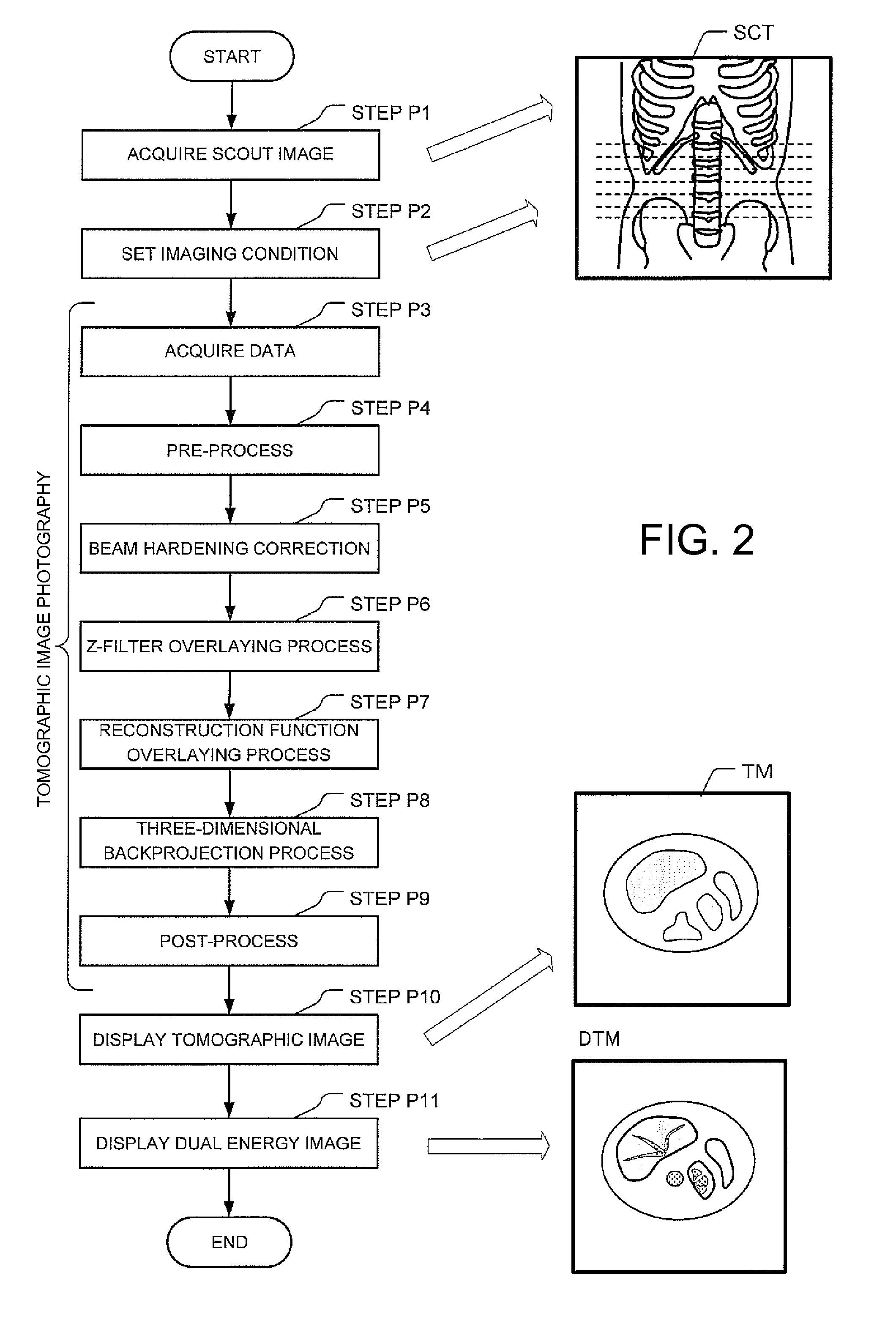
[0052]The process at Step P11 referred to above will next be described in further detail using a
[0053]The present embodiment will explain an example in which a dual energy image is reconstructed in a tomographic image space, using a flowchart shown in FIG. 3.
[0054]At Step 101, a process for determining the ratio between the values of pixels of a tomographic image obtained at an X-ray tube voltage 140 kV and a tomographic image obtained at an X-ray tube voltage 80 kV is performed. Incidentally, the ratio between the pixel values is one example of the difference information between a first X ray and a second X ray with respect to each tomographic images of a subject, based on first X-ray projection data and second X-ray projection data in the present invention.
[0055]FIG. 4(a) is a graph in which, for example, respective pixel values L-HU of a tomographic image at an X-ray tube voltage 80 kV are taken along the vertical axis of the graph, and respective pixel values H-HU of a tomograph...
second embodiment
[0071]A second embodiment will explain an example in which a dual energy image is reconstructed in a projection data space.
[0072]First, a process for determining the ratio between pixels of a tomographic image obtained at an X-ray tube voltage 140 kV and a tomographic image obtained at an X-ray tube voltage 80 kV, which is similar to the process at Step 101 of the first embodiment, is performed.
[0073]Next, each pixel to which the value of the ratio is assigned, is reprojected onto a virtual detector to obtain virtual projection data indicated by a sinogram, which is provided with the coordinate of each pixel and the value of the ratio as data.
[0074]Next, an allowable width (range) of the ratio between pixel values is set by the segment condition setting unit 36 in a manner similar to the process at Step 102 of the first embodiment.
[0075]Next, it is determined at Step 103 of the first embodiment whether the value of a pixel ratio between the respective virtual projection data falls w...
PUM
 Abstract
Description
Claims
Application Information