Endoscopic system for lung biopsy and biopsy method of insufflating gas to collapse a lung

a biopsy system and lung technology, applied in the field of endoscopic biopsy system, can solve the problems of difficult control, difficult to move sharp, scattered barbs, and take a long time to collect superficial layers of tissue from a large surface area, and achieve the effect of a shorter period and a larger surface area

Inactive Publication Date: 2009-05-14
CHEST INNOVATIONS
View PDF17 Cites 14 Cited by
  • Summary
  • Abstract
  • Description
  • Claims
  • Application Information

AI Technical Summary

Benefits of technology

[0012]The sealing, separating and collecting functions of the endoscopic biopsy system of the present invention occur internally within the patient from which the sample is taken. Therefore, there is no exposure of separated biopsy sample materials (i.e. blood, fluid, cells, tissue, DNA, RNA, etc.) to the external environment prior to the time they are ready for analysis, thereby avoiding the risks of sample contamination and degradation. The collection methods and devices of the present invention are sealed and airtight. These include the use of a collection bag and the use of a tubular vacuum suction system that draws separated samples through internal channels to a sealed external collection chamber.
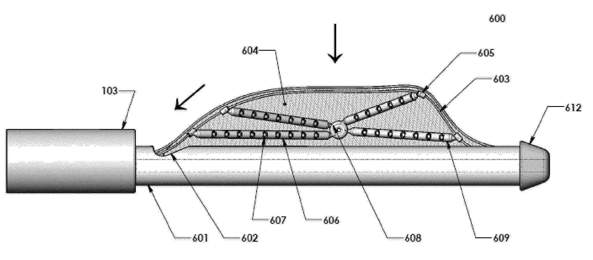
[0017]According to another aspect of the present invention, the endoscopic biopsy system comprises several soft extruded tubes (“octo-arms”), as described in the previous paragraph, joined together by mesh-netting in between adjacent tubes and with a drawstring surrounding their periphery. In this system of tubes (an “octo-mat”), each tube with openings thereupon and one or more wire therein functions the same as it would independently, as described in the previous paragraph. The advantage of this system of tubes is that it has the capacity to expand (via the flexible mesh-netting joints and relaxation of the peripheral drawstring) to cover a larger surface area within a shorter period of time. Further, the network of mesh-netting holding the tubes together secures retrieval of any material missed by the suction system on the first pass. The network of mesh-netting is particularly beneficial when the vacuum pressure is low (i.e. due to equipment constraints or sensitivity at the biopsy site). The network of mesh-netting is also particularly beneficial for use in biopsy sites that are dense with potential sample material because even a high pressure vacuum system is unlikely to be able to keep pace with the rate at which the tubular openings come into contact with sample material. The mesh-netting network affords the vacuum system a second, third, etc. chance to capture material as it rebounds from the netting to approach the tubular openings.

Problems solved by technology

The problem with this device is that it would take a very long time to collect a superficial layer of tissue from a large surface area.
The problem with this device is that the sharp, scattered barbs are not moveable and are difficult to control precisely for grasping a particular area of tissue while leaving its vicinity unharmed.
Further, the barbs are likely to shred the sample into small fragments as it is collected (in a manner similar to a cheese or vegetable grater) which can destroy certain characteristics of the tissue desired for analysis.
Thus, the barb shredder system does not appear to be conducive to the collection of large, intact volumes of specimens.
Unlike nodular or pedunculated sample sites having tissue easily grasped with forceps, planar surfaces are traditionally more difficult to biopsy without destroying the sample or significantly damaging nearby healthy tissue.
Conventional rigid chest tubes are frequently painful and conventional flexible chest tubes frequently kink or buckle requiring painful manipulation or reinsertion.

Method used


Image

  • Endoscopic system for lung biopsy and biopsy method of insufflating gas to collapse a lung
  • Endoscopic system for lung biopsy and biopsy method of insufflating gas to collapse a lung
  • Endoscopic system for lung biopsy and biopsy method of insufflating gas to collapse a lung

Examples


Embodiment Construction

[0049]First, a general procedure for the collection of biopsy samples from a lung according to the systems and methods of this invention will be outlined. Second, various embodiments of the endoscopic biopsy system and various methods for their use will be described in detail with reference to the several drawings. Although the systems and methods of this invention are illustrated with respect to collecting biopsy samples from a lung, the systems and methods are not limited to lung biopsy. One having ordinary skill in the art will recognize that the systems and methods described herein are readily adapted for the collection of biopsy samples from several regions of the body.

General Procedure

[0050]Step One: Consent, Anesthesia, Medical Staff, and Set-Up

[0051]Prior to beginning the procedure, the informed consent of the patient should be obtained.

[0052]One advantage of the present invention, as compared to traditional open-surgery biopsy techniques, is that it is done under local anes...


PUM


Abstract

An endoscopic biopsy system comprising a means for drawing a sample to be sealed, separated, and / or collected towards an instrument, such as a structure that includes one or more of: an extendable wire, an extendable mast, an extruded tube with at least one opening and a vacuum therein, a pivot point, a hinge, and a mounting block. The system should have a means to transfer energy to a sample after a sample is grasped, such as a conducting wire or an anvil. The system should also have an airtight means to remove a separated sample from a biopsy site for analysis, such as a collection bag or an internal vacuum suction system. The endoscopic biopsy system can be used in a method of obtaining a biopsy from the thoracic cavity that includes a step of insufflating gas to induce pneumothorax and collapse a lung.

Description

BACKGROUND OF THE INVENTION[0001]1. Field of the Invention[0002]The present invention relates to an endoscopic biopsy system comprising an instrument that can seal surrounding tissue(s), separate sample(s), and / or collect sample(s) from the interior of a subject for removal and analysis and to a method of obtaining biopsy samples using the system in accordance with minimally invasive surgical techniques.[0003]2. Description of the Related Art[0004]Several systems, devices, and methods have been proposed for obtaining biopsy samples. Recent developments in the art have focused on the successive collection and removal of multiple samples during one procedure without reinserting the biopsy instrument into the body between samples. U.S. Patent Application Publication No. 2007 / 0213632 (Okazaki et al.) and U.S. Pat. Appl. Pub. No. 2006 / 0258955 (Hoffman et al.) disclose such systems.[0005]Another focus area for recent developments has been on methods used to separate tissue from its surrou...

Claims


Application Information

Patent Timeline
Patent Type & Authority Applications(United States)
IPC IPC(8): A61B10/02A61B1/018
CPCA61B10/0266A61B10/0283A61B2018/1407A61B2017/32004A61B10/04
Inventor CHIN, ROY SING-FATTDHARAN, MURALI
Owner CHEST INNOVATIONS
Why Eureka
Try Eureka