Typically, primary palatal
surgery does not restore correct positioning of this
muscle, which may contribute to poor auditory tube functioning and decreased ventilation of the
tympanic cavity (Abe et al., 2004).
This would suggest that even a repaired palatal cleft may be functioning at a
disadvantage compared to that of individuals with normal velopharyngeal
anatomy.
However, it appears to have little significant
impact on movements during speech production.
Aside from abnormal
muscle positioning found in individuals with a cleft palate, complications are also present due to the existence of a cleft palate.
Problems related to feeding, maxillary facial growth,
dentition, hearing, and speech are just a few of the obstacles these children and their families will encounter throughout the child's life.
Even after
surgical repair of the cleft palate, children often continue to have an inadequate speaking mechanism.
However, in individuals with a cleft palate, the anterolateral levator fibers, if left attached to the nasal mucosa, appear to tether levator movement and compromise the movement of a retropositioned levator
muscle.
Even after the primary palatoplasty, the velopharynx may function abnormally resulting in a
coupling effect between the oral and nasal cavities at the level of the velopharyngeal port.
During speech production, this effect results in the presence of hypernasality or abnormal nasal
resonance during oralized vowels and consonants.
The existence of hypernasality may also have devastating effects on the child's
quality of life.
Velopharyngeal inadequacy results when the velum is not able to create a tight seal against the posterior
pharyngeal wall, thus the mechanism is functioning inadequately.
A third cause of VPI is due to faulty positioning or improper
dissection of the levator muscle during primary palatoplasty.
In such cases, the levator muscle fibers may not be fully dissected off the
hard palate.
Another cause may be a disparity of levator sling fibers through the body of the velum.
In such cases, the levator sling may have been surgically positioned correctly, however, with time the muscles may have drifted more anteriorly towards an unfavorable position.
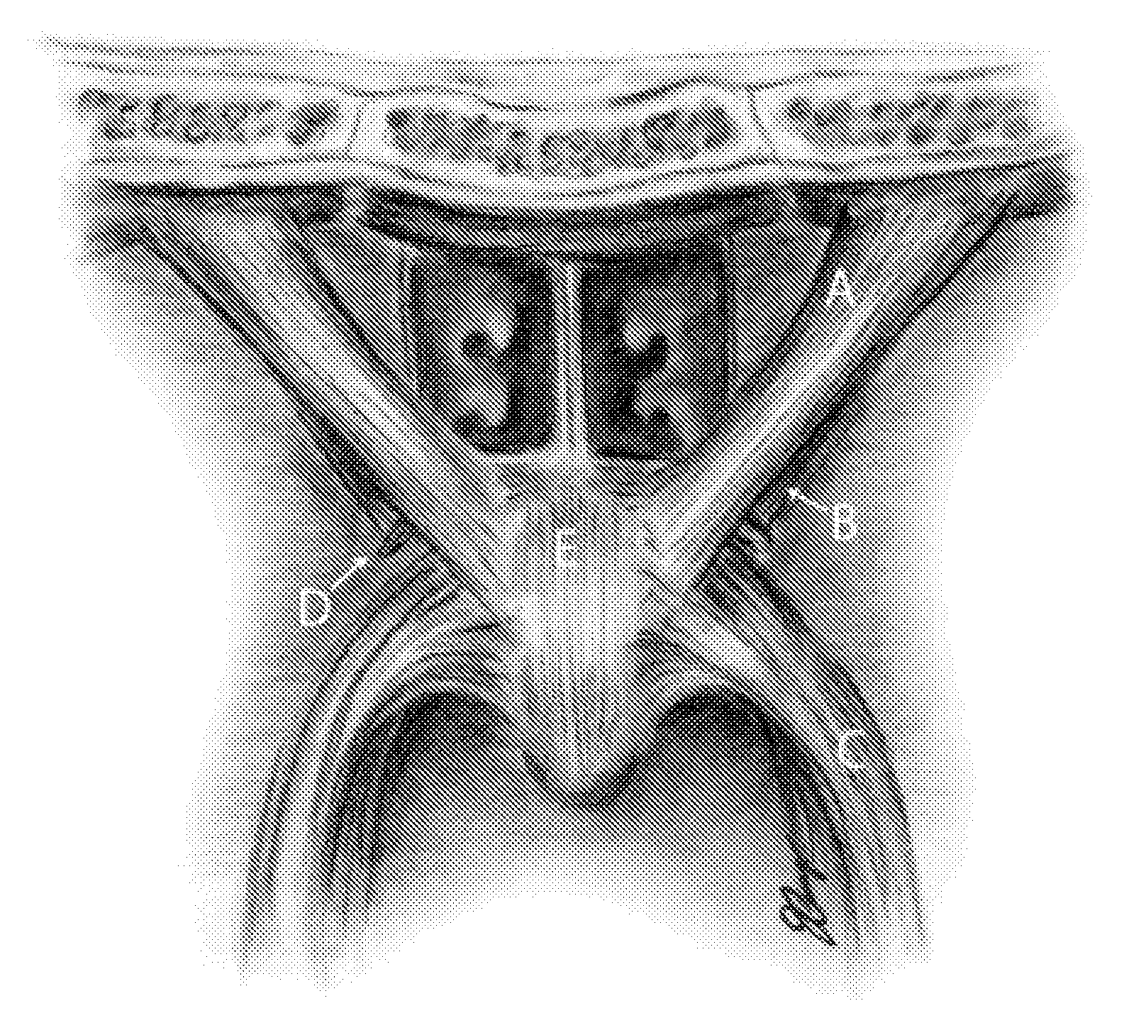
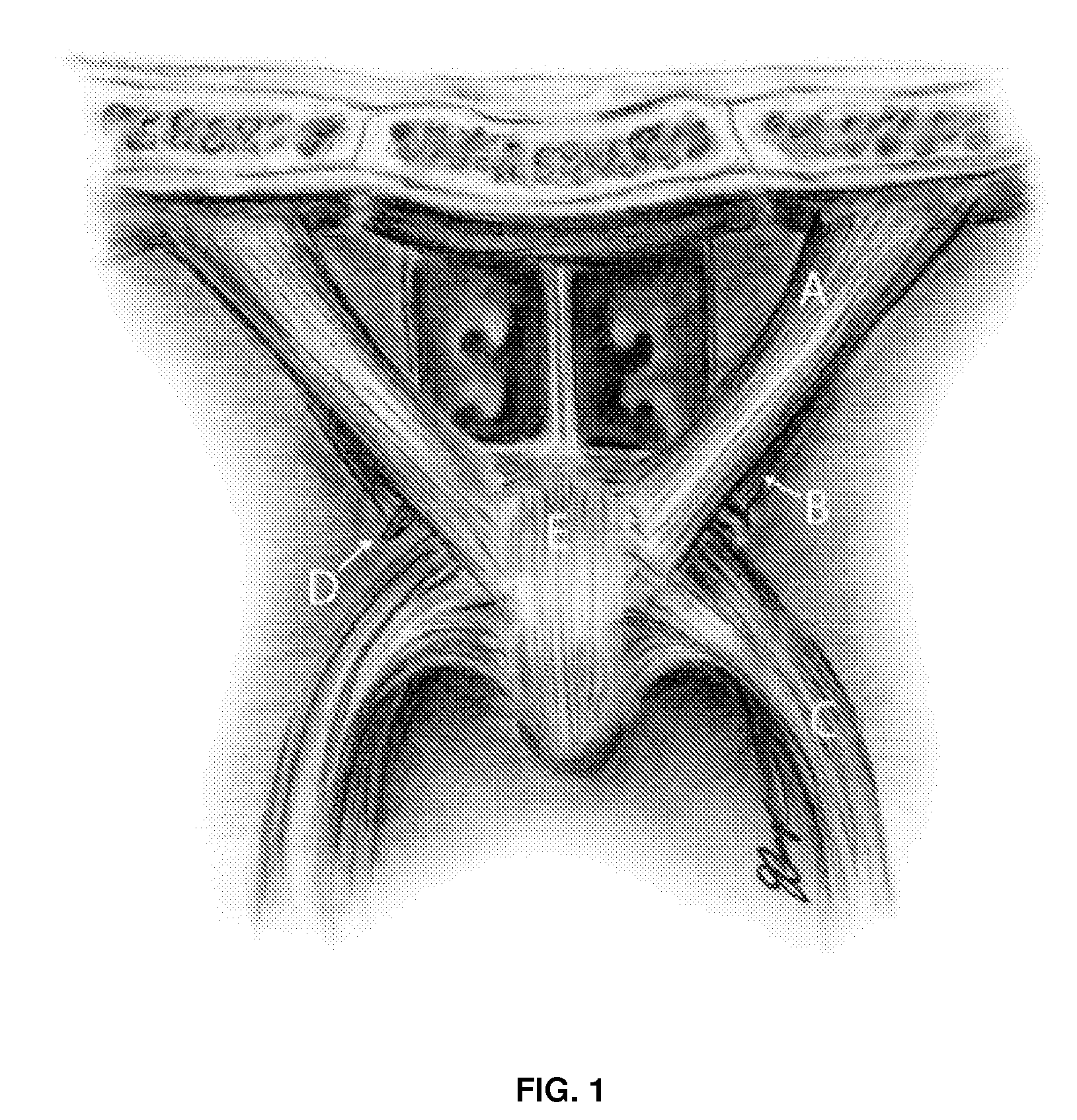
Unless
visual inspection of the levator muscle through medical images (such as magnetic
resonance images) is used during a patient's
clinical treatment, the surgeon cannot always be certain that all the fibers have been relocated to the appropriate position.
Even if the muscle is repositioned correctly, adequate levator
muscle mass through the midline of the velum may or may not be achieved.
There is a lack of data regarding what constitutes adequate
mass through the midline.
Secondly, when viewing an oblique coronal MR image, it is difficult to differentiate the levator muscle from the musculus uvulae.
No studies have examined how the reconstructed soft tissue responds to external forces or whether the muscle might with time migrate towards a less favorable position.
A major drawback to current surgical practices is that there is limited presurgical planning.
Traditional
lateral view x-
ray does not allow the surgeon to visualize the levator muscle before surgery.
There are disadvantages to using these methods to examine muscle function.
Dissection presents complications due to the relative inaccessibility of the region such that tissue must be removed in order to
gain access to other areas of interest (Ettema, Kuehn, Perlman, & Alperin, 2002).
Histologic processes also add complications secondary to the extraction, preservation, and
staining of the human tissue.
Furthermore, both methods are destructive in nature and can only be done on cadaveric material.
Lateral view x-
ray and CT allow for study
in vivo, however, are considered to place the patient at risk of
exposure to
radiation and muscle cannot be visualized in contrast to adjacent soft tissue structures.
Neither of these procedures, however, allows the examiner to visualize underlying muscles including the levator muscle fibers.
Although nasoendoscopy does not
expose the patient to
radiation, unlike CT and x-ray, it is not always tolerable for young patients.
In addition, inspection of the mechanism is limited to the velopharyngeal port (e.g. closure patterns, closure gaps, and
pharyngeal wall movement).
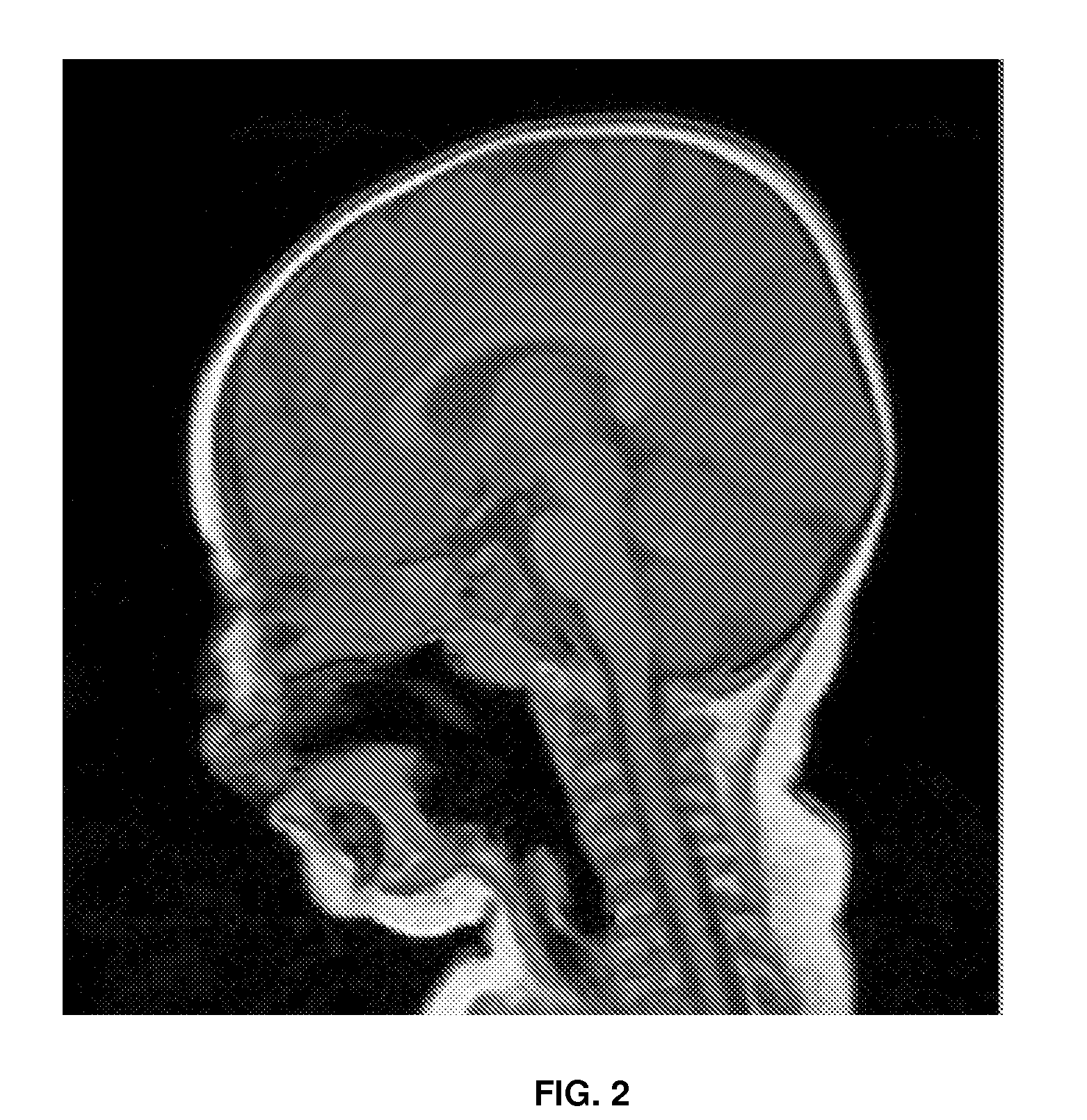
There is a limited amount of previous work in using MRI to study and measure the velopharyngeal mechanism.
Some noted limitations of the study include the use of 2D verses 3D acquisition, static imaging verses
dynamic imaging, and only two MRI sessions versus three or more to provide information related to possible muscle relapse.
In the field of speech science, limited research has been done to image and construct a 3D model of the velopharyngeal mechanism.
However, a limitation of computer generated models is the difficulty in simulating soft tissue deformations.
As a result, the computer modeling that is based on CT data only demonstrates preoperative structures.
The velopharyngeal mechanism is often difficult for students to conceptualize.
While exploration of the velopharyngeal mechanism through
cadaver dissection would provide the most rewarding learning experience, it is not always available due to time and money constraints (Perry, Kuehn, & Langlois, 2007).
However, this does not allow the surgeon to visualize the levator muscle before surgery.
No studies have examined how the reconstructed soft tissue responds to external forces or whether the muscle might with time migrate towards a less favorable position.
Subject 3 was supposed to be imaged using the 3 Tesla
system, however, the
scanner did not become available to Carle Foundation Hospital until after the subject's scheduled surgery date.
This is due to the presence of the musculus uvulae through the midline of the velum, which is difficult to separate from the levator fibers when taking midline thickness measurements.
Therefore, even after
surgical repair of the hard and soft tissue structures, this muscle still may not contribute to midline thickness measurements.
In addition, following palatal repair, the levator muscle may not be a cohesive
muscle mass in the midline making it difficult to determine the true thickness of the muscle in that location.
Amira uses several
smoothing techniques, however, this can lead to a loss of data particularly when the object has relatively few voxels selected as in the case of the levator muscle.
As a result, the surfaces tend to be rigid and irregular, lacking realism (FIG. 17).
For example, the styloid process, pterygoid plates, and hamulus are not easily detected on the original
MR images.
However, this region does not have major implications on the structures and functions of the velopharyngeal mechanism.
This problem was not resolved and appears to be a result of the MR
image acquisition rather than a result of conversion between Amira and Maya since the problem was observed in both programs.
However, before this could be done, the general
anesthesia wore off and the child was not compliant.
However, measures were not consistently similar between subjects with a similar weight.
Velopharyngeal function is generally adequate for vegetative purposes (
swallowing), however, may be inadequate for speech even after primary repair of the palate.
A muscle morphology that appears normal yet does not create adequate posterior and superior movement of the velum will have substantial negative effects on speech.
Hypernasal speech can, in turn, have negative effects on a child's psychosocial development.
This presents a particular challenge when assessing the speech outcome following surgery.
None of these methods allow the
surgical team to view the levator muscle before or after surgery.
However, if the muscle dimensions are not met, it might be assumed that velopharyngeal function would be compromised.
It is not possible to determine if the variation found in the muscle length and distance between origins is statistically significant due to the
small sample size.
A muscle bundle that converges very sharply would display a small axial angle of origin and would be disadvantageous for drawing the velum posteriorly.
The angle of origin in the axial
image plane is more difficult to obtain compared to the previous angles of origin measures.
Measurements and reconstructions of the levator muscle were conducted using two dimensional images, which makes analysis of the third angle of origin difficult.
However, the muscle was difficult to delineate from the surrounding tissue structures in this particular region.
Although the
surgical planning tool designed in this research is not refined or sophisticated enough for some of these proposed surgical applications, it is a step closer.

