Automatic Segmentation of the Heart and Aorta in Medical 3-D Scans Without Contrast Media Injections

a technology of computed tomography and heart and aorta, which is applied in the field of medical imaging using computed tomography, can solve the problems of inability to perform stress tests, limited noninvasive methods of detection, and added, although manageable, complexity, etc., and achieves improved reproducibility of measurements, improved precision, and reduced number of interactions between the operator and the image.

Inactive Publication Date: 2009-05-28
ARNOLD BEN A
View PDF34 Cites 36 Cited by
  • Summary
  • Abstract
  • Description
  • Claims
  • Application Information

AI Technical Summary

Benefits of technology

[0012]Methods are disclosed which provide automatic location and quantification of arterial calcium in the major arteries of the body. The preferred embodiments disclose methods for coronary and aortic calcium analysis using computerized tomography. The methods preferably use calibrated images for improved precision, but can operate without calibration. The number of interactions by the operator with the images is greatly reduced and in some cases approach zero. The reproducibility of measurements is improved since the operator's subjective judgment is essentially eliminated.
[0013]In one preferred embodiment, calcium in the coronary arteries of living patients can be quantified reproducibly by automatic methods. The heart is segmented using preferably calibrated images. The heart is located and its boundaries are identified in 3-D space. In specific patients, it may be necessary for the operator to mark the inferior boundary of the heart to further aid the software in separating heart tissue from liver or sternum. The mode of a histogram analysis of the 3-D segmented heart provides a best measure of the blood / heart tissue density. This value is used to smooth the heart image to remove streak artifacts. The histogram mode is also incorporated into the calibration equation. The aorta is located automatically by using shape, size and density constraints. The aorta is segmented off from the heart and is also used as a beginning location in the search for coronary artery locations.
[0016]The methods improve reproducibility by use of the calibration methods and automatic location and quantification. Reproducibility is also improved due to the use of machine defined objective and reproducible criteria. The automatic algorithm greatly speeds the exam as each individual CT slice and each individual calcification need not be identified and analyzed.
[0020]In a second preferred method to automatically quantify calcium in the aorta, a different auto search algorithm is used. This algorithm has advantages in older patients who may have rotated vertebral bodies.
[0021]Either search algorithm will operate on any major artery of the body, including the carotids. Either algorithm can operate with or without calibration. However, performance is improved with external calibration using a phantom and internal calibration using blood as a surrogate internal calibration sample.

Problems solved by technology

While early detection and prevention of atherosclerotic plaque in coronary arteries is desirable, coronary calcium screening is not available in most communities of the U.S. or in the remainder of the world.
Conventional noninvasive methods of detection, such as stress tests, are limited by poor performance.
Current cardiac CT images are acquired with ECG gating, which adds some, although manageable, complexity.
Although the correlation was high, the regression equation relating the actual mass to the mass estimates indicated that the CT mass estimates consistently underestimate actual coronary calcium mass.

Method used


Image

  • Automatic Segmentation of the Heart and Aorta in Medical 3-D Scans Without Contrast Media Injections
  • Automatic Segmentation of the Heart and Aorta in Medical 3-D Scans Without Contrast Media Injections
  • Automatic Segmentation of the Heart and Aorta in Medical 3-D Scans Without Contrast Media Injections

Examples


Embodiment Construction

[0049]A premise of certain embodiments described herein is that the atherosclerotic process is systemic, affecting all of the arteries of the body in a similar way. Although plaque build-up may occur and / or progress at different rates in different arteries, this process occurs throughout the body. Others have studied the close relationship of cardiac calcium and extracoronary plaque by comparing ultrasound measurements of carotid, aortic, and femoral plaque with ultrafast CT measurements of coronary calcifications. These studies have shown that patients with coronary calcifications had a higher prevalence of aortic and femoral plaque.

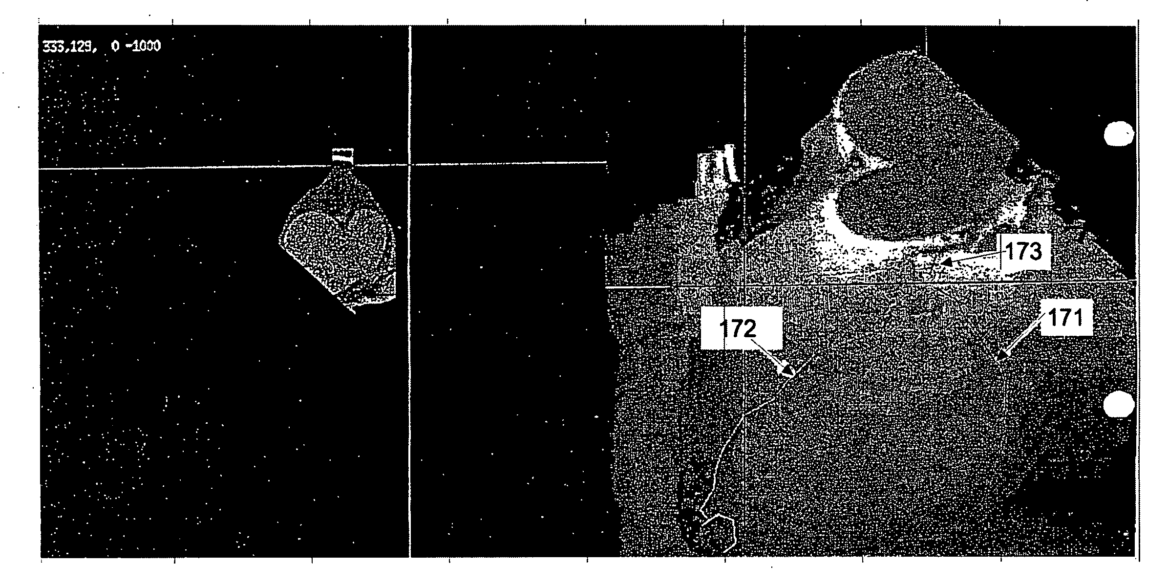
[0050]The abdominal aorta is a site of atherosclerotic plaque containing calcifications. The aorta has minimal motion, thus allowing easy imaging using conventional CT scanners. Although aortic calcifications have been associated subjectively with atherosclerotic disease, aortic calcium has not been quantified or proposed as a quantitative diagnostic te...


PUM


Abstract

A method automatically segments the heart and abdominal aorta from volumetric images without the need to inject iodine contrast media into the subject. The method automatically quantifies arterial plaque (hard plaque, soft plaque or both) in the cardiovascular system. Plaque definitions include subject specific in vivo blood / muscle density measurements, subject specific voxel statistical parameters and 2-D and 3-D voxel connectivity criteria, which are used to automatically identify the plaques. The locations and outlines of the major arteries are determined in a 3-D coordinate system and the specific coordinates of the detected plaques are displayed in a plaque map for follow-up exams or ease in plaque review and reporting the results.

Description

RELATED APPLICATIONS[0001]This application is a continuation of U.S. patent application Ser. No. 10 / 303,663, filed on Nov. 23, 2002, which claims priority to U.S. Provisional Patent Application No. 60 / 333,223, filed Nov. 24, 2001, both of which are incorporated in their entireties by reference herein.BACKGROUND OF THE INVENTION[0002]1. Field of the Invention[0003]The present invention relates to the field of medical imaging using computed tomography (“CT”), and in particular, to measurements of calcium in the vascular system of a living body.[0004]2. Description of the Related Art[0005]Cardiovascular disease, including heart attacks and strokes, is caused by atherosclerotic plaque build-up from calcification of the arteries of the body, including the coronary arteries, cerebral arteries, renal arteries, etc., and is the leading cause of death in the Western world. Coronary artery disease (“CAD”), the leading cause of death in the United States, is receiving a great amount of attenti...

Claims


Application Information

Patent Timeline
Patent Type & Authority Applications(United States)
IPC IPC(8): G06K9/00A61B5/05G06K9/34G06T7/00G06T7/60
CPCG06T7/0012G06T7/0083G06T7/60G06T7/602G06T2207/10081A61B6/583G06T2207/20168G06T2207/30048G06T2207/30101Y10S128/922G06T2207/20104G06T7/12G06T7/62
Inventor ARNOLD, BEN A.REED, JUDD E.
Owner ARNOLD BEN A
Why Eureka
Try Eureka