Methods and compositions for the repair and/or regeneration of damaged myocardium

a technology of myocardium and composition, applied in the field of cardiac surgery, can solve problems such as limited practicality, and achieve the effect of restoring the structural and functional integrity of the infar

Inactive Publication Date: 2009-06-04
NEW YORK MEDICAL COLLEGE
View PDF22 Cites 31 Cited by
  • Summary
  • Abstract
  • Description
  • Claims
  • Application Information

AI Technical Summary

Benefits of technology

[0044]It has surprisingly been found that the implantation of somatic stem cells into the myocardium surrounding an infarct following a myocardial infarction, migrate into the damaged area, where they differentiate into myocytes, endothelial cells and smooth muscle cells and then proliferate and form structures including myocardium, coronary arteries, arterioles, and capillaries, restoring the structural and functional integrity of the infarct.
[0045]It has also surprisingly been found that following a myocardial infarction, the administration of a cytokine to the patient, stimulates the patient's own resident and / or circulating stem cells, causing them to enter the blood stream and home to the infarcted area. It has also been found that once the cells home to the infarct, they migrate into the damaged tissue, where they differentiate into myocytes, endothelial cells and smooth muscle cells and then proliferate and form structures including myocardium, coronary arteries, arterioles and capillaries, restoring structural and functional integrity to the infracted area.
[0047]The identification of c-Met on hematopoietic and hepatic stem cells (89, 90, 91) and, most importantly, on satellite skeletal muscle cells (92) has prompted the determining of whether its ligand, hepatocyte growth factor (HGF), has a biological effect on CSCs. Assuming that HGF mobilize and promote the translocation of CSCs from anatomical storage areas to the site of damage acutely after infarction. HGF positively influences cell migration (93) through the expression and activation of matrix metalloproteinase-2 (94, 95). This enzyme family destroys barriers in the extracellular matrix thereby facilitating CSC movement, homing and tissue restoration.

Problems solved by technology

However, this possibility had been limited by our lack of understanding of CSC colonization, proliferation and differentiation in new organized, functioning myocardium (61,87).

Method used


Image

  • Methods and compositions for the repair and/or regeneration of damaged myocardium
  • Methods and compositions for the repair and/or regeneration of damaged myocardium
  • Methods and compositions for the repair and/or regeneration of damaged myocardium

Examples


example 1

Hematopoietic Stem Cell (HSC) Repair of Infarcted Myocardium

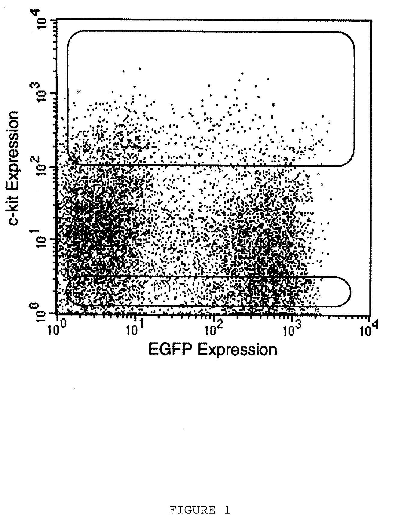
[0235]A. Harvesting of Hematopoietic Stem Cells

[0236]Bone marrow was harvested from the femurs and tibias of male transgenic mice expressing enhanced green fluorescent protein (EGFP). After surgical removal of the femurs and tibias, the muscle was dissected and the upper and lower surface of the bone was cut on the surface to allow the collecting buffer to infiltrate the bone marrow. The fluid containing buffer and cells was collected in tubes such as 1.5 ml Epindorf tubes. Bone marrow cells were suspended in PBS containing 5% fetal calf serum (FCS) and incubated on ice with rat anti-mouse monoclonal antibodies specific for the following hematopoietic lineages: CD4 and CD8 (T-lymphocytes), B-220 (B-lymphocytes), Mac-1 (macrophages), GR-1 (granulocytes) (Caltag Laboratories) and TER-119 (erythrocytes) (Pharmingen). Cells were then rinsed in PBS and incubated for 30 minutes with magnetic beads coated with goat anti-rat immuno...

example 2

Mobilization of Bone Marrow Cells to Repair Infarcted Myocardium

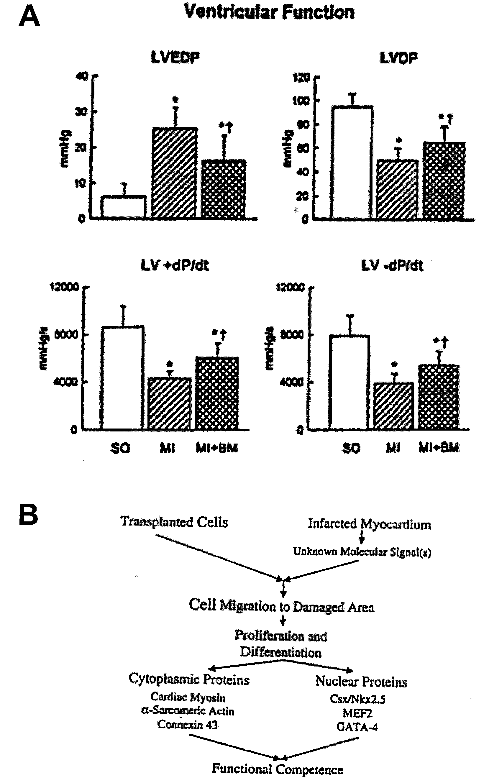
[0252]A. Myocardial Infarction and Cytokines.

[0253]Fifteen C57BL / 6 male mice at 2 months of age were splenectomized and 2 weeks later were injected subcutaneously with recombinant rat stem cell factor (SCF), 200 μg / kg / day, and recombinant human granulocyte colony stimulating factor (G-CSF), 50 μg / kg / day (Amgen), once a day for 5 days (Bodine et al., 1994; Orlic et al., 1993). Under ether anesthesia, the left ventricle (LV) was exposed and the coronary artery was ligated (Orlic et al., 2001; Li et al., 1997; Li et al., 1999). SCF and G-CSF were given for 3 more days. Controls consisted of splenectomized infarcted and sham-operated (SO) mice injected with saline. BrdU, 50 μg / kg body weight, was given once a day, for 13 days, before sacrifice; mice were killed at 27 days. Protocols were approved by New York Medical College. Results are mean±SD. Significance was determined by the Student's t test and Bonferroni method (Li e...

example 3

Migration of Primitive Cardiac Cells in the Adult Mouse Heart

[0270]To determine whether a population of primitive cells was present in the adult ventricular myocardium and whether these cells possessed the ability to migrate, three major growth factors were utilized as chemoattractants: hepatocyte growth factor (HGF), stem cell factor (SCF) and granulocyte monocyte colony stimulating factor (GM-CSF). SCF and GMCSF were selected because they have been shown to promote translocation of hematopoietic stem. cells. Although HGF induces migration of hematopoietic stem cells, this growth factor is largely implicated in mitosis, differentiation and migration of cardiac cell precursors during early cardiogenesis. On this basis, enzymatically dissociated cells from the mouse heart were separated according to their size. Methods for dissociating cardiac cells from heart tissue are well-known to those skilled in the art and therefore would not involve undue experimentation (Cf U.S. Pat. No. 6,2...


PUM

PropertyMeasurementUnit
volume fractionaaaaaaaaaa
volume percentaaaaaaaaaa

Abstract

Methods, compositions, and kits for repairing damaged myocardium and / or myocardial cells including the administration of cytokines are disclosed and claimed.

Description

RELATED APPLICATIONS / PATENT & INCORPORATION BY REFERENCE[0001]This application is a continuation application of U.S. patent application Ser. No. 10 / 162,796, filed Jun. 5, 2002, which is a continuation in part of U.S. patent application Ser. No. 09 / 919,732 filed Jul. 31, 2001, now abandoned, and which claims priority from Provisional U.S. Patent Application Ser. Nos. 60 / 295,807, 60 / 295,806, 60 / 295, 805, 60 / 295,804, and 60 / 295,803 filed Jun. 6, 2001, the disclosures of all of which are herein incorporated by reference in their entirety. This application claims only subject matter disclosed in the parent application and therefore presents no new matter.[0002]Each of the applications and patents cited in this text, including each of the foregoing cited applications, as well as each document or reference cited in each of the applications and patents (including during the prosecution of each issued patent; “application cited documents”), and each of the PCT and foreign applications or pat...

Claims


Application Information

Patent Timeline
Patent Type & Authority Applications(United States)
IPC IPC(8): A61K38/19A61K35/12A61K38/00A61K38/18A61K38/22A61K38/30A61P9/00A61P9/04C12N5/02C12N5/077C12N5/0789
CPCA61K38/18A61K38/1833A61K38/30A61K38/193A61K2300/00A61P21/00A61P43/00A61P9/00A61P9/04A61P9/10
Inventor ANVERSA, PIERO
Owner NEW YORK MEDICAL COLLEGE
Why Eureka
Try Eureka