
Centrifugal device and method for ova detection

a centrifugal device and ova technology, applied in the field of centrifugal device and method for ova detection, can solve the problems of low parasite population, inability to detect, difficult direct examination, etc., and achieve the effect of simple, rapid and highly accura

Inactive Publication Date: 2009-10-15
STATSPIN
View PDF48 Cites 18 Cited by
  • Summary
  • Abstract
  • Description
  • Claims
  • Application Information

AI Technical Summary

Benefits of technology

[0009]A centrifugal fecal analyzer device and centrifugal floatation method are provided for the separation of ova from human and animal fecal specimens and the subsequent collection of ova onto a microscope slide for microscopic examination. The device and method are simple, rapid and highly accurate and can be used on a routine basis in veterinary and clinical laboratories. The device and method overcome the limitations and problems of the prior art noted above.
[0010]More specifically, the centrifugal fecal analyzer device and method provide increased test efficiency and improved accuracy. The device and method allow rapid separation of ova from fecal matter and allow testing and diagnosis within a typical one-half hour patient appointment. The number of steps required in the analysis are limited, and only minimal handling of feces is required, allowing collection of parasitic ova without significant contamination of laboratory personnel, equipment or facilities. Collection of parasite ova can be accomplished with minimal interference from fecal debris and other buoyant material that may be contained in the fecal sample, such as vegetation.
[0013]In operation, a user adds flotation fluid into the rotor assembly. The user cores a fecal sample with the coring assembly and inserts the coring assembly into the opening in the rotor assembly. The user rotates and / or reciprocates the coring assembly to break up the sample and mix it with the flotation fluid. Optionally, the user may cap the device and shake it to further mix the sample. The user removes the detachable handle from the coring assembly, leaving the coring element in the rotor assembly. Sufficient fluid may be added at this stage to completely fill the rotor assembly. The rotor assembly is placed in a centrifuge and spun for an appropriate time, during which fecal matter and debris that are more dense than the flotation fluid are forced outwardly from the center of rotation into the sediment chamber, while the less dense ova are forced inwardly to the surface of the liquid in the mixing chamber. After centrifugation, the rotor assembly is removed from the centrifuge and placed on a level surface, and more flotation fluid is added, if necessary, through the central opening, or through a fill port if present, until a meniscus is created at the opening of the rotor assembly. A coverslip is placed over the rotor assembly opening and after a short period of time, as needed, the ova rise up in the flotation fluid and adhere to the coverslip. The coverslip can then be removed and transferred to a microscope slide for analysis using standard microscopy methods.
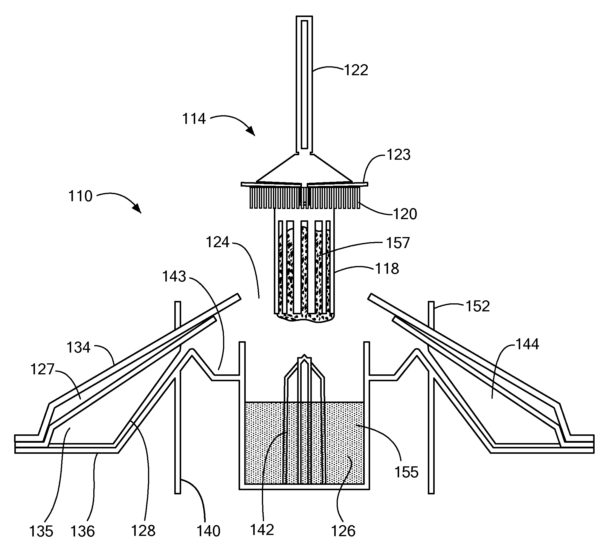
[0014]In another aspect of the present invention, a centrifugal device is provided in which the ova are delivered through centrifugation to a pipette tip for dispensing onto a microscope slide or coverslip. In this manner, the wait for ova to rise through a flotation fluid is also minimized.

Problems solved by technology

This technique is rarely used in modern laboratories because the presence of debris in the fecal sample makes direct examination extremely difficult and prone to error.
Also, the small sample size used makes it likely that a low population of parasites, such as during the early stages of an infestation, may not be detected.
This prior art process has been improved over the years, but still is not optimized and suffers from several limitations including the risk of exposure of laboratory personnel to potentially dangerous pathogens, complexity, unpleasant odor and also a degree of unreliability or inaccuracy.
While this latter technique proved to be more accurate and reliable, the requirement for a relatively large and costly centrifuge, the multiple transfer steps involved and the high potential for spills and aerosol generation limited the acceptance of the technique as a routine laboratory procedure.
While the FECALYZER and other similar devices such as the OVASSAY are widely used today, particularly in companion animal veterinary practices, the devices of this technique have several key deficiencies including a) the time required for complete floatation of ova to the surface of the liquid can be excessive thereby making it impossible to provide a diagnosis during a typical one-half hour patient appointment and b) without the benefit of prefiltration or pre-separation by centrifugation, the straining method of these devices under natural gravity may trap a portion of the ova thereby contributing to inaccuracy of the diagnosis.

Method used


Image

  • Centrifugal device and method for ova detection
  • Centrifugal device and method for ova detection
  • Centrifugal device and method for ova detection

Examples


Embodiment Construction

[0064]The disclosures of PCT International Application No. US2007 / 019639, filed Sep. 10, 2007, and U.S. Provisional Patent Application No. 60 / 843,347, filed Sep. 8, 2006, U.S. Provisional Patent Application No. 60 / 843,176, filed Sep. 8, 2006, and U.S. Provisional Patent Application No. 60 / 861,993, filed Nov. 30, 2006, are incorporated by reference herein.

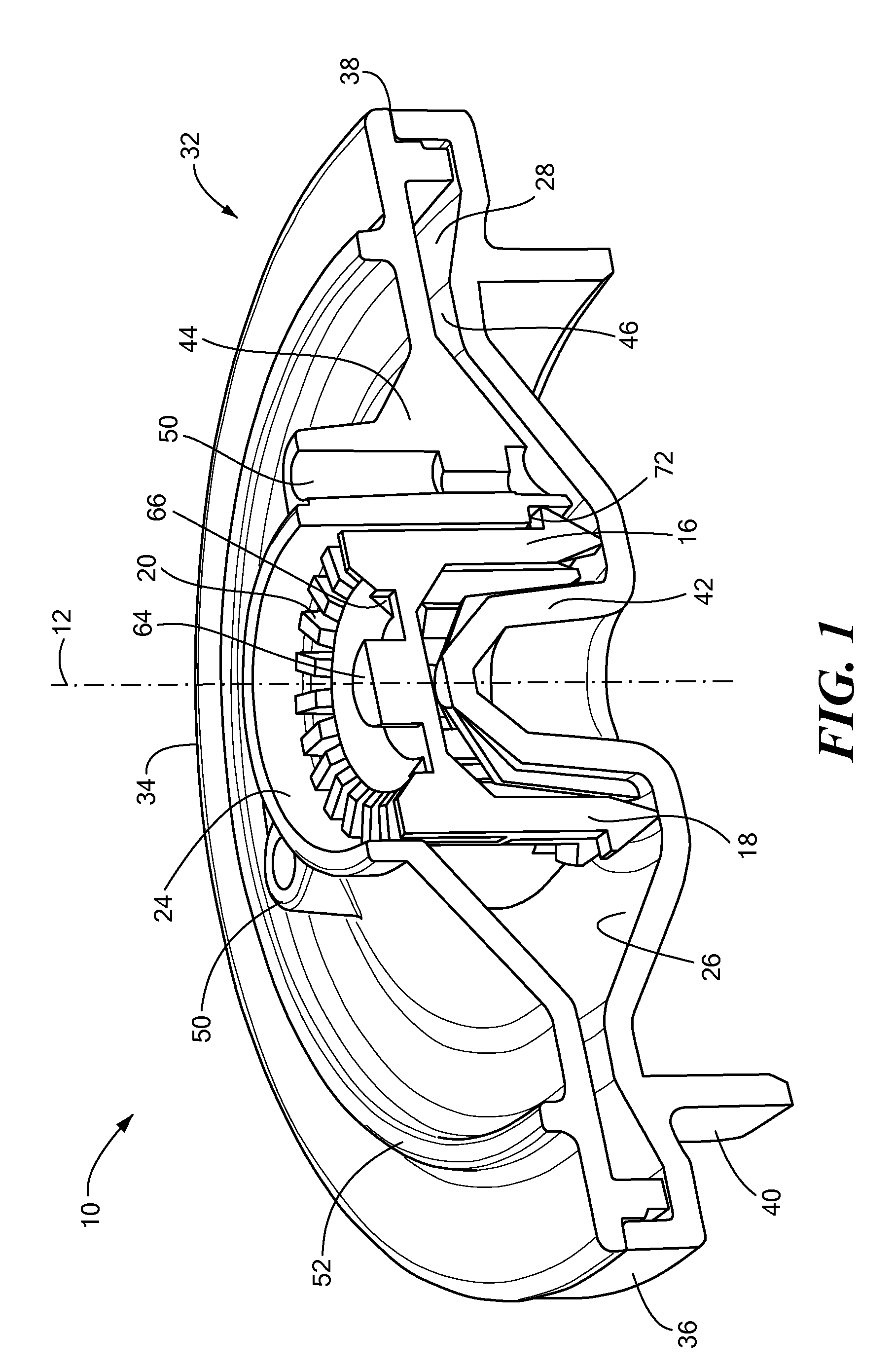
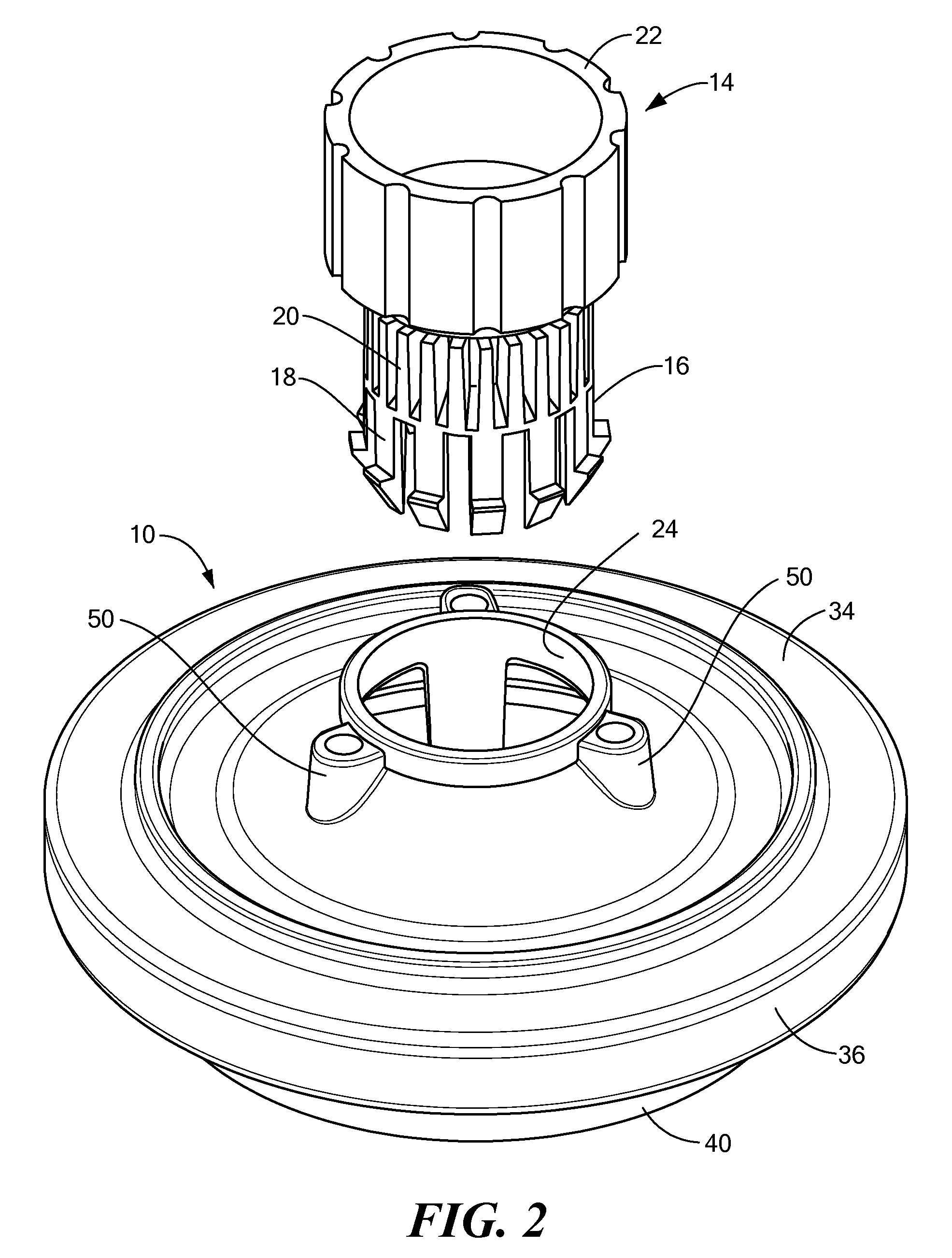
[0065]A first embodiment of a centrifugal device is illustrated in FIGS. 1-10. The device includes a rotor assembly 10 for rotation about its central axis 12 in a centrifuge (not shown) and a coring assembly 14. The coring assembly includes a lower portion 16 having a coring element 18 and optionally a filter element 20, and a removable upper portion or handle 22. The coring assembly is used to retrieve a fecal sample and insert it into the rotor assembly through an opening 24 in the top. The rotor assembly includes a mixing or receiving chamber 26, in which the fecal sample is initially mixed with a flotation fluid, and a sediment ...


PUM


Abstract

A centrifugal device and method are provided for the separation of buoyant material such as parasitic ova from fecal matter. A rotor assembly, rotatable about its central axis in a centrifuge, includes a housing with a centrally located top opening leading to a centrally located mixing chamber. An annular sediment chamber is provided, also coaxial about the central axis, connected by a passage with the mixing chamber. A coring assembly is used to retrieve and insert a fecal sample into the mixing chamber for mixing with a flotation fluid. During centrifugation, heavier fecal components pass radially outwardly to the sediment chamber while the ova collect on the inward surface of the flotation fluid. After centrifugation, more flotation fluid is added, if needed, until a meniscus forms at the top opening. A coverslip is placed over the top opening and the ova float to the surface of the fluid and adhere to the coverslip. The coverslip is removed and the ova detected using standard microscopy procedures. In another aspect, a centrifugal device is provided in which the ova are delivered through centrifugation to a pipette tip for dispensing onto a microscope slide or coverslip.

Description

CROSS REFERENCE TO RELATED APPLICATIONS[0001]This application is a continuation-in-part of and claims the benefit under 35 U.S.C. § 120 of PCT International Application No. US2007 / 019639, filed Sep. 10, 2007, which claims priority under 35 U.S.C. § 119(e) of U.S. Provisional Patent Application No. 60 / 843,347, filed Sep. 8, 2006, U.S. Provisional Patent Application No. 60 / 843,176, filed Sep. 8, 2006, and U.S. Provisional Patent Application No. 60 / 861,993, filed Nov. 30, 2006, the disclosures of all of which are incorporated by reference herein.STATEMENT REGARDING FEDERALLY SPONSORED RESEARCH OR DEVELOPMENT[0002]N / ABACKGROUND OF THE INVENTION[0003]It has long been recognized that analyzing fecal specimens for parasite ova by microscopy is a simple and effective method for identifying parasites afflicting a patient. This method is routinely used in clinical and veterinary laboratories around the world to identify specific parasites in fecal specimens from animals and humans so that the...

Claims


Application Information

Patent Timeline
IPC IPC(8): C12N1/02C12M1/00
CPCA61B10/0038B01L3/0272B01L3/5021B01L3/5029B01L2300/0681B04B2005/0478B04B5/0414B04B7/02B04B11/04B04B11/06B04B2005/0464B04B5/04
Inventor PETITHORY, HENRYKELLAND, JAMESSULLIVAN, KEVINMARTIN, LINDA
Owner STATSPIN
Learn More