Method for the analysis of tissue sections

a tissue section and analysis method technology, applied in the field of histologic classification of tissue sections, can solve the problem of not being able to achieve a classification of sufficient quality withou

Inactive Publication Date: 2009-11-26
BRUKER DALTONIK GMBH & CO KG
View PDF5 Cites 67 Cited by
  • Summary
  • Abstract
  • Description
  • Claims
  • Application Information

AI Technical Summary

Benefits of technology

[0022]The lower spatial resolution of the mass spectrometric image is balanced by the high molecular information content of the mass spectra. The mass spectrometric information can be a local mass spectrum assigned to the subarea and its surroundings, or differences between the local mass spectrum and mass spectra that are assigned to other subareas of the tissue section or to mass spectra from other sources of data (e.g., databases). Moreover, mass spectrometric information preferably may include the local mass spectrum being assigned to one or several classes by statistical analysis. To this end, the local mass spectrum is generally compared with mass spectra from other subareas of the tissue section and / or with mass spectra from other data sources. Suitable statistical analyses are, for example: support vector machines (SVM), genetic algorithms for cluster analysis, principal component analysis (PCA), decision trees, nearest neighbor classification (k-nearest neighbor (k-NN)) or neuronal networks (e.g., linear vector quantization (LVQ), neural gas (NG), self-organizing map (SOM), et cetera). The classes correspond to those of histologic classification and relate to the tissue types that occur in the tissue section, tissue differentiation, pathogens and disease statuses, and presence of foreign matter.
[0023]So-called peak lists, or reduced mass spectra, are also considered as mass spectra in this case. A peak list is a list of value pairs that are extracted from a measured raw spectrum and which each contain the mass and the signal strength of a signal peak in the measured raw spectrum. A reduced mass spectrum contains only the signals of one or more mass windows, which are usually specified by the user.
[0024]In order to histologically classify a tissue section with sufficient quality, it may be necessary to combine the relevant light-optical and mass spectrometric information from several subareas of the tissue section. The histologic classification can be limited to one region or several regions of the tissue section and can relate in particular to single cells or several single cells.
[0025]These and other objects, features and advantages of the present invention will become more apparent in light of the following detailed description of preferred embodiments thereof, as illustrated in the accompanying drawings.

Problems solved by technology

In contrast to the prior art, both types of information are therefore used for the histologic classification; in some cases, a classification of sufficient quality would not be possible without this combination.

Method used


Image

  • Method for the analysis of tissue sections
  • Method for the analysis of tissue sections
  • Method for the analysis of tissue sections

Examples


Embodiment Construction

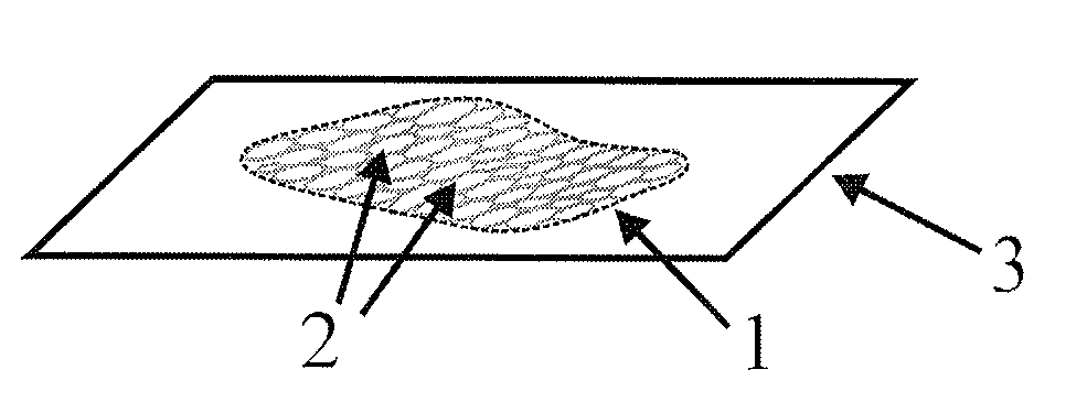
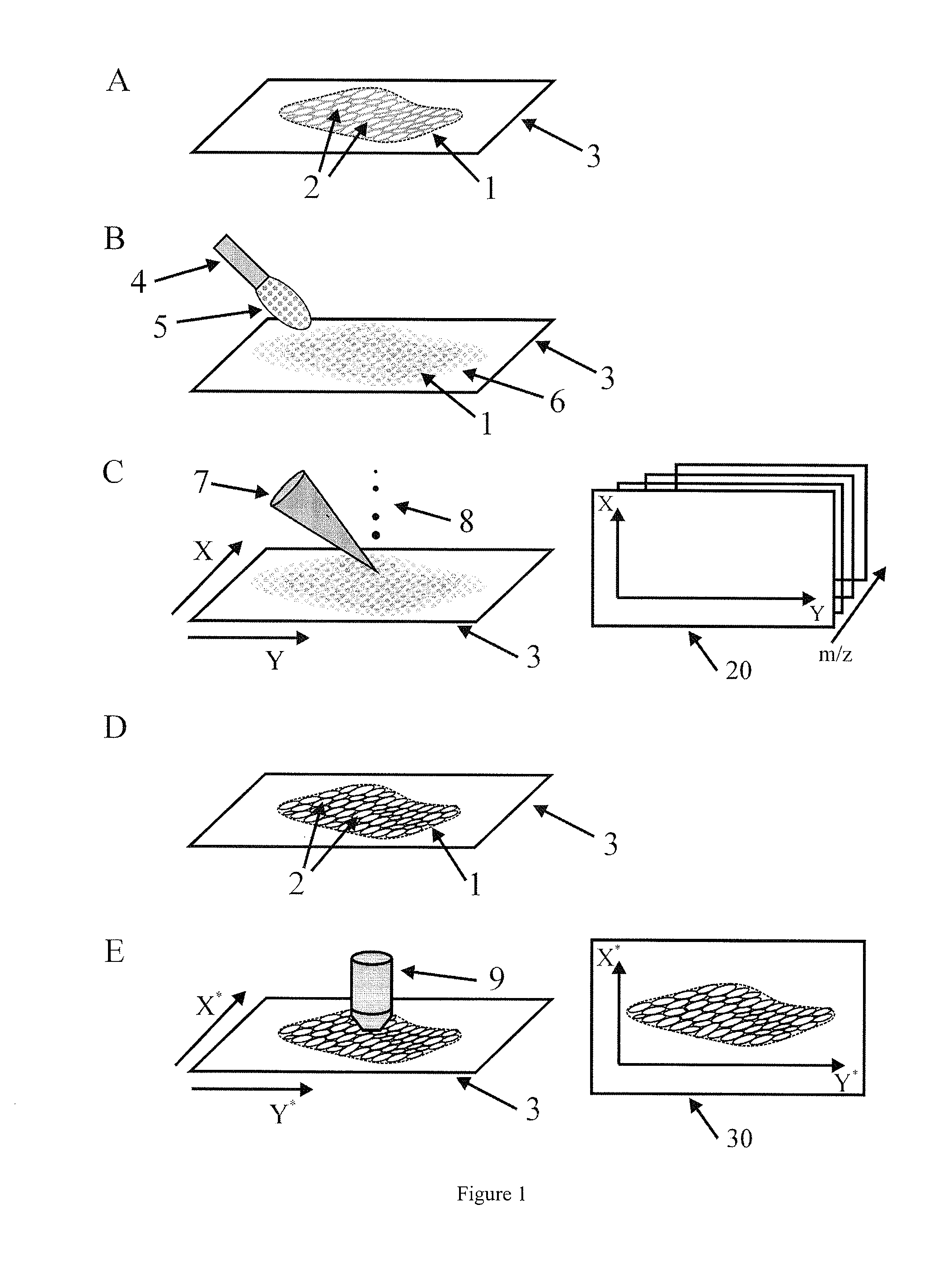
[0028]FIGS. 1 and 2 comprise steps A to G and show a preferred method for histologic classification of a tissue section.

[0029]In Step A, a tissue section 1 some ten micrometers thick is provided on a specimen slide 3. This involves first freezing a tissue sample to stabilize it before cutting it with a microtome.

[0030]In Step B, a matrix layer 6 is applied to the tissue section 1. A device 4 uses vibrations to produce a mist 5 of small droplets from a dissolved matrix substance; these droplets deposit on the tissue section 1 and start to dry. Such a device is disclosed in published U.S. patent application 2007 / 0278400, which is hereby incorporated by reference. Nebulization and subsequent partial drying of the matrix droplets on the tissue section 1 are repeated cyclically until the matrix layer 6 is in an optimum state for an imaging mass spectrometric analysis.

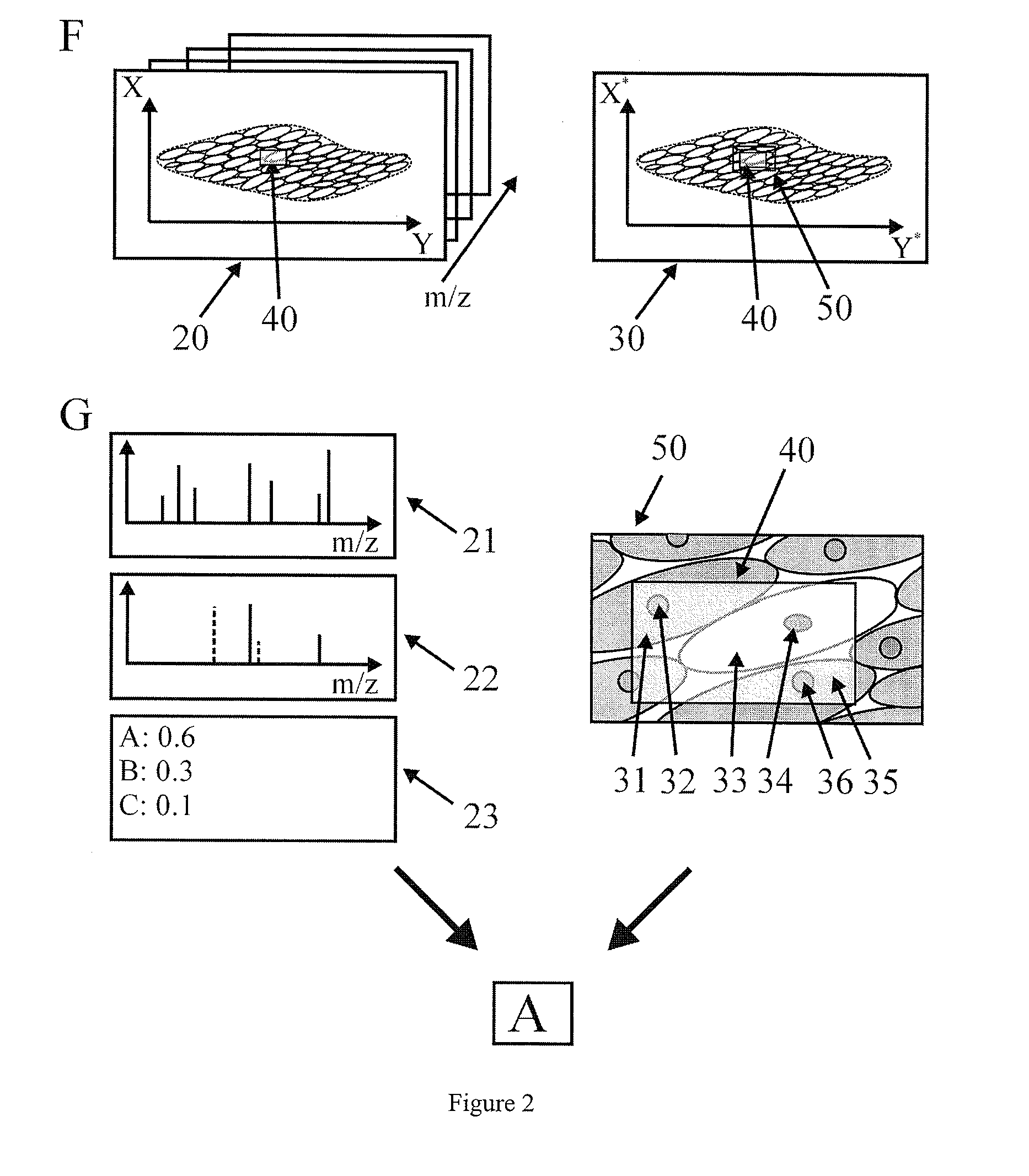
[0031]Step C involves taking a mass spectrometric image 20 of the tissue section 1 prepared in Step B. The tissue section ...


PUM


Abstract

The present invention relates to a method for the histologic classification of a tissue section. The method includes acquiring a mass spectrometric image and a light-optical image of the same tissue section (the optical image having a higher spatial resolution than the mass spectrometric image) and combining optical information on the structures of a subarea of the tissue section with mass spectrometric information on the subarea (the structures not being spatially resolved in the mass spectrometric image).

Description

PRIORITY INFORMATION[0001]This patent application claims priority from German patent application 10 2008 023 438.9 filed May 14, 2008, which is hereby incorporated by reference.FIELD OF THE INVENTION[0002]The present invention relates to a method for the histologic classification of tissue sections.BACKGROUND OF THE INVENTION[0003]Histology is the study of human, animal and plant tissues, in particular their structure and function. Histologic classification is generally carried out on a stained tissue section a few micrometers thick and concerns the tissue types present, tissue differentiation, bacterial and parasitic pathogens in the tissue, disease statuses of the tissue, and content of foreign matter like pesticides or drugs and their metabolites. The classification can be limited to one or more subareas of a tissue section, or can even apply to only one or more individual cells. The disease statuses of human tissue concern inflammatory disorders, metabolic diseases and the detec...

Claims


Application Information

Patent Timeline
Patent Type & Authority Applications(United States)
IPC IPC(8): B01D59/44H01J49/26
CPCG06T7/0012H01J49/0004G06T2207/30024G06V20/698G06V2201/03G06F2218/08
Inventor DEININGER, SOREN-OLIVERWALCH, AXEL
Owner BRUKER DALTONIK GMBH & CO KG
Why Eureka
Try Eureka