Free-hand three-dimensional ultrasound diagnostic imaging with position and angle determination sensors

a three-dimensional ultrasound and position and angle determination technology, applied in the field of ultrasonic imaging, can solve the problems of inability to consider quantitative imaging tools, inability to use free-hand scanning as a reliable technique, and high operator training requirements, and achieves improved ultrasound scanning accuracy, low cost, and simple construction and operation.

Inactive Publication Date: 2009-12-10
TRUSTEES OF BOSTON UNIV +1
View PDF40 Cites 131 Cited by
  • Summary
  • Abstract
  • Description
  • Claims
  • Application Information

AI Technical Summary

Benefits of technology

[0018]The present invention seeks to provide a free-hand, registration system for ultrasonic imaging, which is characterized by simplicity of construction and operation and relatively low cost. The system may be implemented in original equipment or as a retrofit to existing equipment having only two-dimensional (2D) imaging capabilities. Position tags (the term “position tag” is used inclusively herein to include position data and, where appropriate, orientation / angle data) associated with 2D image planes are computed from a variety of sensor configurations, all of which may be output to ultrasound image display programs for volumetric rendering by known interpolation techniques which typically form a sequence of ultrasound image planes with equal spacing and fixed lateral positioning or other suitable geometries for interpolation. The invention, thus, permits improved ultrasound scanning accuracy by reducing or eliminating variations in the scanning process introduced by a number of factors, including non-uniform scanning by a user, as well as sensor-dependent errors due to manufacturing variation, drift and hysteresis.
[0022]The ultrasound imaging registration systems and methods described are unique relative to registration methods presently available, in that the position and angle sensors acquire their respective data without the assistance of external position or orientation references (i.e., the data sensing is internal to the transducer probe, eliminating the need of some existing systems to perform triangulation with external sources.)
[0026]The position sensor(s) are of a type that acquires data along a single or multiple axes, including, but not limited to, optical sensors, self-contained electromagnetic sensors, and capacitive MEMS devices. In a preferred embodiment the position sensor comprises one or more light source(s) for illuminating the region of interest with sufficient intensity such that light reflects from the region of interest, an optical imaging means including at least one lens disposed in or upon the probe, so as to receive light reflected from the region of interest in the form of an optical image, and a light-sensitive image capture device for converting the optical image output from the lens into said position signal such as, for example a charge coupled device camera and digital signal processor. The light may be coupled to the image capture device through an appropriately designed optical fiber bundle. Several alternative designs of such an optical sensor will be described below. By optically acquiring images of the surface of a region of interest, and thus information regarding the position of the transducer probe relative to the region of interest or, alternatively stated, to reference position, the acquisition of positional information is much less sensitive to noise occurring during movement of the transducer probe. The optical path between the scanned skin surface and the unit in the transducer probe is relatively short and is not easily disturbed. This enhances the accuracy of the detected position of the transducer probe and thus also the quality of the three-dimensional ultrasound image resulting from a composition of two-dimensional slices based on said positional information.

Problems solved by technology

This technique is widely used (such as Sonocubic for Terason), but it requires much operator training and cannot even in such cases be considered a quantitative imaging tool.
Therefore, free-hand scanning is not a reliable technique for the above mentioned applications.
Motor drives must be included within the transducer design, and consequently increase the size of the handle and cost of the probe and require motor driver power and software.
This approach is a quantitative imaging technique, but with several limitations, such as not permitting Doppler imaging, not allowing 4D imaging (real time 3D ultrasound), and typically imaging only a small volume.
Such sensing methods require expensive equipment external to the sensing device for triangulation purposes; these can cause electromagnetic interference with other medical equipment commonly found in hospitals and clinics.
A further disadvantage of these sensor types is the fact that the scanning room must have these sensors installed and the system calibrated, before actual scanning can occur.
However, sparse two-dimensional transducer arrays have reduced resolution due to the reduced number of array elements.
Also, cost of two-dimensional array transducers is another limiting factor along with the small volume that can be imaged (same limitation as the mechanically vibrated transducer).
The method is computationally demanding, cannot work with non-parallel scan planes, and cannot differentiate movement to the left from movement to the right.
Accurate surface rendering and volume rendering are very difficult to achieve with free-hand scanning even by skilled operators.
As this scanning requirement seldom is met, the result of the reconstruction is distorted.
High quality results for these applications cannot be easily achieved with free hand 3D ultrasound with known techniques.

Method used


Image

  • Free-hand three-dimensional ultrasound diagnostic imaging with position and angle determination sensors
  • Free-hand three-dimensional ultrasound diagnostic imaging with position and angle determination sensors
  • Free-hand three-dimensional ultrasound diagnostic imaging with position and angle determination sensors

Examples


Embodiment Construction

Overview

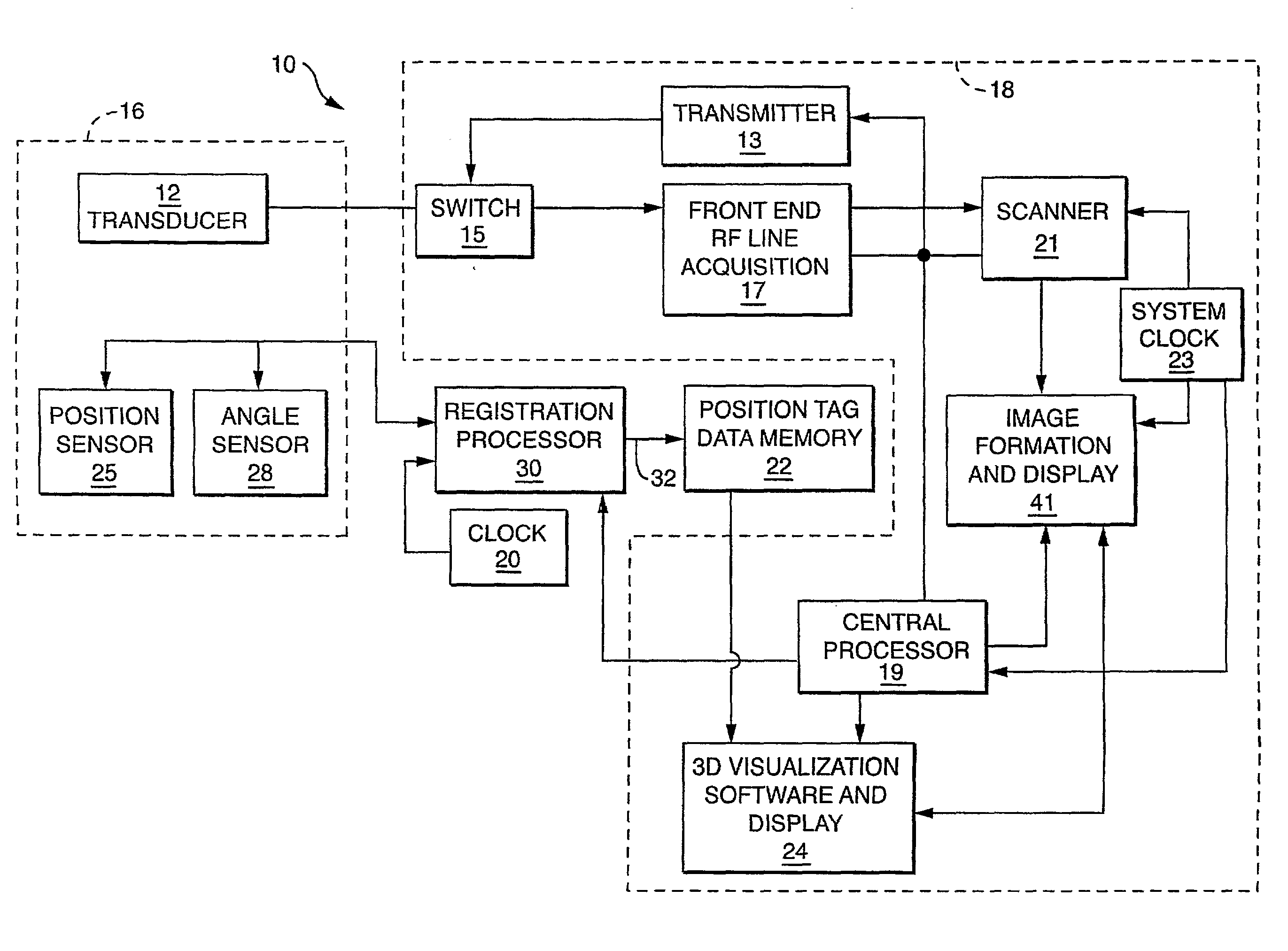
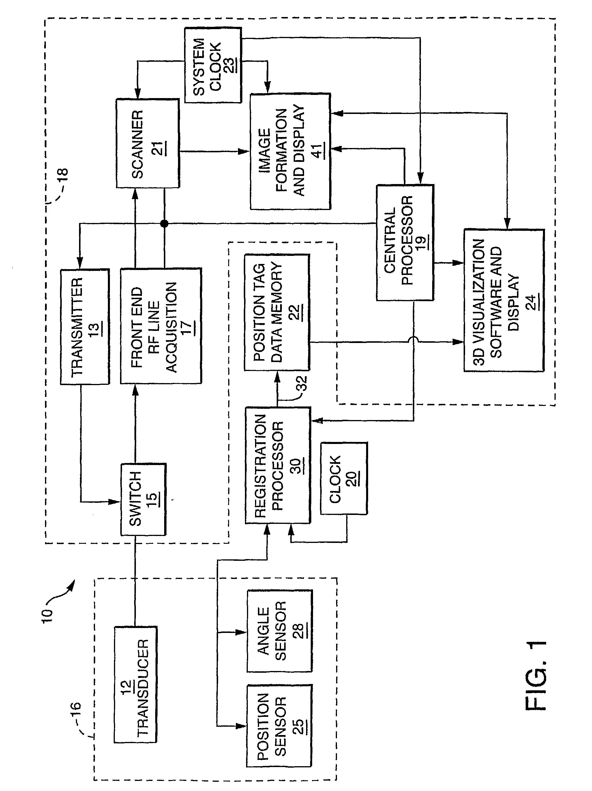
[0034]FIG. 1 shows a free-hand ultrasound medical diagnostic imaging system 10 within which is a first embodiment of an ultrasound registration system. An ultrasound imaging system sends excitation signals through a transmitter 13 through a switch 15 to the transducer 12 operatively disposed in a probe housing 16. The ultrasound array transducer 12 detects response echoes from a region of interest within a patient's anatomy. The imaging system receives echoes from the transducer 12 through the switch 15 that routes the signals to a front end 17 from where they are sent by a central processor 19 in synchronization with a system clock 23 to a scanner 21. From the scanner 21, processed signals are sent to the image formation and display section 41 from which 2D image frames are formed in synchronism with the system clock 23. The registration system includes, preferably, a system clock 20, memory 22 for storing position tags (described below) associated with each 2D ultrasound i...


PUM


Abstract

A freehand 3-D imaging system includes an integrated sensor configuration that provides position and orientation of each 2D imaging plane used for 3-D reconstruction without the need for external references. The position sensors communicate with the imaging system using either wired and wireless means. At least one translational and one angular sensor or three translational sensors acquire data utilized to compute position tags associated with 2D ultrasound image scan frames. The sensors can be built into the ultrasound transducer or can be reversibly connected and therefore retrofitted to existing imaging probes for freehand 3D imaging.

Description

STATEMENT REGARDING FEDERALLY SPONSORED RESEARCH OR DEVELOPMENT[0001]This invention was made with Government support from the U.S. Army Medical Research Acquisition Activity under Contract No. DAMD17-03-2-0006. The Government has certain rights in the invention.BACKGROUND OF THE INVENTION[0002]The present invention relates to ultrasonic imaging generally and more particularly to three-dimensional ultrasonic imaging using conventional two-dimensional ultrasonic imaging apparatus.[0003]Over the last decade, 3D medical imaging has been playing an increasingly important role, in particular in computerized tomography (CT) and magnetic resonance imaging (MRI). The 3D reconstruction ability with these modalities has also improved over the same period of time. Given the method of CT and MRI scanning, the position of scan planes has been well defined. 3D ultrasound is now also finding widespread interest, where the most prominent specialty for 3D medical ultrasound imaging is in obstetrics, ...

Claims


Application Information

Patent Timeline
Patent Type & Authority Applications(United States)
IPC IPC(8): A61B8/14
CPCA61B2019/5276A61B2019/5295G01S15/8936G01S15/8993A61B2019/5255A61B8/483A61B19/5244A61B2019/467A61B2019/5248A61B8/14A61B8/4227A61B8/4254A61B34/20A61B2034/2048A61B2034/2055A61B2090/067A61B2090/367A61B2090/378
Inventor PEDERSEN, PEDER C.SZABO, THOMAS L.
Owner TRUSTEES OF BOSTON UNIV
Why Eureka
Try Eureka