
Kerf cranial closure methods and device

a cranial closure and kerf technology, applied in the field of cranial closure improvements, can solve the problems of bone flap healing, never healing, palpable gap in the bone, etc., and achieve the effects of promoting bone growth and ingrowth, promoting osteogenesis and osteoinduction, and increasing the density of the strip

Inactive Publication Date: 2010-02-11
ALLOSOURCE
View PDF30 Cites 14 Cited by
  • Summary
  • Abstract
  • Description
  • Claims
  • Application Information

AI Technical Summary

Benefits of technology

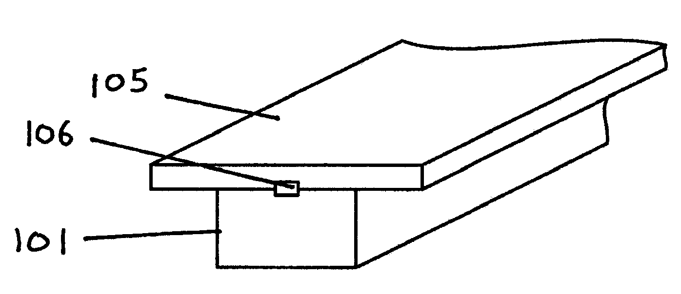
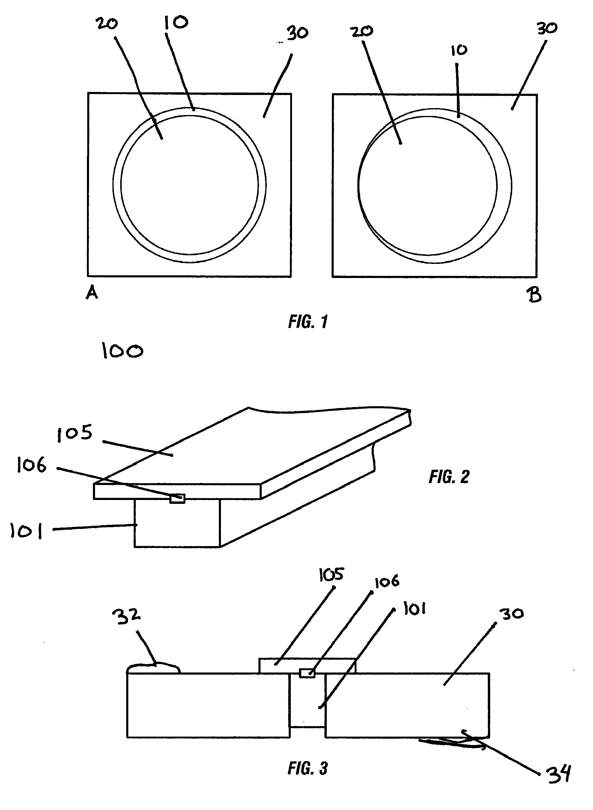
[0007]The present disclosure generally comprises a device, methods for use, and kits including a device used in craniotomies comprising strips alone or strips and / or plugs used to assist with improved cranial closure. The device features a strip for laying into the kerf (gap) left by a craniotome blade; and optionally a plug for filling into a bur hole made by a drill in the craniotomy process. Embodiments of the device feature a strip or plug which leaves a substantially smooth contour with an outer surface of a cranium; wherein said strip or plug is secured by compression forces which reduce the tendency of the strips or plugs to fall into the craniotomy towards the dura or brain.
[0012]An additional embodiment features a device for closing about a 2 mm to about a 5 mm gap wide and about 3 mm to about 1 cm deep in the cranial bone of a subject wherein the strip or plug is about 2 mm to 12 mm wide when in an uncompressed state and capable of being compressed to fill the kerf; where said material is sufficiently elastic to decompress after being compressed to be placed in the gap so that said material expands to the width of the gap and results in a substantially secure placement of said material within said gap.
[0014]An additional embodiment features a kit for treating a cranial gap associated with a craniotomy in a subject comprising: a preparation of demineralized or partially demineralized bone or bone substitute; wherein said preparation is formed into a malleable strip. The kit also comprises a vial of infusion materials to be added to the strip comprising at least one of the following; a paste, gel, or other moldable or pourable liquid for the purpose of hardening the device into a solid matrix to create a hard surface or a watertight seal calcium-based materials (such as tricalcium phosphate) or demineralized bone matrix to increase the density of the strip and / or provide substrate for further bone growth; blood, blood derivative products, kerf bone, marrow, or stem cells used to promote osteogenesis and osteoinduction; biological growth factors in order to promote bone growth and ingrowth, such as via osteogenesis, osteoconduction, and / or osteoinduction; antibiotics, antibacterial agents and / or antiseptic agents in order to prevent bone flap infection.

Problems solved by technology

The bone flap heals in most cases of benign disease, but may never heal in cases where radiation is administered to the healing bone.
In either case, though the bone may be solidly attached at its edges, there is often a palpable gap in the bone which may be visible below the scalp.
Because many craniotomies are performed below the hairline, this often results in gross external deformity.
Even for craniotomies located off of the forehead, the palpable or visible deformity (particularly for patients who do not have covering hair) is often distressing to the patient.
Unfortunately, a suitable device for assisting cranial reconstruction and decreasing cranial deformities has not yet been described.

Method used


Image

  • Kerf cranial closure methods and device
  • Kerf cranial closure methods and device
  • Kerf cranial closure methods and device

Examples


example 1

[0065]Kerf Cranial Closure Device Design Variations when Used in Strips

[0066]The kerf cranial closure device contemplated will feature many of the following properties which may optimize cranial closure performance.

[0067]1) The design is intended to specifically close the bony defect made in the skull by any the of the common commercially available craniotomes, known as the kerf;

[0068]2) It should compress into a kerf defect;

[0069]3) The bottom side should be narrower or less dense than the top side to ease its introduction into the kerf;

[0070]4) The bottom side in its uncompressed dimensions should be slightly greater than, equal to, or less than the width of the kerf to allow it to be introduced easily into the defect to be filled;

[0071]5) It should be of a compressed width of 1-4.5 mm at the top side, 1-4 mm on the bottom side;

[0072]6) The shape is tapered with a cross-section that is wedge-shaped, trapezoidal, keel, or bullet-shaped, with the narrower end positioned towards the ...

example 2

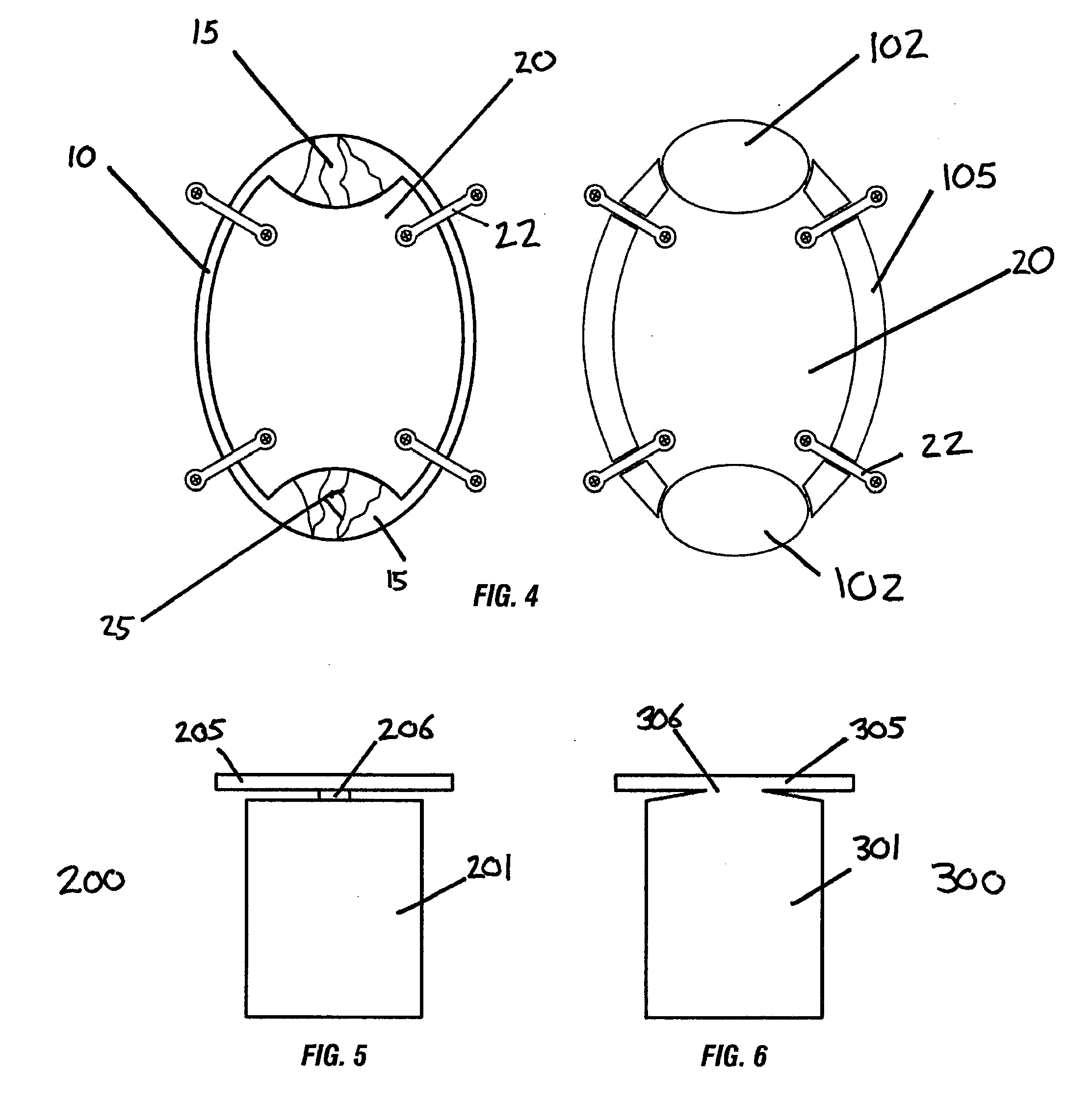
[0082]The kerf cranial closure device contemplated may feature a bur hole plug with many of the following properties which may optimize cranial closure performance.

[0083]1) The design of the device is intended specifically to close the bony defect made in the skull, known as a bur hole (whether round, rectangular, or square), by any of the common commercially available craniotomy drills or craniotome perforators,

[0084]2) The device should compress into the bur hole defect;

[0085]4) The bottom diameter should be smaller or less dense than the top side to ease its introduction into the bur hole;

[0086]5) The bottom side in its uncompressed dimensions should be slightly more than, equal to, or less than the diameter of the bur hole to allow it to be introduced easily into the defect to be filled;

[0087]6) The device should be of a compressed width of diameter of about 10-16 mm at the top side, and 8-15.5 mm on the bottom side;

[0088]7) The shape is tapered with a cross-section that is wedg...

example 3

[0094]Exemplary Method of Using a Cranial Closure Device Such as the CranioFuse™ Cranial Closure Device.

Cranial Defect

[0095]In the creation of a craniotomy, the bone is opened from its external surface to the level of the dura by placement of one or more bur holes, made either freehand with a high-speed drill or with a cranial perforator. The bur holes are connected with a high speed drill router (craniotome footplate attachment), which creates a trough in the bone, known as the kerf.

Closure of Cranium

[0096]At the conclusion of the intracranial part of the operation, the free bone flap is secured to the surrounding cranium with a fixation device. Typically this consists of titanium plates and screws (various manufacturers, e.g. Medtronic, Integra, Codman, Innovasis, Aesculap, W. Lorenz, etc. . . . ) or a disk / post device (Rapid Flap, CranioFix, others).

Application of CranioFuse™ Cranial Closure Device

[0097]The kerf is filled with a sufficient number of CranioFuse™ Cranial Closure De...


PUM


Abstract

The present disclosure is for a device for filling the gap (kerf) left in the repair of a craniotomy and the methods for using and manufacturing such a device. The kerf device may be a preparation of demineralized or partially demineralized bone or bone substitute formed into a malleable strip that can be pressed or molded into the opening in between the skull and bone flap in order to allow bone healing without a gap or indentation.

Description

CROSS REFERENCE TO RELATED APPLICATION(S)[0001]This application claims the benefit of priority under 35 U.S.C. §119(e) of U.S. Ser. No. 61 / 086,764 filed Aug. 6, 2008 and of U.S. Ser. No. 61 / 175,449 filed May 4, 2009 the entire content of both is incorporated herein by reference.BACKGROUND OF THE INVENTION[0002]1. Field of the Invention[0003]The invention relates generally to cranial closure improvements and more specifically to devices and methods used to improve cranial healing and reconstruction and the decrease in palpable or visible deformities often present after a craniotomy.[0004]2. Background Information[0005]Craniotomy is a common operation in the United States. It is performed for a variety of indications, including head trauma, aneurysm repair, and tumor removal, among others. Most craniotomies are performed by drilling one or more bur holes in the skull down to the level of the dura covering the brain and connecting them with a routing bit on a high-speed drill. The bit ...

Claims


Application Information

Patent Timeline
Patent Type & Authority Applications(United States)
IPC IPC(8): A61B17/08
CPCA61B17/688A61B2017/00889A61B2017/00946A61B2017/00933A61B2017/00893A61B17/00A61B17/68A61F2/28A61M37/00
Inventor NAKAJI, PETER
Owner ALLOSOURCE
Learn More