

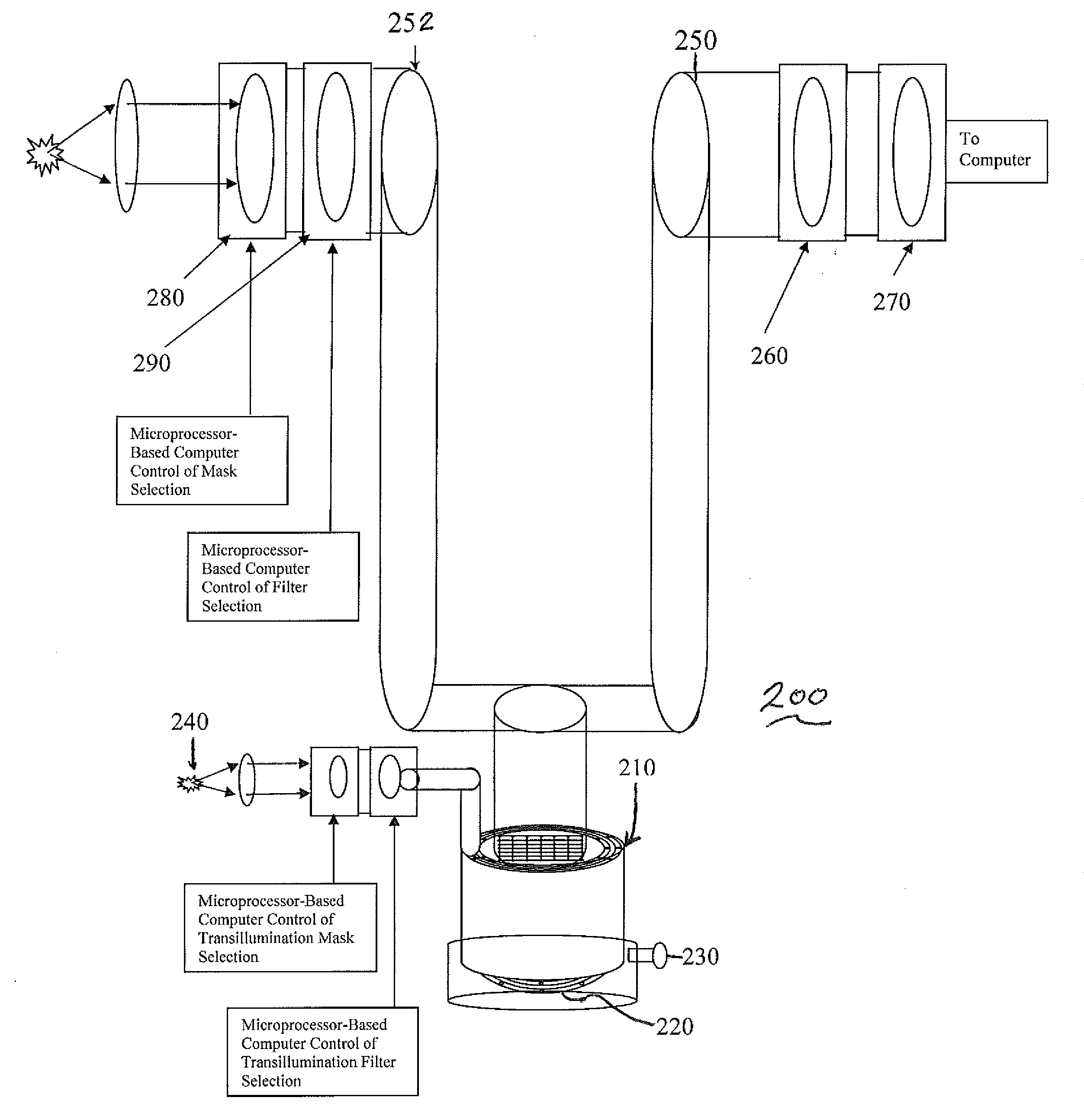
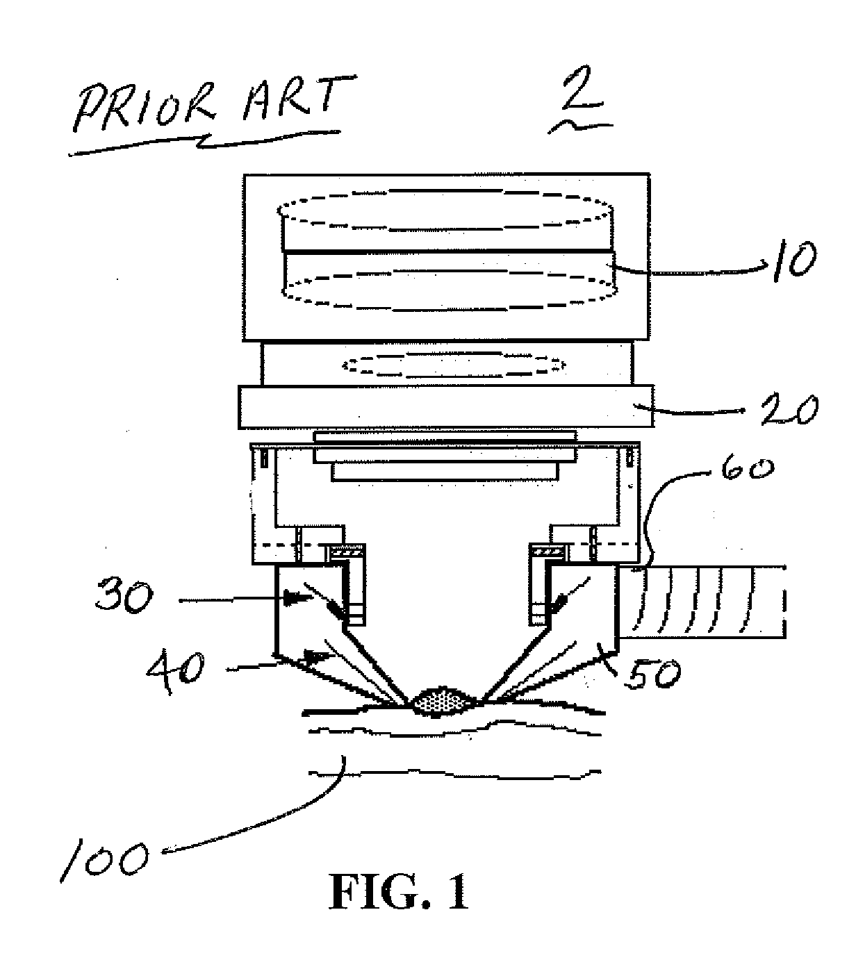
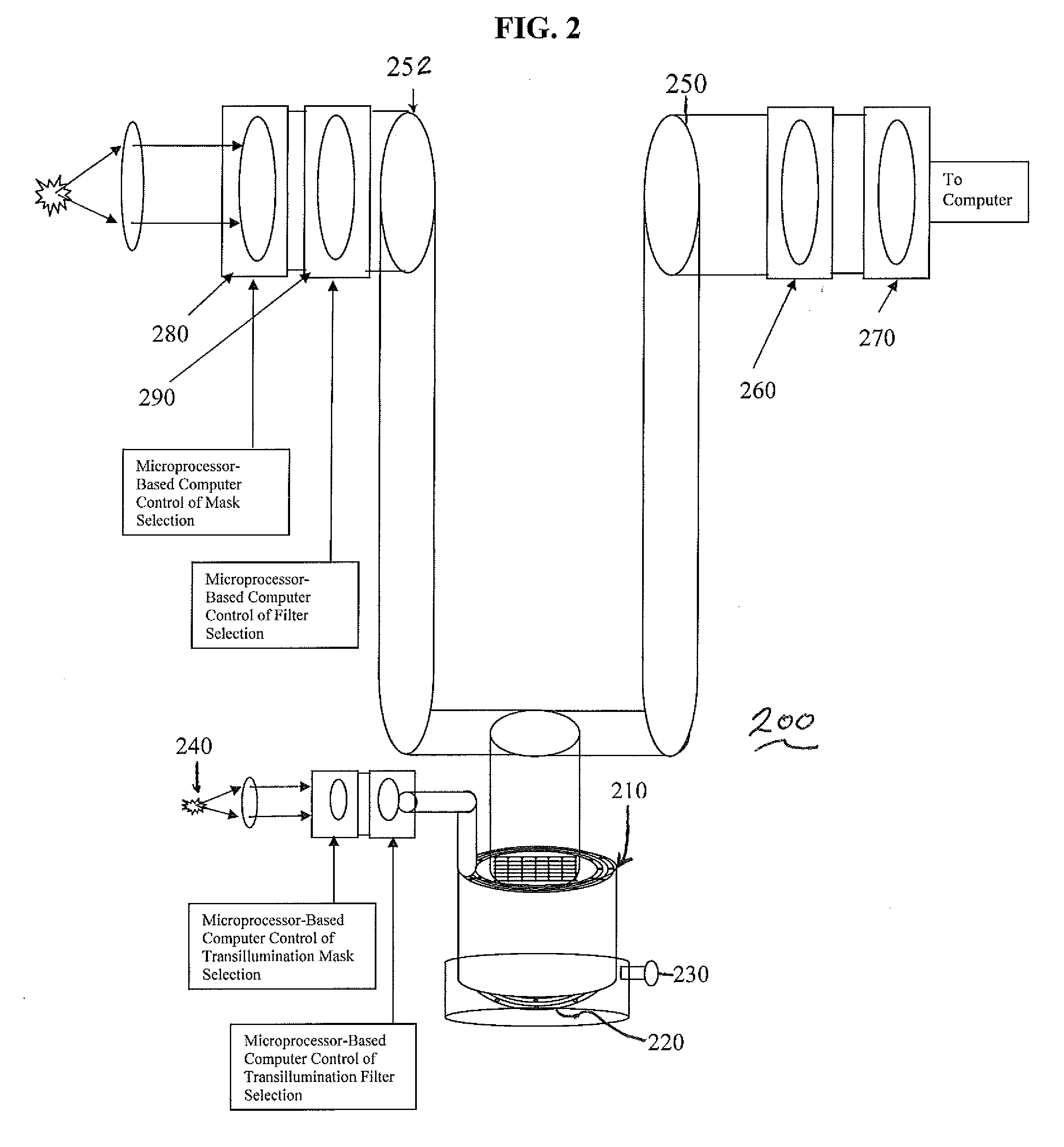
Method and Apparatus for Multi-spectral Imaging and Analysis of Skin Lesions and Biological Tissues
a multi-spectral imaging and analysis technology, applied in the field of methods and apparatus for imaging and analysis of skin lesions and biological tissues, can solve the problems of high mortality of malignant melanoma, inability to accurately characterize skin lesions, and inability to accurately identify malignant melanoma, so as to improve the characterization of skin
- Summary
- Abstract
- Description
- Claims
- Application Information
AI Technical Summary
Benefits of technology
Problems solved by technology
Method used
Image

Examples
example
[0120]It is assumed a skin lesion property (such as melanin, hemoglobin, or de-oxy-hemoglobin concentration) is represented by Fi(x,y,z); i=1, . . . , I where I is the number of skin-lesion properties. It is also assumed that the Monte-Carlo simulation and iterative ART-based reconstruction of skin provides the updated initial weight matrix with 3-D distributions of photon energy density represented by Ej(α,β,γ) with path lengths Pk,i,j((αl,βm,γn)0, Ej;lmn), (αl,βm,γn)1, Ej;lmn), . . . , (αl,βm,γn)Z, Ej;lmn)); k=1, . . . , K where K is the number of paths between source and detector for a specific imaging wavelength j (j=1, . . . , J) for imaging the skin-lesion property Fi(x,y,z). Each path length is a list of voxel coordinates (αl,βm,γn)z; z=1, . . . , Z (where Z is the number of voxels between source and detector) with their respective associated energy level Ej;lmn. The normalized probability distribution of photon energy provides initial weights for reconstructing the object in...
PUM
 Abstract
Description
Claims
Application Information