Method for screening for vitamin d receptor ligands

a technology of vitamin d receptor and ligand, which is applied in the field of screening for compounds participating in osteoblast differentiation, can solve the problems of unclear mechanism of bone mass increase mediated by vitamin dsub>3/sub>derivatives, poor effect of increasing the bone density of lumbar vertebrae or collum femoris, etc., and achieves increased osteoblast differentiation, increased vdr ligand-dependent transcription activity, and enhanced activity

Inactive Publication Date: 2010-03-18
THE UNIV OF TOKYO +1
View PDF2 Cites 0 Cited by
  • Summary
  • Abstract
  • Description
  • Claims
  • Application Information

AI Technical Summary

Benefits of technology

[0106]In order to examine the induction of osteoblast differentiation by 1,25 (OH)2D3 when CDP is expressed by HOS cells, pSPORT6-CDP was transfected to HOS cells by using Lipofectamin2000 (Invitrogen) as a reagent. When the cells reached confluence and after the incubation of 24 hours, culturing was performed in Minimum Essential Medium Alpha Medium, 10% fetal bovine serum, 50 μg / mL ascorbic acid, 1,25 (OH)2D3 at various concentrations or in the presence of the vehicle (ethanol). After three days, cells were recovered, and total RNA was extracted using an RNeasy Mini Kit (QIAGEN, Product No. 74104). Reverse transcription reactions were performed by oligo dT primers using SuperScriptlll RT (Invitrogen, 18080-044) and the amount of expression of various genes was determined using real-time RT-PCR (Applied Biosystems, GeneAmp7000). The amount of gene expression was corrected using GAPDH which is a housekeeping gene and shown as a relative figure of GAPDH and the amount of respective gene expression. The formula used is the same as that used in Example 5.
[0107]In the group into which CDP was introduced, gene expression of alkaline phosphatase (ALP) (FIG. 7B) known as a marker for osteoblast differentiation and osteocalcin (FIG. 7C) was increased in addition to CYP24 (FIG. 7A) resulting from 1,25 (OH)2D3 stimulation. This results suggest that CDP increases osteoblast differentiation by 1,25 (OH)2D3 stimulation.
[0108]In order to determine whether or not CDP expression increases VDR ligand-dependent transcription activity, a dual luciferase assay was performed using transient transfection in the same manner as Example 6. The reporter vector used in the dual luciferase assay was a pGL4 vector containing a sequence of human CYP24 gene promoter or a sequence of a human osteocalcin gene promoter upstream of the luciferase gene. The pGL4 vector was commercially obtained from Promega. Furthermore, a DNA sequence encoding a region of 4-427 amino acid residues for human VDR was introduced into a pTracerCMV2 vector (Invitrogen) in order to construct a human VDR expression plasmid. The expression vector for intracellular expression of the CDP protein was the pSPORT6-CDP vector used in Example 4. HOS cells used in the assay were cultured in DMEM medium containing 10% FBS and were passaged every two or three days. The culturing conditions were 37° C., 5% CO2. DMEM medium containing 10% DCC-FBS (charcoal processed FBS) but not containing phenol red was used in culturing of the HOS cells during the assay. A cell suspension was prepared so that the concentration of HOS cells in a 48 well plate was 8×10⁴ cells / 0.25 mL / well. A mixture was prepared using Lipofectamin 2000 (Invitrogen) as a reagent in order to perform transfection of reporter vectors, various types of expression vectors. The mixture was mixed with the cell suspension and cells in 0.25 mL lots were placed into each well. After two hours, this was replaced by Vehicle (ethanol) or DMEM medium containing 10% DCC-FBS (charcoal processed FBS) containing 1,25 (OH)2D3 (10−8M) but not containing phenol red. The medium was removed after 24 hours and after washing once in PBS(−), luciferase activity was determined by a Dual-Luciferase Assay kit (Promega) using a luminometer (Bethold Japan). The results were expressed as a “transcription activation” being the value represented by (firefly luciferase activity) / (renilla luciferase activity). In FIG. 8A, VDR-mediated transcription activation of a human CYP24 promoter increases in the presence of 1,25 (OH)2D3 (10−8M) and that activity is further increased when CDP is expressed. In the same manner, in FIG. 8B, VDR-mediated transcription activation of a human osteocalcin promoter increases in the presence of 1,25 (OH)2D3 (10−8M) and that activity is further increased when CDP is expressed. These results show that VDR-mediated transcription activation is also increased in CYP24 or an osteocalcin promoter which is a naturally occurring VDR target gene. However, in FIG. 8B, the assay is performed using VDR present in the HOS cell without transfecting a VDR expression vector.
[0109]In order to examine whether or not CDP actually binds to VDR in the cell in the presence of 1,25 (OH)2D3, firstly, A FLAG-human VDR(FLAG-hVDR) expression plasmid was constructed by introducing a DNA sequence formed by connecting a DNA sequence encoding a FLAG tag sequence to the N terminal of a DNA sequence encoding human VDR into a pcDNA3 vector (Invitrogen). A plasmid containing one or both of pSPORT6-CDP and FLAG-hVDR was transfected into HOS cells using Lipofectamin2000 (Invitrogen) as a reagent (the samples in FIG. 9 into which the expression plasmid has been genetically introduced are marked as “+” and those samples without any genetic introduced material are marked “−”). After 24 hours, culturing was performed for 24 hours in the presence of DMEM medium containing 10% FBS and including 1,25 (OH)2D3 at various concentrations or Vehicle (ethanol). Thereafter, the cells were washed in PBS, removed using a scraper and collected by centrifugation. The cells were dissolved in TNE buffer (20 mM HEPES (pH=7.9), 150 mM NaCl, 1% NP40, 1 mM EDTA), centrifuged for 30 minutes at 15000×g at 4° C. and the supernatant used as a cell lysate. Anti-FLAG (registered Trademark) M2 resin (SIGMA, Product No. A2220) was added to the lysate and mixed at 4° C. for three hours in the presence and in the absence of 1,25 (OH)2D3. Thereafter, the resin was washed in TNE buffer three times, SDS sample buffer added to the resin and SDS-PAGE performed. The method of performing SDS-PAGE and western blotting is the same as that in Example 3. The resulting sample was subjected to SDS-PAGE and CDP and FLAG-VDR were detected using western blotting. The primary antibody against the FLAG peptide was anti-FLAG rabbit IgG (SIGMA) (Product No. F7425). Anti-CDP goat IgG (C-20) (Santa Cruz Biotechnology, Product No. sc-6327) was used to detect the CDP protein. The results are shown in FIG. 9. The “Input” for Lanes 1-3 shows the result for the cell lysate prior to mixing with anti-FLAG antibody resin which was used in SDS-PAGE. The “IP-FLAG” used in Lanes 4-6 shows the results of SDS-PAGE and western blotting on the protein obtained by immuno-precipitation of FLAG-VDR together with the protein binding to FLAG-VDR by mixing with the anti-FLAG antibody resin. Although Lane 6 shows a band of the CDP protein, almost none was detected in Lane 5. On the other hand, FLAG-VDR was detected in a similar manner in both Lanes 5 and 6. In other words, in Lane 6 in which 1,25 (OH)2D3 is present, FLAG-VDR binds to CDP and is immunoprecipitated as a complex. In Lane 5 in which 1,25 (OH)2D3 is absent, a complex of FLAG-VDR and CDP is not formed and FLAG-VDR is immunoprecipitated.

Problems solved by technology

On the other hand, in a clinical environment, the effect of increasing the bone density of the lumbar vertebrae or the collum femoris is poor.
However, the mechanism for bone mass increase mediated by administration of active vitamin D3 derivatives remains unclear.
However, a molecular biological mechanism of this action remains unclear.
As a result, since vitamin D3 derivatives increase blood levels of calcium, side effects such as hypercalcemia constitute problems for their clinical application.
However, identification or isolation of complexes forming VDR in osteoblasts has not been reported.

Method used


Image

  • Method for screening for vitamin d receptor ligands
  • Method for screening for vitamin d receptor ligands
  • Method for screening for vitamin d receptor ligands

Examples


example 1

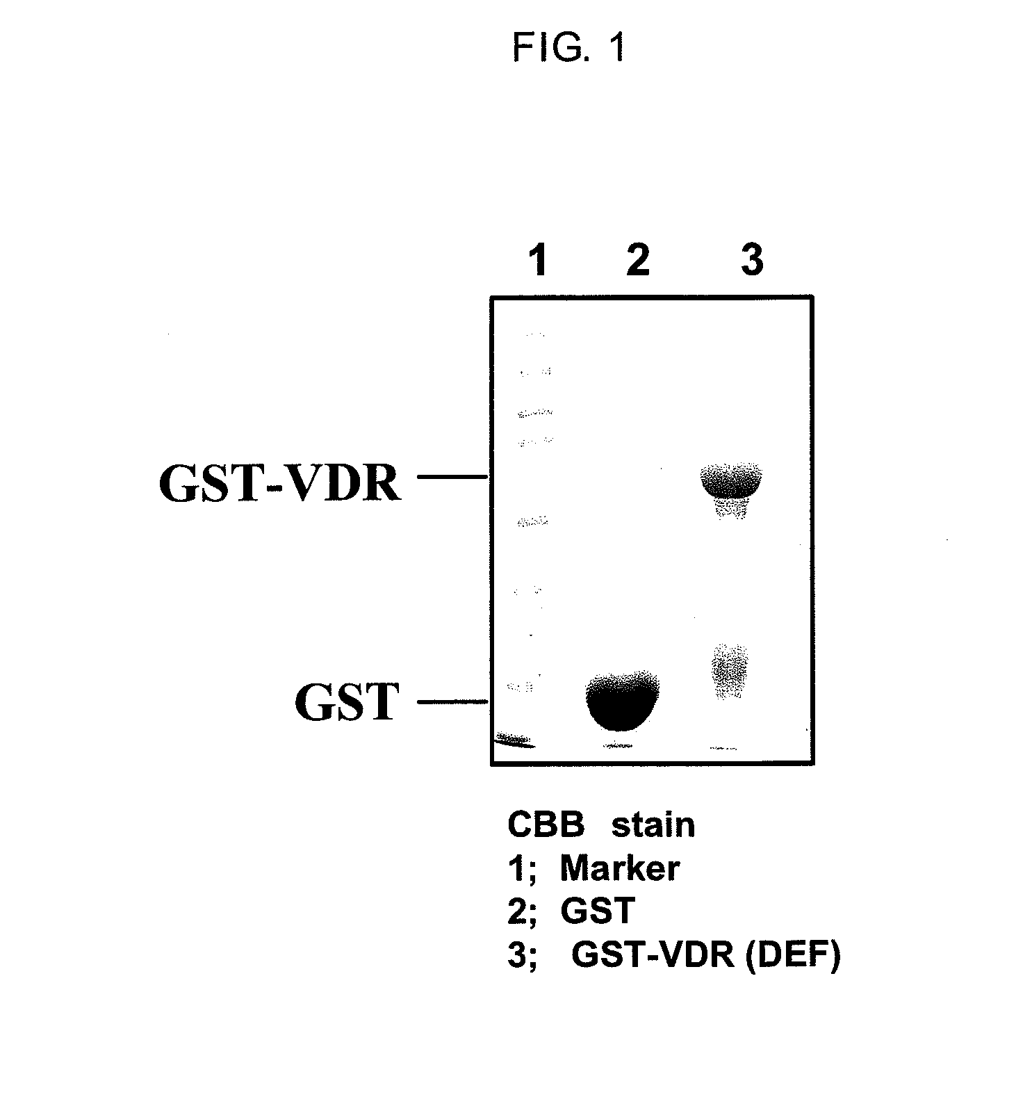
Purification of Protein Complex of Fusion Proteins Between VDR and Glutathione-S-Transferase (GST)

[0099]Firstly, GST-VDR which is a bait protein was expressed in E coli. With respect to rat VDR sequences reported by Kitagawa et al., Cell, Vol. 113, 905-917, 2003, E coli. (DH5alpha strain) transformed using either a pGEX4T-1 vector containing the ligand binding region (Amersham Biosciences) or a pGEX4T-1 vector containing DNA sequences encoding the human VDR ligand binding region (DEF region) (The structure of the VDR domain is explained in J Wesley Pike et al. The Vitamin D Receptor. (2005) Vitamin D, 2nd Edition, Feldman, Pike, Glorieux (eds.) p. 167-191) was cultured in 2 L of LB medium at 37° C., while shaking at 120 revolutions. When the absorbance at 600 nm reached 0.4-0.5, 0.1 mM isopropyl-1-thio-β-D-galactopyranoside (IPTG) was added to the culture and culturing was continued for 3 hours at 27° C. while shaking at 120 revolutions in order to induce GST-VDR protein expression....

example 2

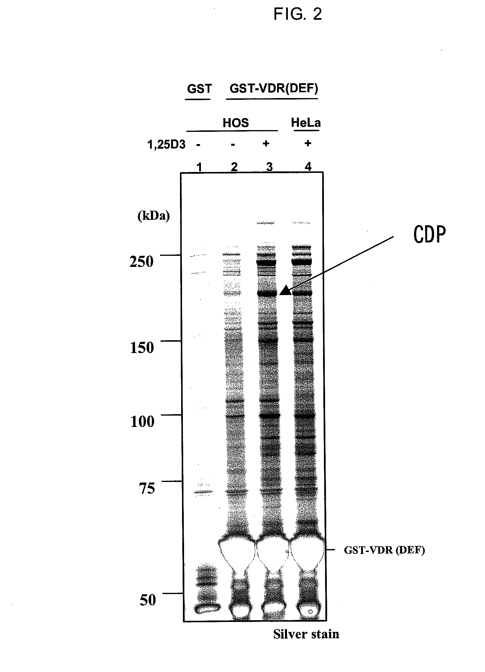
Identification and Purification of Protein Bound to GST-VDR (DEF)

[0100]In accordance with a method described in Dignam J D. et al., Nucleic Acids Research, 1983, Vol. 11, No. 5 1475-1489, a nuclear protein extract was prepared from HOS cells which is a human osteoblast cell strain (obtained from ATCC, Number CRL-1543). The resulting nuclear protein extract was dissolved in buffer D (20 mM HEPES, 10% glycerol, 0.15 M KCl, 0.2 mM EDTA, 0.5 mM PMSF, 0.01% 2-mercaptoethanol (pH=7.9). The nuclear protein extract was mixed for one hour or overnight with GST-VDR resin either in the presence or absence of 1,25(OH)2D3 which is a VDR ligand. The GST-VDR resin was washed and placed in a state in which only a specifically binding protein was bound to the GST-VDR resin. The protein bound to the resin was eluted using a reduced glutathione solution (pH=8.0) having a concentration of 15 mM on the GST-VDR resin. The eluted protein was subjected to SDS-PAGE under reducing conditions in the same mann...

example 3

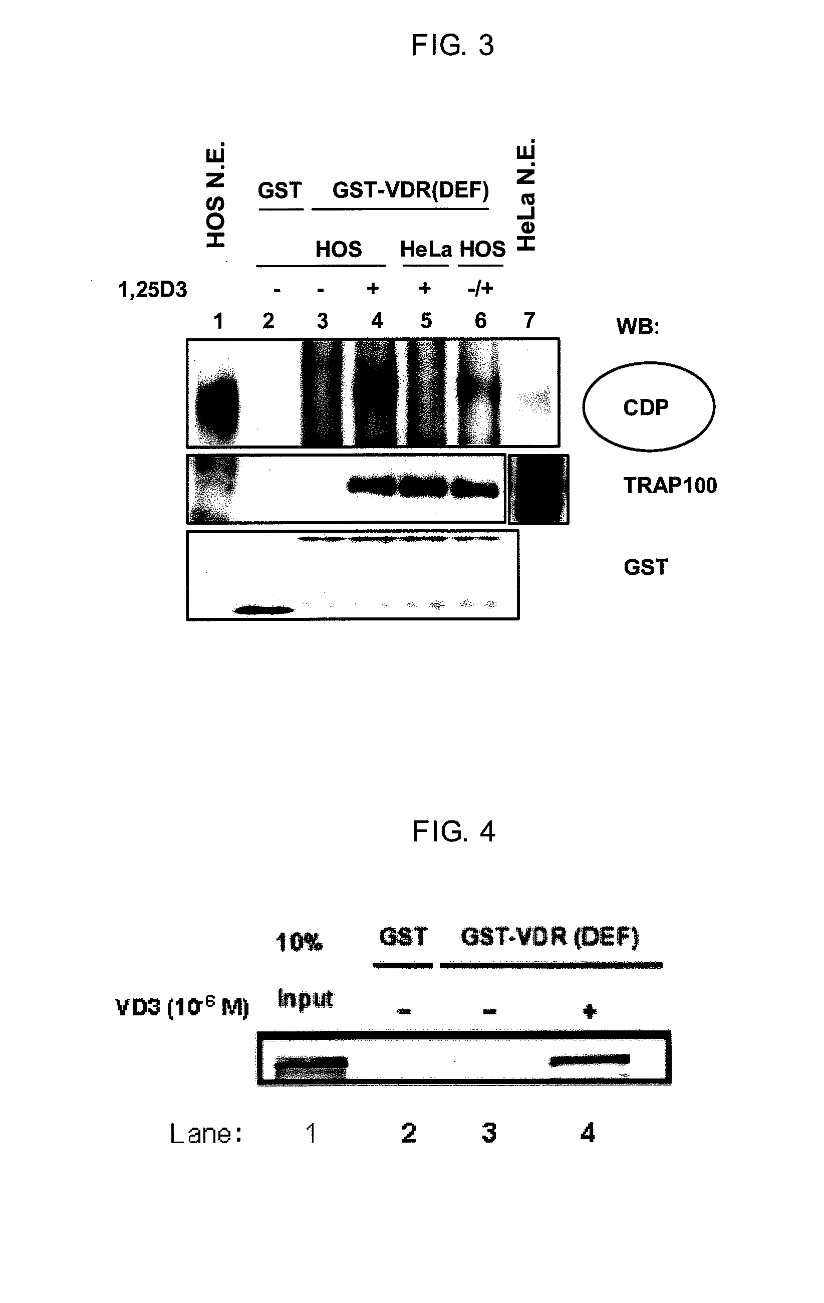
Detection of CDP by Western Blotting

[0101]A part of the eluted liquid from Example 2 was subjected to SDS-PAGE under reducing conditions, blotted to a PVDF membrane (Millipore, Product No. IPVH304F0) and the detection of CDP was performed using standard techniques. The primary antibodies were anti-CDP (M-222) rabbit polyclonal IgG (Santa Cruz Biotechnology, Product No. sc-13024). The secondary antibody was anti-rabbit IgG labeled with HRP (Amersham Biosciences, Product No. NA934V). ECL Plus (GE Healthcare Bioscience, Product No. RPN2132) was used in the detection operation. The results are shown in FIG. 3. Although a CDP band was not detected nearly at all in Lane 3 in the absence of a ligand, a CDP band was strongly detected in Lane 4 in the presence of a ligand. For comparison, a nuclear protein extract from HeLa cells which are uterine cancer-derived cells rather than osteoblasts was obtained in the same manner and mixed with GST-VDR in the presence of a ligand. The elution of th...


PUM

PropertyMeasurementUnit
excitation wavelengthsaaaaaaaaaa

Abstract

The present invention relates to a screening method for a compound and includes a step of detecting binding between a vitamin D receptor and CDP. A novel method enabling screening in vitro of a compound having strong bone forming action and low side effects, a compound participating in osteoblast differentiation and selected by the method of screening, a therapeutic agent containing the compound for diseases intervening in the increase in vitamin D-mediated transcriptional activity by a complex of a vitamin D receptor and CDP, and a kit for performing the screening method are provided.

Description

TECHNICAL FIELD[0001]The present invention relates to a novel method of screening for compounds participating in osteoblast differentiation. In particular, the present invention relates to an in vitro use of VDR (vitamin D receptor) and CDP (CCAAT Displacement Protein) to screen for the above compounds.BACKGROUND ART[0002]Active vitamin D3 derivatives such as 1α,25-dihydroxyvitamin D3 (hereafter “1,25 (OH)2D3 or 1,25D3) or 1α-hydroxy vitamin D3 (hereafter “1α (OH)D3) have extensive clinical applications as therapeutic substances for metabolic bone diseases such as osteoporosis. The physiological effect of these active vitamin D3 derivatives is known to be mediated by VDR. VDR is not only present in tissue such as the small intestine, bone, kidneys and parathyroid but is also present in various types of cells including immune system cells and tumor cells. Consequently active vitamin D3 derivatives are known to have various physiological effects including (1) calcium and bone metaboli...

Claims


Application Information

Patent Timeline
Patent Type & Authority Applications(United States)
IPC IPC(8): C12Q1/68G01N33/53
CPCC12N15/1086C12Q1/025G01N2500/00G01N33/566G01N33/82G01N33/5044Y10S435/81
Inventor OCHIAI, EIJI
Owner THE UNIV OF TOKYO
Why Eureka
Try Eureka